From the free course AP Biology on Lykke
1.6 Nucleic Acids
Key concepts: Nucleotides and nucleic acid synthesis · The 3′ hydroxyl and 5′ phosphate groups of nucleotide sugars · Determining nucleic acid sequences from overlapping fragments · Restriction endonucleases and DNA fragment generation · Determining allele frequencies using electrophoretic analysis · Alignment of the first five bases or amino acids · Gel electrophoresis of nucleic acids and amino acids · Agarose gels, electrical current, and conducting buffer · Bio-Rad modular laboratory research resources · Proteases, including bromelain and papain
A DNA strand grows in one direction: new nucleotides are added to its free $3'$ end, where a $3'$ hydroxyl group can react with the incoming nucleotide’s $5'$ phosphate.
1.6 Nucleic Acids
A DNA strand grows in one direction: new nucleotides are added to its free $3'$ end, where a $3'$ hydroxyl group can react with the incoming nucleotide’s $5'$ phosphate. That directional chemistry explains how cells build nucleic acids, how scientists read unknown sequences, and how DNA fragments become evidence about alleles.
Learning Objective 1.6: Explain the connection between the sequence and the molecular structure of nucleic acids.
Essential Knowledge: Nucleic acids have a linear sequence of nucleotides. During nucleic acid synthesis, nucleotides are added to the $3'$ end of the growing strand, forming covalent bonds between nucleotides. The detailed molecular structures of specific nucleotides are beyond the scope of the AP Exam.
From nucleotide sugars to a growing strand
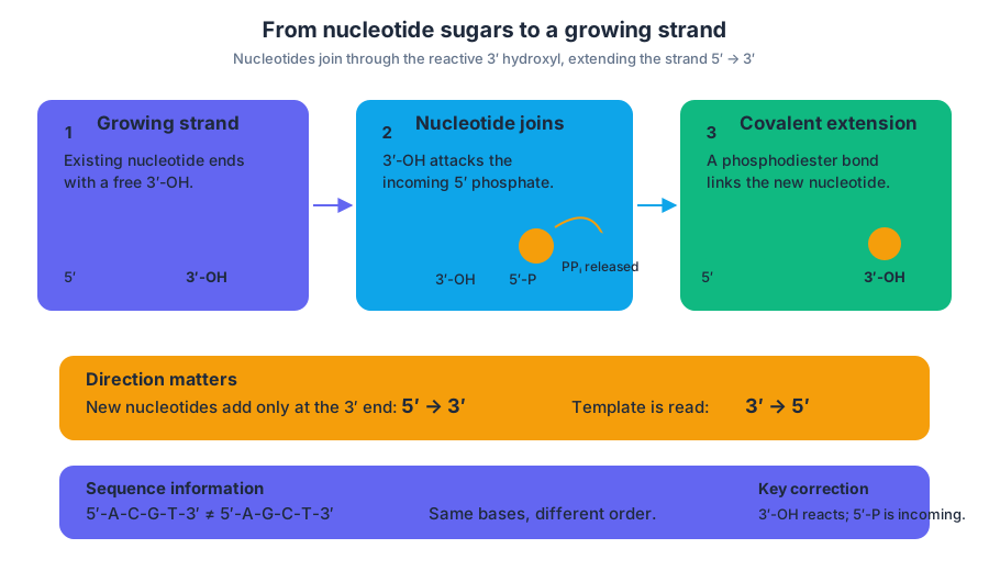
A nucleotide is the repeating subunit of a nucleic acid. In a nucleic-acid strand, each nucleotide contributes a sugar-phosphate portion to the backbone and a nitrogen-containing base that carries sequence information. The base sequence is linear: for example, $5'$-$A$-$C$-$G$-$T$-$3'$ is different from $5'$-$A$-$G$-$C$-$T$-$3'$, even though both strands contain the same kinds of bases.
The two ends of a strand are chemically different. The $3'$ end has a free hydroxyl group, written $3'$-$OH$, on the sugar; the $5'$ end is associated with a phosphate group. During synthesis, the $3'$ hydroxyl of the growing strand attacks the incoming nucleotide’s $5'$ phosphate. A covalent phosphodiester bond forms between the nucleotides, and pyrophosphate, $PP_i$, is released from the incoming nucleoside triphosphate.
The result is not random attachment. Because the new nucleotide is added to the $3'$ end, the strand is synthesized in the $5' \rightarrow 3'$ direction. Its complementary template is read in the opposite direction, $3' \rightarrow 5'$. This orientation is essential when interpreting sequence diagrams or deciding which end of a fragment can extend.
Misconception check: “The $5'$ end adds new nucleotides because it has the phosphate.”
Correction: The incoming nucleotide supplies the phosphate, but the growing strand supplies the reactive $3'$ hydroxyl. Addition occurs at the growing strand’s $3'$ end.
Reading a sequence from overlapping fragments
A long nucleic-acid sequence can be reconstructed from shorter fragments if the fragments overlap. The overlap acts like matching pieces of a biological sentence: align identical internal bases, then extend the sequence across the shared region.
Worked example. Suppose a sequencing procedure produces these fragments:
- Fragment 1: $5'$-$A C G T A$-$3'$
- Fragment 2: $5'$-$G T A C C$-$3'$
- Fragment 3: $5'$-$A C C G A$-$3'$
Fragment 1 and Fragment 2 overlap at $G T A$, giving $A C G T A C C$. Fragment 2 and Fragment 3 overlap at $A C C$, extending the reconstruction to:
$$5' - A C G T A C C G A - 3'$$
The same reasoning can be applied to the first five bases or amino acids in sequence-analysis exercises. Begin by aligning the shortest matching run, verify that the neighboring bases agree, and then report the sequence in the requested orientation. A single misplaced overlap can shift every later position, so sequence alignment is an evidence-matching task, not a guessing task.
Restriction endonucleases and DNA fragments
Restriction endonucleases are enzymes that cut DNA at particular recognition sequences. Their cuts generate fragments of different lengths. Scientists can compare those fragment patterns to create a genetic signature, map restriction sites, or distinguish DNA samples.
A restriction enzyme does not “read” the entire genome indiscriminately. It recognizes a specific DNA sequence and cleaves the phosphodiester backbone at or near that site. A nuclease is the broader category of enzyme that breaks nucleic acids; a restriction endonuclease is a more specific DNA-cutting tool used for controlled fragment generation.
Gel electrophoresis: separating fragments by movement
Gel electrophoresis separates molecules according to how rapidly they migrate through a porous gel when an electrical current is applied. Agarose, a gelatinlike material purified from seaweed, is dissolved in a current-carrying buffer and allowed to solidify around a comb, creating wells for DNA samples.
DNA carries an overall negative charge because of its phosphate groups, so DNA fragments migrate toward the positive electrode. Smaller fragments move more easily through the agarose pores and travel farther than larger fragments. Agarose gels can separate DNA fragments of roughly $200$ to $50{,}000$ base pairs.
Interpretation example: If individual A’s lane contains bands at the same positions as two different reference fragments, individual A may carry two distinguishable DNA variants. The band positions are evidence of fragment length; they are not themselves allele names unless the experiment defines which fragment corresponds to which allele.
Estimating allele frequency from electrophoretic data
Electrophoresis can reveal allele frequencies when enzymes or DNA fragments from multiple individuals produce distinguishable patterns. First classify each individual’s banding pattern as a genotype, then count allele copies—not merely the number of people showing a band.
For a diploid sample of $N$ individuals, there are $2N$ allele copies. If an allele appears $n$ times, its frequency is:
$$p=\frac{n}{2N}$$
For example, if $20$ individuals are analyzed and the target allele is counted $18$ times, then:
$$p=\frac{18}{40}=0.45$$
This calculation develops Science Practice 4: Representing and Describing Data, Science Practice 5: Statistical Tests and Data Analysis, and Science Practice 6: Argumentation. A strong conclusion identifies the observed band pattern, explains how it supports a genotype assignment, and connects the genotype counts to the allele-frequency calculation.
Retrieval check
Why does a strand synthesized from an incoming nucleotide triphosphate extend at its $3'$ end, and why does a smaller DNA fragment travel farther through agarose than a larger one? A complete answer should mention the growing strand’s $3'$ hydroxyl, the incoming nucleotide’s $5'$ phosphate, pyrophosphate release, DNA’s negative charge, and the gel’s porous structure.