From the free course AP Psychology on Lykke
1.4 The Brain
Key concepts: Brain structures and psychological disorders · Cerebral hemispheric specialization · Left and right brain hemispheres · Corpus callosum and split-brain research · Contralateral organization of the hemispheres · Broca’s area and speech production · Wernicke’s area and speech comprehension · Aphasia resulting from brain damage · Prefrontal cortex and higher-order thinking · Motor cortex and skeletal movement
A person can understand a spoken sentence yet be unable to produce fluent speech—or move one side of the body while the other side remains unaffected—because brain functions are organized across specialized but connected regions.
1.4 The Brain
A person can understand a spoken sentence yet be unable to produce fluent speech—or move one side of the body while the other side remains unaffected—because brain functions are organized across specialized but connected regions. The central question is not whether one structure “controls” a behavior by itself, but how specialized systems work together.
A map from structure to function
The frontal lobes, located behind the forehead, support planning, decision-making, impulse control, higher-order thinking, and other executive functions—mental processes used to organize behavior toward a goal. The prefrontal cortex, the frontmost part of the frontal lobes, is especially important for these higher-order processes. The motor cortex, located at the rear of the frontal lobes, controls most types of skeletal movement.
The parietal lobes process touch and help organize information about the body and surrounding space. Other structures contribute specialized functions: the thalamus relays much sensory information, the hippocampus supports memory formation, the amygdala helps process emotionally significant information, the hypothalamus helps regulate motivated biological states, the cerebellum coordinates movement, and the medulla helps regulate vital functions such as breathing and heart activity. These functions are useful predictions, not isolated “buttons” in the brain.
| Structure or region | Core function | If functioning is disrupted |
|---|---|---|
| Prefrontal cortex | Planning, judgment, inhibition, executive functioning | Impaired decision-making or self-control may occur |
| Motor cortex | Most skeletal movement | Movement on the opposite side of the body may be affected |
| Parietal lobes | Touch and spatial organization | Body or spatial processing may be disrupted |
| Hippocampus | Formation of new memories | New learning may become difficult |
| Amygdala | Processing emotionally significant information | Emotional responses may become altered |
| Cerebellum | Coordination and balance | Movement may become poorly coordinated |
| Medulla | Vital autonomic functions | Life-sustaining regulation may be threatened |
Language and aphasia
Language areas are typically located in the left hemisphere. Broca’s area supports speech production: damage can produce aphasia, a language impairment in which a person may struggle to produce speech. Wernicke’s area supports speech comprehension: damage can impair the ability to understand spoken language. Aphasia does not mean that intelligence has disappeared; it means that particular language processes have been disrupted.
Worked example: Suppose a patient speaks slowly, uses very few words, and appears to know what they want to say but cannot produce fluent sentences. The strongest application is damage involving Broca’s area and speech production. If the patient instead speaks fluently but the words lack meaning and they cannot understand questions, Wernicke’s area and speech comprehension are more relevant. A real clinical evaluation would require evidence beyond a short description.
Hemispheres, corpus callosum, and contralateral organization
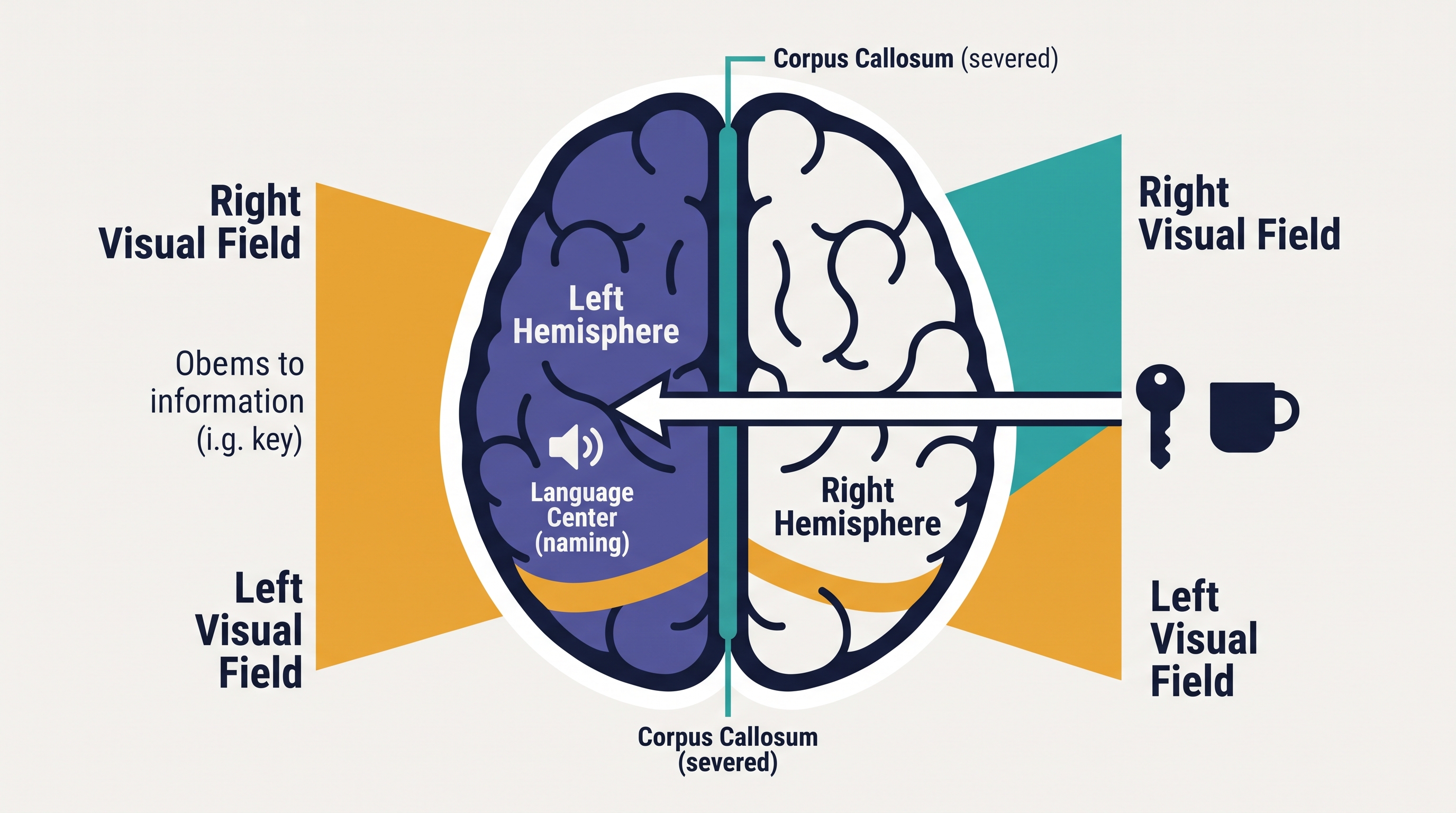
The two cerebral hemispheres may specialize in different activities, but specialization is relative, not absolute. The corpus callosum is a bundle of nerve fibers connecting the hemispheres and allowing information to be shared between them. In split-brain research, surgeons severed the corpus callosum, sometimes as a treatment for severe epilepsy, so researchers could study what happens when communication between hemispheres is greatly reduced.
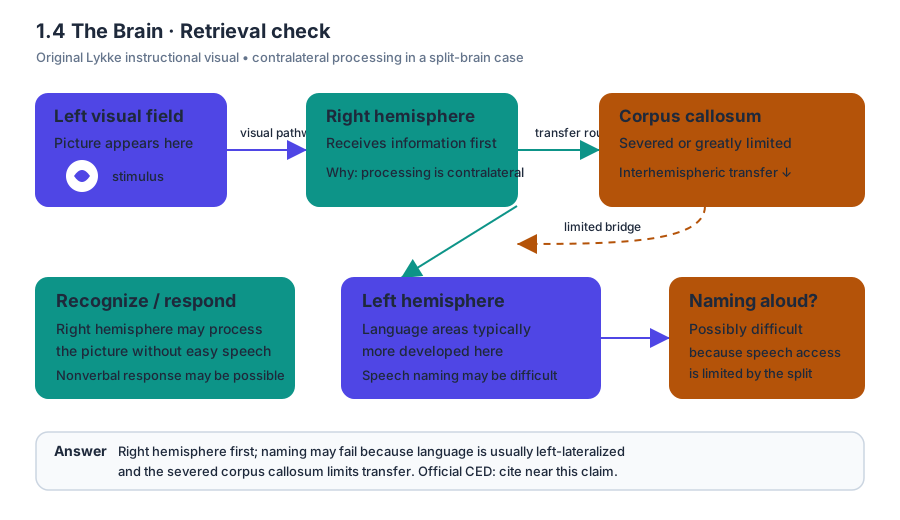
The hemispheres have contralateral organization: each hemisphere primarily processes information from and controls movement on the opposite side of the body. Information presented in the left visual field is initially processed by the right hemisphere; information presented in the right visual field is initially processed by the left hemisphere. Researchers can therefore present an image briefly to one visual field and observe what the person can name, draw, or select.
Worked split-brain example: If a word appears only in the right visual field, it is initially sent to the left hemisphere, where language areas are typically located. The person may be able to say the word aloud. If the same word appears only in the left visual field, it is initially sent to the right hemisphere; a split-brain patient may have difficulty saying it aloud but may still identify or select the object with the left hand. The result supports hemispheric specialization and contralateral organization—it does not prove that one hemisphere works independently in everyday life.
Plasticity, research, and psychological disorders
Brain plasticity is the brain’s ability to rewire itself, modify existing connections, or create new connections throughout development. Plasticity can allow another region to assume some functions after damage, although recovery depends on factors such as the location, extent, and timing of the damage.
Researchers use EEG, which records electrical activity at the scalp; fMRI, which estimates changes in blood oxygen associated with neural activity; case studies; and surgical procedures such as lesioning. Each method answers different questions: a scan can reveal activity or structure, while a lesion or carefully documented case can provide stronger evidence about what changes when a region is damaged. None automatically proves that one location alone causes a complex behavior.
Brain structures and psychological disorders can be connected through patterns of association. For example, differences in prefrontal, amygdala, hippocampal, or broader neural-network functioning have been associated with some anxiety, mood, trauma-related, and cognitive disorders. However, an association does not establish that a single structure caused the disorder: biology, learning, environment, and social context can interact, and the same brain finding may appear across different conditions.
Misconception check: “People are either left-brained or right-brained.” Incorrect. The hemispheres show specialization, especially for some language and perceptual functions, but normal behavior depends on communication across both hemispheres. Contralateral organization and split-brain findings reveal useful processing differences—not two separate personality types.
Retrieval check
A researcher presents a picture only to a split-brain patient’s left visual field. Which hemisphere initially receives the information, and why might the patient have difficulty naming it aloud? Answer: The right hemisphere initially receives it because visual processing is contralateral. Naming may be difficult because language areas are typically more developed in the left hemisphere, and severing the corpus callosum limits transfer to that hemisphere. This applies 1.A by using a psychological concept in an unfamiliar scenario and 2.A by interpreting what a research procedure can establish.
Source Materials
- Ap Psychology Course And Exam Description
- Ap Psychology Course At A Glance
- AP Psychology CED: Student Responses and Scoring Commentary
- AP Psychology Exam Questions
- Free-Response Questions
- Free-Response Questions - Set 1
- Free-Response Questions - Set 2
- exhibits. One of the interactive exhibits featured glass marbles. He grabbed a large marble and tried to bounce it on the ground. At one point during the visit, he became separated from his parents, a