From the free course Anatomy and Physiology on Lykke
Chapter 28: Development and Inheritance
Key concepts: Timing and location of fertilization · Oocyte survival after ovulation · Sperm capacitation · Acrosome reaction and penetration of the oocyte coverings · Block to polyspermy · Formation of the zygote · Monozygotic (identical) twinning · In vitro fertilization (IVF) · Oocyte retrieval and sperm preparation for IVF · Embryo transfer to the uterus
By the end of this chapter the student can: List and explain the steps involved in fertilization; Describe the major events in embryonic development; Describe the major events in fetal development; Discuss the adaptations of the body to pregnancy; Describe the physiologic adjustments that the newborn must make in the first hours of extrauterine life; Summarize the physiology of lactation; Classify and describe the different patterns of inheritance.
Chapter 28: Development and Inheritance
A single fertilized egg can become an infant with trillions of specialized cells in roughly nine months. That transformation depends on coordinated fertilization, cell division, tissue differentiation, organ formation, fetal growth, birth, postnatal adaptation, lactation, and the expression of inherited genetic information. Development is shaped not only by the genes received from the egg and sperm but also by the environment in which those genes are expressed.
The chapter follows development in time: sperm and oocyte meet during fertilization; the resulting zygote becomes an embryo; the embryo becomes a fetus; pregnancy culminates in labor and birth; and the newborn rapidly adjusts to life outside the uterus. The final section explains how genetic information is transmitted and how inheritance patterns produce variation among individuals.
28.1 Fertilization
Fertilization is not simply the moment when a sperm touches an egg. It is a sequence of tightly controlled events that brings together two haploid gametes, restores the diploid chromosome number, activates development, and prevents additional sperm from entering the oocyte. The process normally occurs in the ampulla, the expanded portion of the uterine tube closest to the ovary.
Location and timing
At ovulation, a secondary oocyte leaves the ovary and is swept into the uterine tube by the fimbriae and currents produced by cilia. The oocyte remains capable of being fertilized for approximately $12$ to $24$ hours after ovulation. Sperm, by contrast, may remain viable in the female reproductive tract for several days, although their ability to fertilize depends on successful physiological changes.
For fertilization to occur, viable sperm must reach the oocyte while it is still in the uterine tube. If the oocyte is not fertilized, it degenerates and is eventually eliminated. A common misconception is that fertilization normally occurs in the uterus; in reality, the uterus is the site where the early embryo implants, whereas fertilization usually occurs in the uterine tube.
Sperm transport and capacitation
Sperm travel from the vagina through the cervix and uterus into a uterine tube. Their movement depends partly on the beating of their flagella and partly on muscular contractions and fluid currents in the female reproductive tract. Many sperm never reach the oocyte, and the oocyte is surrounded by layers that create additional barriers.
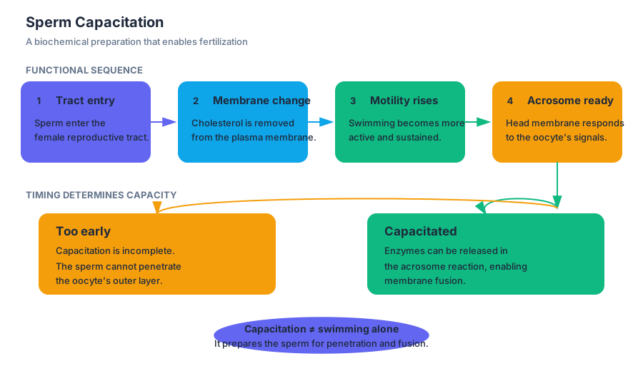
Before a sperm can fertilize an oocyte, it must undergo capacitation, a series of functional changes that occur in the female reproductive tract. Capacitation removes or alters molecules acquired during sperm maturation and changes the sperm plasma membrane. These changes increase sperm motility and prepare the sperm to release enzymes from its acrosome.
Capacitation is the final maturation process that gives sperm the capacity to penetrate and fertilize an oocyte.
If sperm reach the oocyte before capacitation is complete, they cannot penetrate the oocyte’s thick outer covering. Capacitation therefore acts as a physiological timing mechanism: sperm must survive long enough to reach the oocyte, but they must also become functionally competent at the correct place and time.
Penetration of the oocyte coverings
The oocyte is surrounded by the corona radiata, a layer of follicular cells, and the zona pellucida, a glycoprotein-rich extracellular layer. A sperm must move through the corona radiata, bind to the zona pellucida, and ultimately fuse with the oocyte plasma membrane.
The sperm head contains the acrosome, a cap-like structure filled with hydrolytic enzymes. When capacitated sperm bind to the zona pellucida, they undergo the acrosome reaction. The acrosomal membrane changes and releases enzymes that help digest a path through the zona pellucida. This is not uncontrolled destruction: sperm-oocyte recognition molecules help ensure that the sperm binds appropriately before enzyme release.
The sequence can be summarized as follows:
- Sperm are transported through the female reproductive tract.
- Sperm undergo capacitation.
- A capacitated sperm contacts the corona radiata.
- The sperm binds to the zona pellucida.
- The acrosome reaction releases enzymes.
- The sperm penetrates the zona pellucida.
- The sperm and oocyte plasma membranes fuse.
Only the sperm nucleus and certain other components enter the oocyte. The sperm flagellum and other structures are generally broken down. Fusion activates the oocyte and initiates the mechanisms that prevent polyspermy.
The block to polyspermy
Polyspermy is the penetration of an oocyte by more than one sperm. It must be prevented because the union of one haploid oocyte nucleus with two haploid sperm nuclei would produce a cell with three sets of chromosomes, or a triploid cell. Triploidy is incompatible with normal human development.
The oocyte uses two related mechanisms:
- The fast block occurs almost immediately after the first sperm fuses with the oocyte. Sodium ion permeability changes, causing depolarization of the oocyte plasma membrane. This electrical change prevents additional sperm from fusing with the membrane.
- The slow block, also called the cortical reaction, begins when the first sperm activates the oocyte. Secretory vesicles called cortical granules release their contents beneath the zona pellucida. The zona pellucida changes chemically and becomes impenetrable to other sperm.
The fast block acts within moments, while the cortical reaction provides a more durable barrier. Together, they ensure that one oocyte is normally fertilized by one sperm.
Formation of the zygote
After sperm entry, the secondary oocyte completes meiosis II, producing the mature ovum and a second polar body. The genetic material from the sperm forms the male pronucleus, and the genetic material from the oocyte forms the female pronucleus. The pronuclei move toward each other, replicate their DNA, and prepare to combine.
The resulting cell is the zygote, a diploid cell with a complete set of chromosomes. The zygote begins cleavage, a series of rapid mitotic divisions. The daughter cells, called blastomeres, become progressively smaller because the overall size of the zona-enclosed conceptus does not initially increase.
A zygote can sometimes separate and form monozygotic, or identical, twins. Separation can occur as early as the two-cell stage, although it most commonly occurs during the early blastocyst stage, when approximately $70$ to $100$ cells are present. Separation at the two-cell stage produces embryos with individual placentas, whereas later separation can result in twins sharing a placenta and chorionic cavity.
Clinical connection: in vitro fertilization
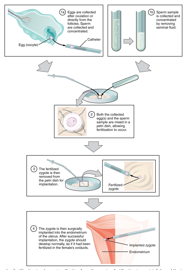
In vitro fertilization, or IVF, is an assisted reproductive technology in which fertilization occurs outside the body. It can help when uterine tubes are blocked, sperm count or motility is low, sperm morphology is abnormal, or sperm cannot penetrate the zona pellucida. IVF is also used with donor gametes, gestational carriers, and other reproductive arrangements.
A typical IVF cycle uses gonadotropins to stimulate the development of multiple follicles rather than the single dominant follicle of an ordinary ovulatory cycle. Follicle-stimulating hormone analogs support follicular development, and a luteinizing hormone analog triggers ovulation. Approximately $10$ to $20$ oocytes may be collected, fertilized in a laboratory, and monitored before an embryo is transferred to the uterus.
For example, a person with healthy oocytes but blocked uterine tubes may produce oocytes normally while preventing sperm and oocyte from meeting naturally. IVF bypasses the blocked tubes by collecting the oocytes, combining them with sperm outside the body, and transferring an early embryo into the uterus.
28.2 Embryonic Development
A fertilized egg does not grow by simply becoming a larger version of itself. It divides, rearranges its cells, establishes body axes, and differentiates into tissues with distinct functions. Embryonic development refers especially to the first eight weeks after fertilization, when the basic body plan and organ systems are established.
Cleavage and implantation
During the first several days, the zygote undergoes cleavage as it travels through the uterine tube toward the uterus. The solid ball of cells is called a morula. Fluid then enters the morula, creating a cavity and producing a blastocyst.
The blastocyst has two major cell populations:
- The outer trophoblast contributes to the embryonic portion of the placenta and helps the conceptus attach to the uterine lining.
- The inner cell mass, or embryoblast, becomes the embryo itself.
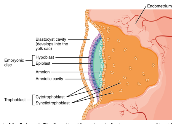
Around the end of the first week, the blastocyst begins implantation in the endometrium. Trophoblast cells invade the uterine lining and differentiate into the cytotrophoblast and the multinucleated syncytiotrophoblast. The syncytiotrophoblast secretes human chorionic gonadotropin, or hCG, which maintains the corpus luteum early in pregnancy. The corpus luteum continues secreting progesterone, preserving the endometrium.
Implantation is the attachment and embedding of the blastocyst into the uterine endometrium.
Germ layers and gastrulation
During the second week, the embryoblast forms a bilaminar embryonic disc composed of the epiblast and hypoblast. Cavities and membranes develop around this disc, including the amniotic cavity and the yolk sac.
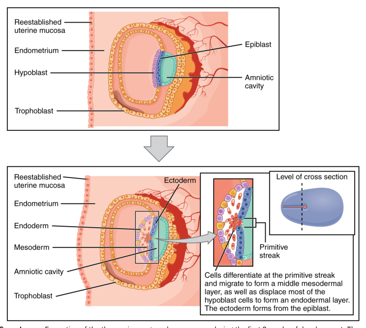
During the third week, gastrulation transforms the bilaminar disc into a trilaminar disc. Cells migrate through the primitive streak and form three germ layers:
- Ectoderm forms the nervous system and epidermis.
- Mesoderm forms connective tissues, muscle, bone, blood, the cardiovascular system, and many urogenital structures.
- Endoderm forms the epithelial lining of the digestive and respiratory tracts and contributes to organs such as the liver and pancreas.
Gastrulation is crucial because it establishes the fundamental cellular sources of the body’s tissues. For example, the spinal cord develops from ectoderm, while vertebrae develop from mesoderm. The tissues lie next to each other in the adult body, but they originate from different germ layers.
Neurulation and body folding
The ectoderm overlying the notochord thickens to form the neural plate. The neural plate bends, creating neural folds and a neural groove. The folds fuse to form the neural tube, which becomes the brain and spinal cord. Cells that detach from the neural folds form the neural crest, which contributes to peripheral nerves, pigment cells, facial structures, and other tissues.
At the same time, the flat embryonic disc folds laterally and longitudinally. This folding turns the disc into a more three-dimensional body form and incorporates part of the yolk sac into the primitive gut. The embryo becomes attached to the developing placenta by the connecting stalk, which develops into the umbilical cord.
Extraembryonic membranes and placenta
Four important extraembryonic membranes support the embryo:
- The amnion encloses the embryo in amniotic fluid, which cushions against physical trauma, permits movement, and helps maintain a stable environment.
- The chorion forms the outer membrane and contributes to the fetal portion of the placenta.
- The yolk sac contributes to early blood formation and supplies early germ cells.
- The allantois contributes to umbilical blood vessels and waste-related structures.
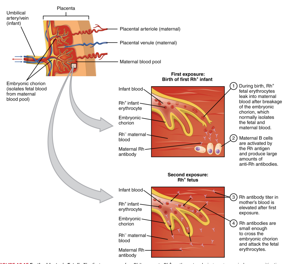
The placenta is a temporary organ formed from fetal and maternal tissues. It permits exchange of oxygen, nutrients, wastes, and hormones between maternal and fetal circulations without normally allowing the two blood supplies to mix directly. The placenta also produces hormones, including hCG, progesterone, estrogens, and human placental lactogen.
The umbilical cord usually contains two umbilical arteries and one umbilical vein. The umbilical arteries carry relatively deoxygenated blood from the fetus toward the placenta, while the umbilical vein carries relatively oxygenated blood toward the fetus.
Organogenesis
During organogenesis, the germ layers fold, interact, and differentiate to produce organ systems. The heart begins forming early and starts beating during the embryonic period. Limb buds appear, the face develops, and the neural tube becomes more specialized. By the end of the eighth week, the embryo has recognizable human features, although the organs are still immature.
The embryonic period is especially vulnerable to teratogens, environmental agents that can disrupt development. Examples include certain drugs, alcohol, infections, radiation, and nutritional deficiencies. The effect depends on the dose, timing, and tissue exposed. A harmful exposure during organ formation can produce structural abnormalities, whereas exposure later may impair growth or functional maturation.
For example, inadequate folate during early development increases the risk of neural tube defects. Folate supports nucleic acid synthesis and cell division, processes required as the neural tube forms and closes.
28.3 Fetal Development
At the beginning of the ninth week after fertilization, the developing human is called a fetus rather than an embryo. The embryonic period established the body plan; the fetal period emphasizes growth, refinement, movement, and functional maturation of organs.
Growth from the ninth week to birth
During the fetal period, the head remains proportionally large at first, but the rest of the body catches up as growth continues. The face becomes more defined, the limbs lengthen, bones begin to ossify, and muscles become capable of producing movement. Although fetal movements may occur earlier, they are often first perceived by the pregnant person during the second trimester.
The fetus receives nutrients and oxygen through the placenta and returns carbon dioxide and metabolic wastes to the maternal circulation. The placenta is not an absolute barrier: some drugs, alcohol, pathogens, and antibodies can cross it. Maternal immunoglobulin $G$ provides passive immune protection to the fetus and newborn, while certain harmful substances can interfere with development.
By the end of the first trimester, most major organ systems are present in a basic form. During the second trimester, the fetus grows rapidly, the skeleton becomes more substantial, hair and nails develop, and the fetus becomes increasingly active. During the third trimester, the fetus gains substantial mass, accumulates fat, and undergoes maturation of the nervous, respiratory, digestive, and immune systems.
Fetal circulation
Fetal circulation is adapted to an environment in which the lungs are not yet exchanging gases with air. Oxygenated blood comes from the placenta through the umbilical vein. Much of this blood bypasses the fetal liver through the ductus venosus and enters the inferior vena cava.
Three fetal shunts redirect blood away from organs that are not yet functioning in their postnatal roles:
- The ductus venosus bypasses much of the liver.
- The foramen ovale allows blood to pass from the right atrium to the left atrium.
- The ductus arteriosus allows blood to pass from the pulmonary trunk into the aorta.
These shunts are necessary because the placenta, not the fetal lungs, oxygenates blood. After birth, lung expansion lowers pulmonary resistance, pressure changes close the foramen ovale, and increased oxygen concentration promotes closure of the ductus arteriosus. The fetal vessels and shunts later become anatomical remnants.
Maturation of organ systems
The fetal lungs produce increasing amounts of surfactant, a substance that reduces surface tension inside the alveoli. Adequate surfactant helps prevent alveolar collapse during expiration. Premature birth can leave the lungs with insufficient surfactant, making breathing difficult.
The fetal liver stores glycogen and produces blood cells during much of development. Bone marrow gradually assumes the major role of hematopoiesis. The kidneys produce urine that contributes to amniotic fluid, although the placenta remains the primary route for removing fetal metabolic wastes.
The fetal nervous system continues maturing throughout pregnancy and after birth. Neural connections, reflexes, sensory responses, and sleep-wake patterns develop progressively. The timing of these changes matters clinically because survival and long-term function depend on the maturity of several systems, not simply on body size.
For example, a fetus born very prematurely may have a functioning heart and kidneys but still face respiratory difficulty because the lungs have not produced enough surfactant and the respiratory control centers are immature.
28.4 Changes During Pregnancy, Labor, and Birth
Pregnancy transforms nearly every major body system. The uterus enlarges from a small pelvic organ into an abdominal organ, blood volume rises, the heart works harder, and the respiratory and digestive systems adapt to the growing fetus. These changes support fetal nutrition and prepare the body for labor and lactation.
Maternal changes during pregnancy
Early pregnancy hormones maintain the endometrium and suppress uterine contractions. Progesterone helps maintain the uterine lining and reduces uterine smooth muscle activity. Estrogens stimulate uterine growth, breast development, and increased blood flow. Human placental lactogen alters maternal metabolism so that more glucose remains available to the fetus while maternal tissues use more fatty acids.
The cardiovascular system responds with increased blood volume and cardiac output. Plasma volume rises substantially, while the increase in red blood cell mass may be proportionally smaller. This can produce a physiological dilution of blood sometimes called physiological anemia of pregnancy. The growing uterus can compress pelvic veins, contributing to lower-extremity edema and varicose veins.
Respiratory changes include increased tidal volume and minute ventilation. The enlarging uterus elevates the diaphragm, but hormonal effects and increased oxygen demand help maintain adequate gas exchange. Digestive changes can include nausea, vomiting, reflux, constipation, and slowed gastrointestinal motility.
The breasts enlarge as ducts and secretory tissue develop. The areolae darken, and the breasts may begin producing colostrum, the first secretion of the mammary glands. Colostrum is rich in protein and immunoglobulins and prepares the infant for feeding before mature milk production becomes established.
Initiation of labor
Labor is the process by which the fetus and placenta leave the uterus. Its exact initiation involves interactions among fetal signals, placental hormones, maternal hormones, and mechanical stretching of the uterus. Near term, increasing estrogenic effects and local prostaglandin activity promote uterine excitability and cervical changes.
The hormone oxytocin stimulates uterine contractions. Cervical stretching caused by the descending fetus increases sensory signals to the hypothalamus, which promotes oxytocin release from the posterior pituitary. Oxytocin intensifies contractions, causing more cervical stretching. This is a positive-feedback loop.
Cervical stretching $\rightarrow$ oxytocin release $\rightarrow$ stronger uterine contractions $\rightarrow$ greater cervical stretching.
Stages of labor
Labor is commonly divided into three stages:
- Dilation begins with regular contractions and ends when the cervix is fully dilated. The cervix becomes thinner and opens sufficiently for the fetus to pass.
- Expulsion begins with complete cervical dilation and ends with delivery of the infant. Maternal abdominal muscles may contract along with the uterus.
- Placental or afterbirth begins after delivery of the infant and ends when the placenta is expelled.
During labor, the fetus typically rotates and changes position as it passes through the birth canal. The head usually presents first because this orientation offers the smallest effective diameter for passage. After birth, continued uterine contraction helps compress blood vessels at the placental attachment site and limits maternal bleeding.
For example, when the fetal head presses against the cervix, the resulting stretch strengthens oxytocin release and contractions. The same feedback mechanism that begins as a subtle hormonal response can produce the powerful coordinated contractions needed for delivery.
28.5 Adjustments of the Infant at Birth and Postnatal Stages
Birth abruptly changes the infant’s physical environment. The newborn leaves a warm, fluid-filled uterus and must breathe air, regulate body temperature, obtain nutrients through the mouth and digestive tract, and circulate blood through lungs that were previously bypassed.
Respiratory adjustment
The first breath requires expansion of the lungs and establishment of a pressure gradient that draws air into the alveoli. Fluid in the fetal airways is displaced or absorbed, and surfactant helps keep the alveoli open. Sensory stimulation, cooling of the skin, and changes in blood gases contribute to activation of the respiratory centers.
The newborn’s first effective breaths lower pulmonary vascular resistance. More blood flows through the pulmonary arteries, where it becomes oxygenated. Increased left atrial pressure functionally closes the foramen ovale, while increased oxygen concentration and reduced prostaglandin influence promote closure of the ductus arteriosus. The ductus venosus also closes after umbilical blood flow stops.
Cardiovascular adjustment
Clamping the umbilical cord removes the low-resistance placental circulation. Systemic vascular resistance rises, while expansion of the lungs lowers resistance in the pulmonary circulation. These pressure changes redirect blood through the lungs and away from the fetal shunts.
The newborn must also maintain adequate blood glucose. The continuous maternal supply of glucose ends when the umbilical cord is clamped, so hepatic glycogen breakdown and feeding become important. Infants with inadequate energy stores or difficulty feeding are at risk for hypoglycemia.
Temperature regulation and feeding
The newborn loses heat through evaporation, conduction, convection, and radiation. A large surface-area-to-mass ratio, thin skin, and limited insulation increase this risk. Nonshivering thermogenesis in brown adipose tissue helps produce heat, but drying, skin-to-skin contact, warm surroundings, and appropriate clothing are also important.
The digestive system must coordinate sucking, swallowing, and breathing. Newborn reflexes support feeding: rooting turns the head toward stimulation near the mouth, and sucking draws milk into the mouth. The gastrointestinal tract then absorbs nutrients and water while the kidneys gradually assume independent regulation of fluid and electrolyte balance.
Postnatal developmental stages
The neonatal period comprises approximately the first month after birth. Infancy follows and includes rapid growth, maturation of the nervous system, and increasing motor and sensory abilities. Childhood and adolescence bring continued body growth, cognitive development, sexual maturation, and reproductive capability.
Development depends on both inherited potential and environmental conditions. Nutrition, infection, toxic exposure, caregiving, sleep, and opportunities for movement can influence growth and function. Development is not a perfectly uniform schedule; individuals differ in timing while still following recognizable biological sequences.
For example, a newborn who begins breathing effectively must still complete several separate adjustments: pulmonary blood flow must increase, fetal shunts must close, body temperature must be protected, glucose must be maintained, and feeding must become coordinated. A normal birth therefore represents the beginning of several physiological transitions rather than the end of development.
28.6 Lactation
Lactation is the production and secretion of milk by the mammary glands. It supplies energy, water, nutrients, immune factors, and signaling molecules during early postnatal life. Lactation depends on mammary development during pregnancy and on hormonal and neural reflexes after birth.
Mammary preparation
During pregnancy, estrogens stimulate growth of the mammary ducts, while progesterone promotes development of the secretory portions of the glands. Prolactin supports milk synthesis, but high levels of estrogen and progesterone during pregnancy inhibit full milk secretion. After the placenta is delivered, these steroid hormone levels fall sharply, allowing prolactin to act more effectively.
The first secretion is colostrum, a thick, yellowish fluid produced during the first days after birth. Colostrum contains less fat and lactose than mature milk but is especially rich in protein and antibodies. Its immunoglobulin concentration is approximately $19\ \mathrm{g/L}$ compared with about $0.1\ \mathrm{g/L}$ in mature milk. Colostrum therefore provides concentrated immune protection while the newborn’s own immune system is still developing.
Milk production and milk ejection
The hormone prolactin, released by the anterior pituitary, stimulates milk synthesis by mammary secretory cells. Suckling activates sensory receptors in the nipple and areola. Nerve signals reach the hypothalamus, which reduces inhibitory dopamine signaling and permits prolactin release.
Suckling also stimulates the posterior pituitary to release oxytocin. Oxytocin contracts myoepithelial cells surrounding the alveoli and ducts, forcing milk toward the nipple. This is the milk ejection reflex, or let-down reflex.
The process occurs step by step:
- The infant suckles.
- Sensory receptors in the nipple are stimulated.
- Hypothalamic signals increase prolactin and oxytocin release.
- Prolactin stimulates production of additional milk.
- Oxytocin contracts myoepithelial cells.
- Milk is ejected into the ducts and becomes available to the infant.
Milk production follows a supply-and-demand pattern. Frequent and effective removal of milk promotes continued production, whereas prolonged milk accumulation suppresses secretion. Psychological stress can interfere with oxytocin-mediated ejection even when milk synthesis continues.
Composition and functions of milk
As lactation progresses, mature milk increases the relative contribution of lactose and fat to meet the infant’s energy needs. It also contains proteins, water, vitamins, minerals, enzymes, hormones, and immune factors. Human milk includes antibodies such as secretory immunoglobulin $A$, which helps protect mucosal surfaces in the infant’s digestive tract.
Human milk is adapted to human infant physiology. Cow’s milk has a different balance of proteins and minerals and is not an appropriate sole food for young infants. Breast milk also changes during a feeding: early milk is relatively more watery, while later milk generally contains more fat and provides greater energy density.
For example, colostrum’s high immunoglobulin content helps protect the newborn intestine during the first days, while mature milk supplies increased lactose and fat as the infant’s energy demands and feeding capacity grow.
28.7 Patterns of Inheritance
Inheritance explains how biological information passes from parents to offspring, but genes do not act in isolation. A person’s genotype is the genetic composition carried in the DNA, whereas the phenotype is the observable result of genotype, development, and environmental influence.
Genes, alleles, and chromosomes
A gene is a segment of DNA that contributes to a functional product or regulates a biological process. Different versions of the same gene are called alleles. Humans normally have pairs of homologous chromosomes, one inherited from each parent. The two alleles at a locus may be identical, making the individual homozygous, or different, making the individual heterozygous.
A dominant allele is expressed in the phenotype when present in one or two copies, whereas a recessive allele is generally expressed only when two copies are present. Dominant does not mean stronger, healthier, or more common, and recessive does not mean weak. These terms describe how alleles interact in determining a particular phenotype.
Mendelian inheritance
In autosomal dominant inheritance, one copy of a dominant allele can produce the trait in either sex. An affected heterozygous parent has a probability of approximately $\frac{1}{2}$ of passing the allele to each child, assuming the other parent does not carry it.
In autosomal recessive inheritance, two recessive alleles are required for the phenotype. Two unaffected heterozygous carriers can have an affected child. Their expected outcomes are approximately $\frac{1}{4}$ affected, $\frac{1}{2}$ unaffected carriers, and $\frac{1}{4}$ homozygous unaffected, although actual families may not match these proportions because probability applies across many conceptions rather than guaranteeing a particular result in a small family.
A concrete example is cystic fibrosis, an autosomal recessive disorder. If both parents are heterozygous carriers, each child independently has a $\frac{1}{4}$ probability of inheriting two disease-associated alleles.
Sex-linked inheritance
Sex-linked inheritance involves genes located on the sex chromosomes. Because males typically have one $X$ chromosome and one $Y$ chromosome, a recessive allele on the $X$ chromosome is more likely to be expressed in males. Females typically have two $X$ chromosomes and may be homozygous or heterozygous for an $X$-linked allele.
An $X$-linked recessive trait is not passed from father to son because fathers give sons a $Y$ chromosome. A father passes his $X$ chromosome to all daughters. A carrier mother may pass an $X$-linked recessive allele to sons, who can express the trait if they receive no corresponding normal allele on another $X$ chromosome.
For example, red-green color vision deficiency commonly follows an $X$-linked recessive pattern. A son who inherits the associated allele from his mother can express the condition because he has only one $X$ chromosome.
Non-Mendelian patterns
Some traits do not fit simple dominant-recessive categories. In incomplete dominance, the heterozygote has an intermediate phenotype. In codominance, both alleles are expressed distinctly. Human blood group $AB$ is an example of codominance because the $I^A$ and $I^B$ alleles are both expressed.
Multiple alleles occur when more than two allele forms exist in a population, even though each person still carries only two alleles at a locus. The ABO blood group system has the alleles $I^A$, $I^B$, and $i$. The $I^A$ and $I^B$ alleles are codominant, while $i$ is recessive to either.
Polygenic inheritance occurs when several genes contribute to one trait. Height and skin pigmentation illustrate polygenic patterns, often producing continuous variation rather than a few sharply separated categories. Environmental influences can further modify the phenotype.
Pleiotropy occurs when one gene influences multiple traits. A mutation in one gene may therefore affect several organ systems. Epistasis occurs when one gene influences or masks the expression of another gene. These interactions explain why phenotype cannot always be predicted by examining one allele in isolation.
Mitochondrial inheritance and genetic variation
Mitochondria contain their own DNA, and mitochondrial genes are usually inherited from the mother because the oocyte contributes most of the cytoplasm to the zygote. Mitochondrial disorders can affect tissues with high energy demands, such as muscle and nervous tissue.
Genetic variation arises through mutation, independent assortment of chromosomes, crossing over during meiosis, and random fertilization. These processes generate combinations of alleles that differ among siblings. Environmental factors then influence how genetic potential is expressed.
A final example is phenylketonuria, or PKU, in which an inherited enzyme deficiency disrupts phenylalanine metabolism. Early dietary treatment can prevent severe neurological damage. The genotype creates a risk, but environmental intervention changes the phenotype, demonstrating why inheritance and development must be considered together.
Development and inheritance are therefore inseparable themes. Genes provide instructions and regulatory possibilities, while fertilization, cellular interactions, hormones, nutrition, placental function, and postnatal conditions determine how those possibilities unfold.
Content adapted from OpenStax Anatomy and Physiology 2e by J. Gordon Betts et al., licensed CC BY 4.0.

Source Materials
Study Anatomy and Physiology on Lykke
This course page is free to open: signed out, you can read its first two sections, and signing in, on any plan, opens the rest. The free Lykke Chrome extension syncs your own Canvas courses, so ChatGPT, Claude and Muse can answer from your classes and deadlines. It only reads Canvas.
Add the free extensionSign in to open every sectionAll sections of Anatomy and Physiology · Browse more course pages