From the free course Anatomy and Physiology on Lykke
Chapter 16: The Neurological Exam
Key concepts: Higher neurological functions and cerebral function · Cerebral cortex organization into gyri and sulci · Cerebral hemispheric specialization · Prefrontal lobe function · Central and peripheral nervous system divisions · Ascending sensory pathways in the spinal cord · Somatosensory pathways from the face · Meninges and meningitis · Frequency coding in the cochlea · Foveal vision and photopigment sensitivity
By the end of this chapter the student can: Describe the major sections of the neurological exam; Outline the benefits of rapidly assessing neurological function; Relate anatomical structures of the nervous system to specific functions; Diagram the connections of the nervous system to the musculature and integument involved in primary sensorimotor responses; Compare and contrast the somatic and visceral reflexes with respect to how they are assessed through the neurological exam.
Chapter 16: The Neurological Exam
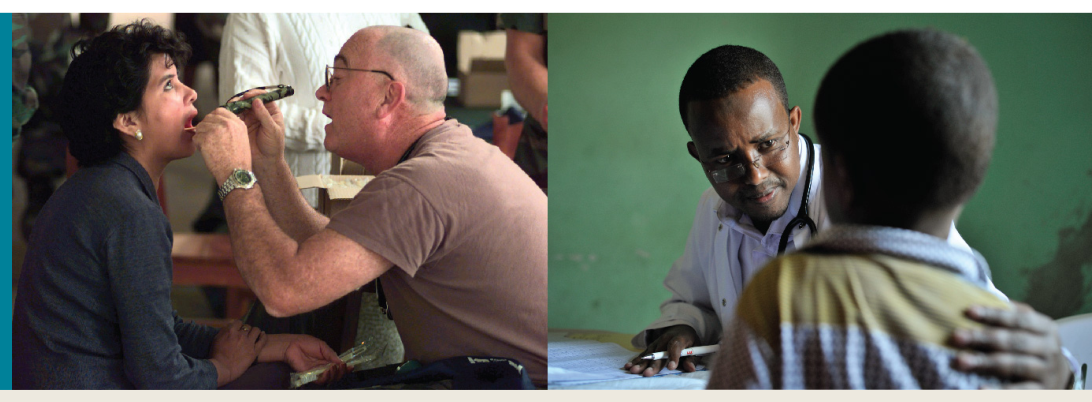
A neurological examination localizes dysfunction by matching an observable loss—such as impaired speech, sensation, movement, or balance—to the nervous-system structure that normally produces it. A patient’s answers, eye movements, reflexes, gait, and ability to identify touch are not isolated facts; together, they form an anatomical map of nervous-system function.
The five divisions of the neurological exam
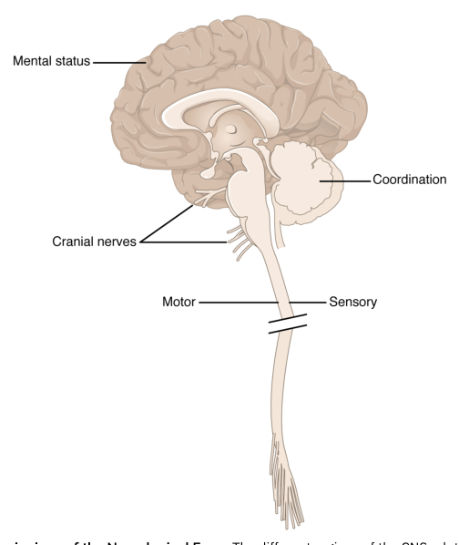
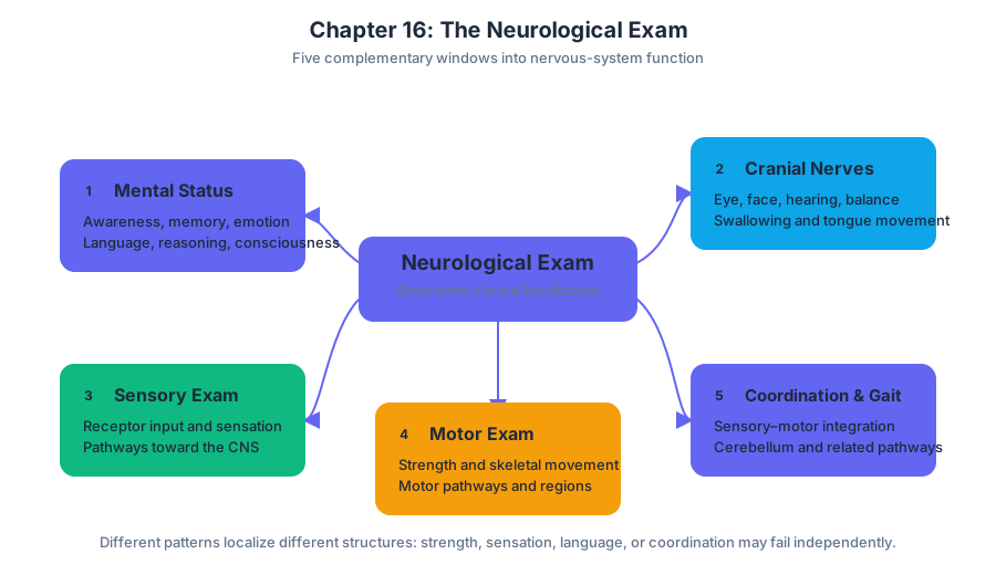
The neurological exam is organized into five major parts: the mental status exam, cranial nerve exam, sensory exam, motor exam, and coordination and gait exam. Each part emphasizes a different level of nervous-system organization.
- The mental status exam evaluates higher neurological functions associated with the cerebrum, including awareness, memory, emotion, language, reasoning, and consciousness.
- The cranial nerve exam assesses structures of the brain and cranial nerves by testing functions such as eye movement, facial sensation, hearing, balance, swallowing, and tongue movement.
- The sensory exam tests the pathways that carry information from receptors toward the central nervous system.
- The motor exam evaluates the pathways and regions responsible for skeletal-muscle movement.
- The coordination and gait exam examines the integration of sensory input with motor control, especially the contribution of the cerebellum and related pathways.
This organization makes the exam clinically powerful: a patient may have normal strength but impaired coordination, or normal sensation in the limbs but difficulty understanding language. Those different patterns suggest damage in different anatomical locations rather than a single generalized failure of the nervous system.
Higher neurological functions and cerebral function
The cerebrum supports the highest levels of nervous-system activity. Memory is the ability to retain and retrieve information; emotion includes subjective feelings and the behavioral responses associated with them; and consciousness is the state of awareness of oneself and the environment. These are called higher neurological functions because they depend on complex integration within the brain rather than on a single simple sensory or motor pathway.
The mental status exam may therefore assess whether a person is alert, oriented, able to communicate, able to remember information, and capable of appropriate reasoning. A disturbance in consciousness can range from confusion to unresponsiveness. Because these functions depend heavily on cerebral activity, an abnormal mental status may indicate dysfunction of the cerebral cortex or of systems that maintain arousal and attention.
Gyri, sulci, and cortical organization
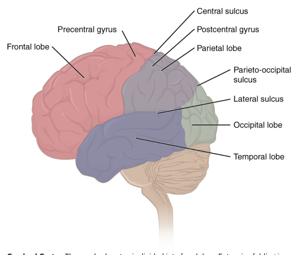
The outer layer of the cerebrum is the cerebral cortex, a thin layer of gray matter containing neuronal cell bodies, dendrites, and synaptic connections. The cortex is folded rather than smooth, allowing a large amount of neural tissue to fit within the limited space of the skull.
A gyrus is a ridge of folded cerebral tissue, whereas a sulcus is the groove between adjacent gyri.
These folds create recognizable anatomical landmarks and help clinicians describe the location of cortical regions. The cerebral cortex is not functionally uniform. Specialized areas are associated with particular abilities, including sensation, voluntary movement, language, memory, spatial reasoning, and interpretation of sensory information. A deficit in one of these functions can therefore help localize a lesion to a particular cortical region.
Language and nonverbal reasoning
Language function is predominantly associated with the left cerebral hemisphere. Important language-related integration areas include Broca’s area, associated with the production of speech, and Wernicke’s area, associated with the comprehension of language. These regions communicate through deep white matter, including the arcuate fasciculus.
The right cerebral hemisphere is predominantly associated with spatial and other nonverbal reasoning abilities. For example, it contributes to interpreting spatial relationships and organizing information that does not depend primarily on spoken or written language.
“Predominantly” does not mean “exclusively.” Language is not confined to the left hemisphere, and spatial reasoning is not confined to the right. The hemispheres work together through their connecting white-matter pathways. Hemispheric specialization describes a tendency for certain functions to rely more heavily on one side, not a complete division into two independent brains.
The prefrontal lobe
The prefrontal lobe, also called the prefrontal cortex, is a specific region of the frontal lobe. It lies anterior to the more specific motor-function areas and contributes to planning movements and intentions before action occurs. It is also associated with personality-type functions, judgment, and the organization of purposeful behavior.
Damage to this region can leave basic sensory and motor abilities relatively intact while changing personality, decision-making, impulse control, or the ability to plan. This distinction explains why a patient can move the limbs normally yet show major changes in behavior or goal-directed action. The classic case of Phineas Gage illustrates the clinical importance of the prefrontal region: an injury passing through the frontal area was associated with profound changes in personality despite survival and substantial preservation of basic physical abilities.
Central and peripheral nervous systems
The nervous system has two anatomical divisions: the central nervous system, or CNS, and the peripheral nervous system, or PNS.
| Division | Main components | Role in neurological examination |
|---|---|---|
| Central nervous system | Brain and spinal cord | Integrates information, generates conscious functions, and contains major sensory and motor pathways |
| Peripheral nervous system | Nerves and neural structures outside the brain and spinal cord | Connects receptors and effectors in the body with the CNS |
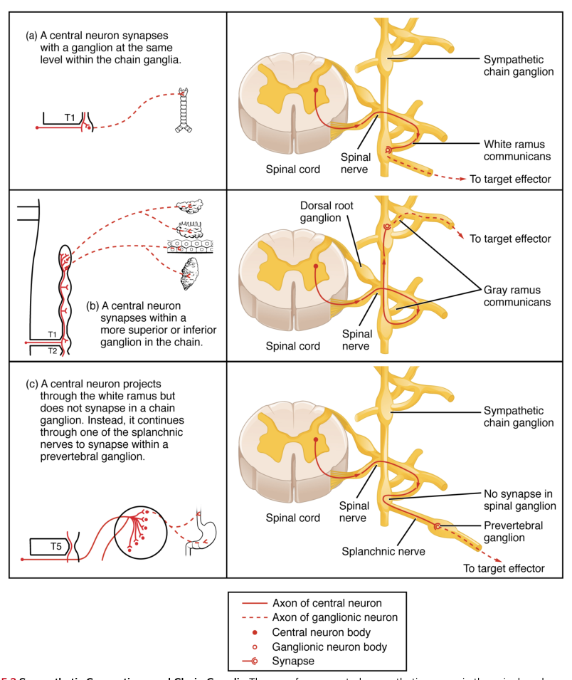
The distinction is anatomical, not a statement that one division works independently. Sensory receptors and peripheral nerves carry information toward the CNS, while motor commands leave the CNS through peripheral pathways to reach effectors. A neurological exam uses this relationship to determine whether a deficit is more consistent with a brain lesion, spinal-cord lesion, or peripheral-nerve problem.
Ascending sensory pathways of the spinal cord
The spinal cord contains two major groups of ascending pathways, meaning neural tracts that carry sensory information toward the brain. Their different functions allow the sensory exam to test more than “feeling” in a general sense.
The dorsal column–medial lemniscus pathway carries discriminative touch, vibration, and conscious proprioception—the awareness of body position and movement. The anterolateral pathway, which includes the spinothalamic pathway, carries pain, temperature, and less precisely localized touch. Sensory information often crosses from one side of the nervous system to the other along these routes, so the location and side of a deficit can help identify where a pathway has been interrupted.
Somatosensory information from the face follows a pathway distinct from the major ascending pathways of the spinal cord. Facial sensation travels primarily through the trigeminal nerve and its associated brainstem nuclei before reaching higher sensory regions. This is why testing facial sensation separately from limb sensation is clinically useful: normal sensation in the legs does not prove that the facial sensory pathway is intact.
Special sensory information in the cranial exam
The cranial nerve exam also tests hearing and vision, which depend on specialized receptor systems. In the cochlea, sound begins as movement of the oval window. That movement produces waves in the cochlear fluids and deflects the basilar membrane. Different regions of the basilar membrane respond preferentially to different sound frequencies, allowing the nervous system to encode pitch.
The visual system similarly contains specialized retinal regions. The fovea is a central area of the retina used for the sharpest vision and has distinct photopigment sensitivity compared with more peripheral retinal regions. Testing visual acuity and visual fields therefore examines not only the eye but also the optic pathways and the cortical regions that interpret visual information.
Clinical localization and meningitis
The neurological exam is especially valuable when nervous-system damage must be identified quickly, as after trauma or a stroke. A language deficit points toward dominant cortical language regions; a personality or planning change suggests prefrontal dysfunction; a selective loss of vibration or pain suggests involvement of different ascending pathways; and an abnormal facial sensory finding raises concern for the trigeminal system or its central connections.
The meninges are protective membranes surrounding the brain and spinal cord. Meningitis is inflammation of the meninges and can have severe effects because inflammation occurs around structures essential for consciousness, sensory processing, movement, and autonomic regulation. A patient with serious meningeal disease may show abnormalities across several portions of the neurological exam, making rapid assessment and localization clinically important.
The exam therefore links structure to function at several scales: cortical regions support higher functions, cranial nerves connect the brain with specialized sensory and motor systems, spinal pathways carry information between the body and brain, and peripheral nerves connect those central structures with receptors and effectors. The pattern of preserved and impaired abilities is the central evidence used to locate neurological dysfunction.
Content adapted from OpenStax Anatomy and Physiology 2e by J. Gordon Betts et al., licensed CC BY 4.0.
Source Materials
Study Anatomy and Physiology on Lykke
This course page is free to open: signed out, you can read its first two sections, and signing in, on any plan, opens the rest. The free Lykke Chrome extension syncs your own Canvas courses, so ChatGPT, Claude and Muse can answer from your classes and deadlines. It only reads Canvas.
Add the free extensionSign in to open every sectionAll sections of Anatomy and Physiology · Browse more course pages