From the free course Anatomy and Physiology on Lykke
Chapter 11: The Muscular System
Key concepts: Skeletal muscle structure and striations · Sarcomeres, actin–myosin cross-bridge formation, and muscle contraction · Concentric, eccentric, isotonic, and isometric muscle contractions · Muscle tension and joint movement · Tendons and their role in attaching muscles to bones · Nervous-system control of muscle tension · Muscle fiber types and fast glycolytic fibers · ATP use during muscle contraction and shivering · Shivering and muscular heat production · Effects and risks of anabolic steroids on muscle performance and mass
By the end of this chapter the student can: Describe the actions and roles of agonists and antagonists; Explain the structure and organization of muscle fascicles and their role in generating force; Explain the criteria used to name skeletal muscles; Identify the skeletal muscles and their actions on the skeleton and soft tissues of the body; Identify the origins and insertions of skeletal muscles and the prime movements.
Chapter 11: The Muscular System
Skeletal muscles turn chemical energy into tension, and that tension becomes movement only when muscles, tendons, bones, joints, and the nervous system act together. Every step, facial expression, breath, and change in posture depends on this coordinated system. A muscle does not push a bone; it pulls on it through a tendon, a strong band of dense regular connective tissue that attaches muscle to bone.
The visible bulge of a flexed biceps brachii illustrates this arrangement. The central muscle belly contains the contractile tissue, while tendons extend from its ends to the upper arm and forearm. When the muscle contracts, tension passes through the tendons and moves the skeleton at the elbow joint.
A useful misconception to correct immediately is that a muscle acts alone. Skeletal muscles operate in functional groups: one muscle or group may produce the main movement, another may oppose or reverse it, and others stabilize the joint or provide a supporting base. The sections below connect the microscopic organization of muscle fibers to the large muscle groups that move the human body.
11.1 Interactions of Skeletal Muscles, Their Fascicle Arrangement, and Their Lever Systems
Bending the elbow demonstrates the basic logic of skeletal muscle action. The biceps brachii contracts and produces elbow flexion, while the triceps brachii relaxes enough to permit the movement. To extend the elbow, the triceps becomes the principal contracting muscle and the biceps acts as its opponent. Muscles can therefore be classified by how they participate in a movement rather than simply by their location.
Agonists, antagonists, synergists, and fixators
The agonist, or prime mover, is the muscle chiefly responsible for producing a particular movement. During elbow flexion, the biceps brachii is an important agonist. An antagonist produces the opposite action. The triceps brachii is an antagonist to the biceps during flexion because its contraction extends the elbow.
A muscle can be an agonist for one movement and an antagonist for another. The relationship depends on the movement being analyzed. Synergists assist the agonist by adding force, reducing unwanted movement, or helping produce a more precise action. Fixators are specialized synergists that stabilize the origin of the agonist. For example, muscles that stabilize the scapula allow muscles attached to the humerus to move the arm efficiently.
The sequence of a controlled movement is therefore:
- The nervous system activates the prime mover.
- Synergists assist the movement or prevent unnecessary motion.
- Fixators hold a bone or joint steady.
- The antagonist is inhibited or carefully activated to control the movement.
- Tendons transfer the resulting muscle tension to bones.
A common misconception is that antagonists are always completely inactive. In reality, the nervous system can activate antagonist muscles during a movement to brake motion, stabilize a joint, or provide precise control. The opposing muscles of the body function more like a coordinated team than a simple on-off pair.
Muscle fascicles and force production
A fascicle is a bundle of skeletal muscle fibers. Connective tissue surrounds the entire muscle, each fascicle, and each individual muscle fiber. The epimysium surrounds the whole muscle, the perimysium surrounds a fascicle, and the endomysium surrounds an individual muscle fiber. These connective-tissue layers merge into tendons or broad sheets called aponeuroses, allowing force generated inside the muscle to reach bone.
The arrangement of fascicles determines the shape of the muscle and influences the balance between force, speed, and range of motion. The major patterns are:
- Parallel muscles have fascicles running parallel to the long axis of the muscle. They can usually shorten over a relatively large distance and produce a broad range of movement. The biceps brachii is a parallel, fusiform muscle.
- Convergent muscles have a broad origin and fascicles that converge toward a single tendon. Different portions can contract separately, allowing the muscle to produce movement in more than one direction. The pectoralis major is a convergent muscle.
- Pennate muscles have fascicles attached obliquely to a tendon. Unipennate muscles attach on one side of a tendon, bipennate muscles on both sides, and multipennate muscles have several tendon branches. Pennate muscles pack many fibers into a relatively small area and generally produce substantial force, although their fibers shorten through a smaller distance.
- Circular muscles surround openings and act as sphincters. The orbicularis oris and orbicularis oculi are examples.
- Triangular or fan-shaped muscles have a broad origin and narrow insertion; the temporalis is an example.
The key trade-off is simple: longer, more parallel fibers favor greater shortening distance and movement range, whereas more densely packed pennate fibers favor force production. Muscle size also matters. Increasing the number of myofibrils and contractile proteins makes a fiber thicker, producing hypertrophy, the enlargement of a muscle.
From sarcomere shortening to whole-muscle tension
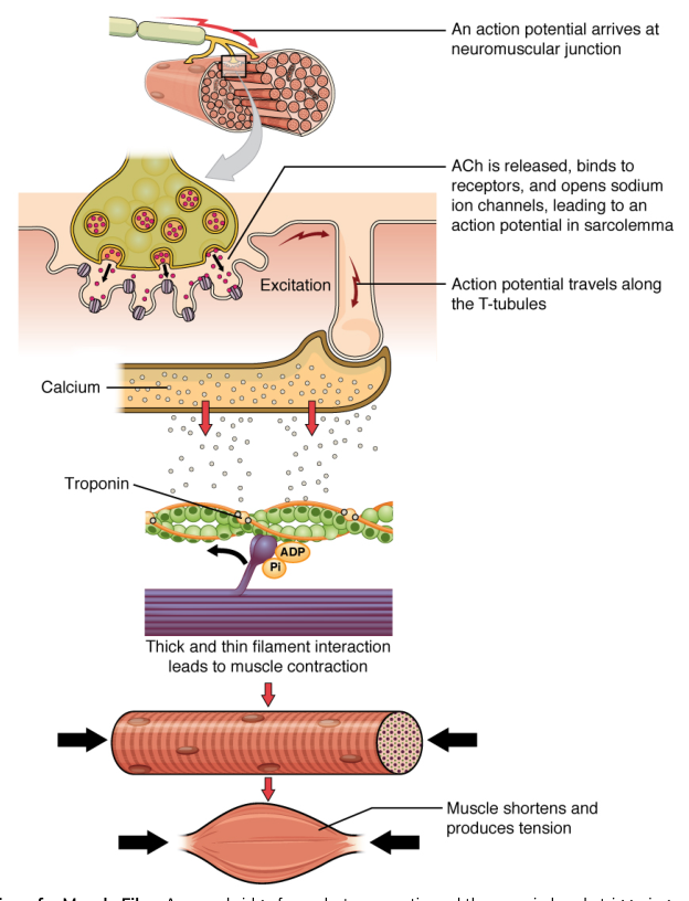
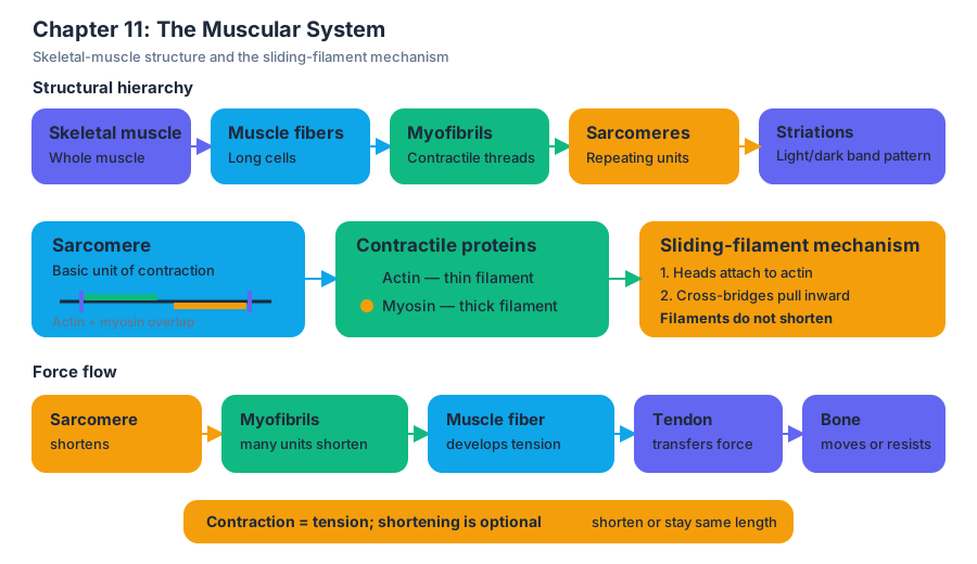
A skeletal muscle fiber is packed with myofibrils, and each myofibril is made of repeating sarcomeres. A sarcomere is the basic contractile unit of striated muscle. It contains thin actin filaments and thick myosin filaments arranged in an orderly pattern. The alternating arrangement of these filaments produces the striated appearance of skeletal muscle.
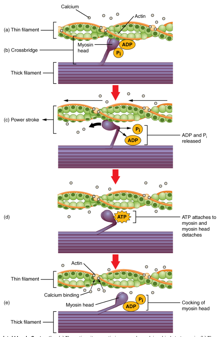
Contraction follows the sliding-filament mechanism:
- A motor neuron releases a neurotransmitter at the neuromuscular junction.
- The muscle fiber membrane generates an electrical signal.
- The signal causes calcium ions to be released inside the fiber.
- Calcium exposes binding sites on actin.
- Myosin heads attach to actin, forming cross-bridges.
- Myosin heads pull actin toward the center of the sarcomere.
- ATP binds to myosin, allowing the head to detach.
- ATP is hydrolyzed, which re-cocks the myosin head for another cycle.
- As many sarcomeres shorten together, the whole fiber develops tension.
- Tension is transmitted through connective tissue and the tendon to the bone.
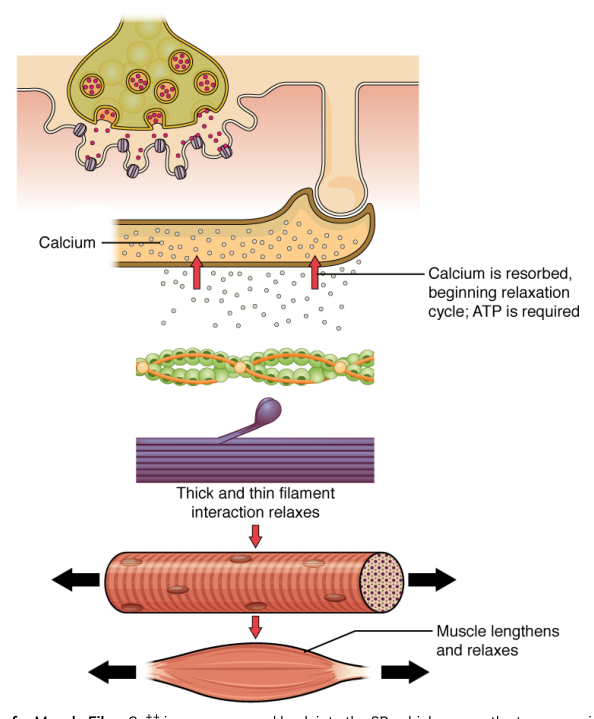
The filaments themselves do not become shorter; they slide past one another. When stimulation stops, calcium is returned to its storage sites, actin binding sites are covered, cross-bridge cycling stops, and the muscle relaxes. ATP is required both for contraction and for detaching myosin from actin, which helps explain why the absence of ATP after death produces rigor mortis.
Lever systems and joint movement
Bones act as levers, joints act as fulcrums, and muscle contractions provide the applied force. A lever can favor force or speed and range of motion, but its arrangement determines the mechanical advantage. In the body, the muscle’s attachment relative to the joint is especially important.
In a first-class lever, the fulcrum lies between the force and the load, as in a seesaw. In a second-class lever, the load lies between the fulcrum and the force, as when rising onto the toes. In a third-class lever, the force lies between the fulcrum and the load. Elbow flexion by the biceps brachii is a third-class lever: the elbow is the fulcrum, the biceps applies force to the forearm between the elbow and the hand, and the hand carries the load.
Third-class levers sacrifice mechanical advantage but allow the hand to move rapidly and through a large distance. This is why lifting a weight with the hand may require considerable muscle force even though the external load is modest.
Types of muscle contraction
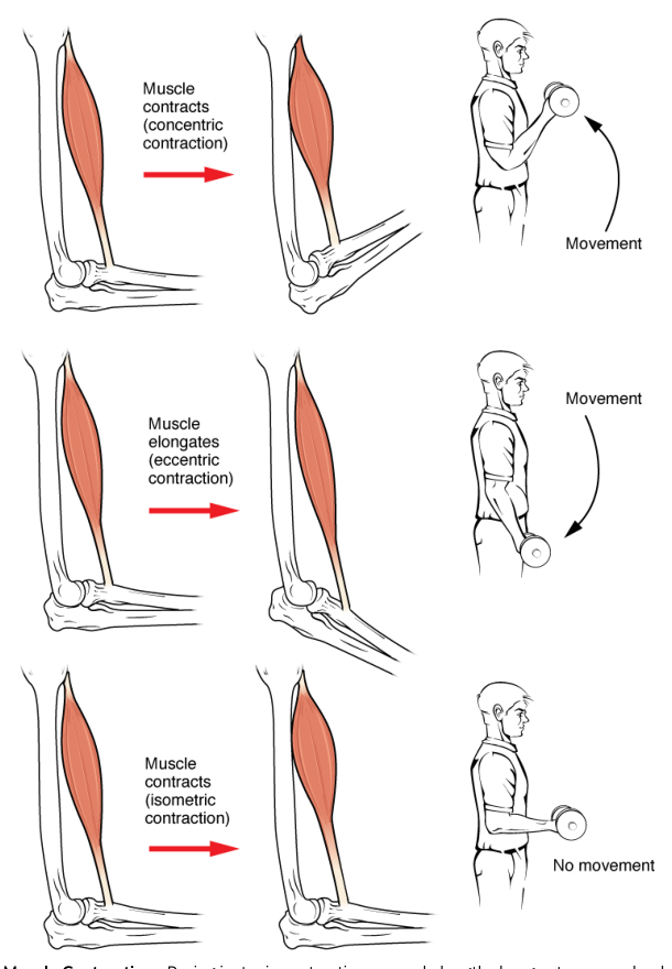
A concentric contraction occurs when a muscle produces tension while shortening. Raising a dumbbell during elbow flexion is concentric contraction of the biceps brachii. An eccentric contraction occurs when a muscle produces tension while lengthening. Lowering the dumbbell in a controlled manner uses eccentric contraction of the biceps to resist gravity.
Both concentric and eccentric contractions are isotonic contractions, meaning that the muscle changes length while moving a load. An isometric contraction produces tension without changing muscle length. Holding a heavy object motionless, or pushing against an immovable wall, requires isometric contraction. The load exceeds the tension needed to shorten the muscle, or the joint position prevents visible movement.
These contractions explain why descending stairs can be demanding even though the body is moving downward. The quadriceps muscles lengthen while remaining active, producing eccentric contractions that control knee flexion and prevent an uncontrolled fall.
Nervous-system control of muscle tension
The nervous system regulates whether a muscle contracts, how strongly it contracts, how long it remains active, and whether it shortens, lengthens, or holds its length. A motor unit consists of one motor neuron and all the skeletal muscle fibers it controls. Small motor units permit precise movements, while large motor units generate powerful movements.
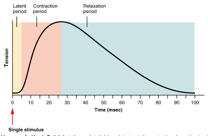
Increasing muscle tension occurs through motor-unit recruitment. The nervous system activates a small number of motor units first and then recruits additional units as greater force is needed. It can also increase the frequency of stimulation. When stimuli arrive close together, individual twitches combine into greater tension; sustained stimulation can produce tetanus, a smooth, maintained contraction.
A practical example is lifting a cup. The nervous system recruits relatively few motor units because the load is light. Lifting a heavy suitcase requires more motor units and greater stimulation frequency. Holding the suitcase still requires ongoing isometric activity, while raising it uses concentric contraction and lowering it uses eccentric contraction.
11.2 Naming Skeletal Muscles
Skeletal muscle names are anatomical clues. A name may reveal where a muscle is located, what it looks like, how large it is, the direction of its fibers, the number of origins it has, or its attachments. Learning these naming rules turns unfamiliar terms into descriptions rather than arbitrary labels.
Location and action
Some muscles are named for the bone or body region they occupy. The temporalis lies over the temporal bone, the intercostal muscles lie between the ribs, and the brachialis lies in the arm. The tibialis anterior is located along the anterior tibia.
Other names describe action. A muscle named flexor decreases the angle at a joint, an extensor increases it, an adductor moves a body part toward the midline, and an abductor moves it away from the midline. The supinator turns the forearm so the palm faces anteriorly or upward, whereas the pronator turns it posteriorly or downward.
Shape, size, direction, and attachments
Shape-based names include deltoid, meaning triangular; trapezius, referring to a trapezoid-like shape; rhomboid, referring to a diamond-like shape; and serratus, meaning saw-toothed because of its digitated edges. Size terms include maximus for largest, minimus for smallest, major for larger, minor for smaller, longus for long, and brevis for short.
Fiber direction may appear in the name. Rectus means straight and usually indicates fibers running parallel to a body region’s longitudinal axis. Transversus means transverse, while oblique means diagonal. The rectus abdominis, transversus abdominis, and external and internal obliques illustrate these directional terms.
Names may also indicate the number of origins. A biceps has two heads or origins, a triceps has three, and a quadriceps has four. Attachment names combine bones or landmarks: the sternocleidomastoid attaches to the sternum, clavicle, and mastoid process; the brachioradialis extends between the arm and forearm.
For example, extensor carpi radialis longus can be decoded as a long muscle that extends the wrist and lies on the radial side of the forearm. The name predicts its location and action before the muscle is memorized individually.
Origin, insertion, and prime movement
The origin is generally the attachment that remains relatively fixed during a movement. The insertion is generally the attachment that moves toward the origin when the muscle contracts. These terms are relative to a particular movement; in some actions, the usual insertion can become fixed and the origin can move.
The muscle’s prime movement is its principal action at a joint. A complete anatomical description therefore links four facts: origin, insertion, action, and the joint crossed. For the biceps brachii, the proximal attachment is on the scapula, the distal attachment is on the radius, and its major actions include elbow flexion and forearm supination.
A name should not be treated as a complete functional description. The attachment angle, joint position, neighboring muscles, and nervous-system activation all influence the movement. Nevertheless, names provide a reliable first set of clues and help organize the large number of skeletal muscles in the body.
11.3 Axial Muscles of the Head, Neck, and Back
Axial muscles arise from or attach to the axial skeleton and help position the head, vertebral column, and trunk. They also move facial structures, the eyes, the tongue, and the jaw. Unlike limb muscles, many axial muscles maintain posture continuously while producing smaller, highly controlled movements.
Muscles of facial expression and mastication
The muscles of facial expression insert into the skin rather than only into bones. The occipitofrontalis raises the eyebrows and wrinkles the forehead. The orbicularis oculi closes the eyelids, while the orbicularis oris closes and protrudes the lips. The zygomaticus elevates the corners of the mouth, producing a smile. The buccinator compresses the cheek and helps keep food between the teeth during chewing.
The principal muscles of mastication move the mandible. The masseter elevates the mandible and closes the jaw. The temporalis elevates and retracts the mandible. The medial and lateral pterygoid muscles assist with elevation, protraction, retraction, and side-to-side movements of the jaw.
A concrete example is chewing bread. The masseter generates powerful jaw elevation, the temporalis helps retract the mandible, the pterygoids shift the jaw from side to side, and the buccinator prevents the food from collecting between the cheek and teeth.
Muscles of the eye, tongue, and neck
The extrinsic eye muscles move the eyeball. The superior rectus elevates the eye, the inferior rectus depresses it, the medial rectus moves it medially, and the lateral rectus moves it laterally. The superior and inferior oblique muscles rotate the eye and contribute to its directional positioning. These muscles work in coordinated pairs so that both eyes focus on the same target.
The tongue contains intrinsic muscles that alter its shape and extrinsic muscles that move it. The genioglossus protrudes the tongue, the hyoglossus depresses it, and the styloglossus retracts and elevates it. Tongue muscles are essential for speech, swallowing, and manipulating food.
The platysma tenses the skin of the neck and can depress the mandible. The sternocleidomastoid flexes the neck when both sides contract; unilateral contraction rotates the head toward the opposite side and tilts it toward the contracting side. The scalenes flex and laterally bend the neck and elevate the first two ribs during forced inspiration.
Muscles of the vertebral column and back
The deep back muscles stabilize and move the vertebral column. The erector spinae group extends the vertebral column and maintains upright posture; unilateral contraction contributes to lateral flexion. The transversospinalis group, including the semispinalis, multifidus, and rotatores, assists extension, rotation, and stabilization of individual vertebrae.
The splenius muscles extend and rotate the head and neck. The quadratus lumborum laterally flexes the vertebral column and fixes the twelfth rib during respiration. The multifidus stabilizes vertebrae and contributes to extension and rotation.
The large superficial muscles of the back also connect the axial skeleton to the pectoral girdle and upper limb. The trapezius elevates, retracts, depresses, and upwardly rotates the scapula; its upper fibers can extend the neck. The latissimus dorsi extends, adducts, and medially rotates the arm. The rhomboid major and minor retract and downwardly rotate the scapula and help hold it against the thoracic wall.
The levator scapulae elevates the scapula and can laterally flex the neck. The serratus posterior superior elevates ribs, whereas the serratus posterior inferior depresses lower ribs. These muscles show why regional categories overlap functionally: a back muscle may move the spine, ribs, scapula, or upper limb depending on its attachments.
When a person lifts a suitcase from the floor, the erector spinae and multifidus stabilize and extend the vertebral column, the trapezius and rhomboids stabilize the scapula, and the latissimus dorsi can assist movement of the upper limb. A failure of these stabilizing actions would allow excessive spinal or scapular motion even if the prime movers were strong.
11.4 Axial Muscles of the Abdominal Wall, and Thorax
The abdominal and thoracic muscles form a mobile protective cylinder around the trunk. They support and compress the abdominal viscera, move the vertebral column, stabilize the pelvis, and change the volume of the thoracic cavity during breathing.
Muscles of the abdominal wall
The rectus abdominis runs vertically along the anterior abdominal wall. It flexes the vertebral column, compresses the abdominal contents, and helps stabilize the pelvis. Its tendinous intersections create the segmented appearance commonly called a “six-pack.”
The external oblique is the most superficial lateral abdominal muscle. It compresses the abdomen, flexes the vertebral column bilaterally, and rotates the trunk to the opposite side when one side contracts. The internal oblique lies deep to it and compresses the abdomen, flexes the trunk, and rotates the trunk toward the same side.
The deepest broad abdominal muscle, the transversus abdominis, has fibers running horizontally. It compresses the abdominal contents and contributes strongly to trunk stability. The quadratus lumborum forms part of the posterior abdominal wall, laterally flexes the trunk, and helps fix the twelfth rib.
The abdominal muscles coordinate rather than operate independently. During a sit-up, the rectus abdominis flexes the vertebral column while the obliques assist and control rotation. During forced expiration, the abdominal wall compresses the abdominal cavity and pushes the diaphragm upward.
Muscles of the thorax and respiration
The diaphragm is the principal muscle of inspiration. It is a dome-shaped sheet separating the thoracic and abdominal cavities. When it contracts, it flattens and moves inferiorly, increasing thoracic volume and lowering pressure inside the lungs. Air flows inward. When it relaxes, it returns to its domed shape, decreasing thoracic volume and helping drive expiration.
The external intercostals lie between the ribs and elevate them during inspiration. The internal intercostals depress the ribs during forced expiration. The innermost intercostals assist the internal intercostals. The intercostal muscles also stabilize the spaces between the ribs so that the thoracic wall moves as a unit.
The pectoralis minor can draw the scapula anteriorly and inferiorly and can assist forced inspiration by elevating the ribs when the scapula is fixed. The serratus anterior protracts the scapula, holds it against the thoracic wall, and upwardly rotates it; it can also assist inspiration.
The pectoralis major is a large chest muscle that flexes, adducts, and medially rotates the arm. Its clavicular portion assists arm flexion, while its sternocostal portion can extend the arm from a flexed position. The subclavius depresses and stabilizes the clavicle.
Consider a deep breath. The nervous system activates the diaphragm and external intercostals. The diaphragm flattens, the ribs elevate, thoracic volume increases, and air enters the lungs. During vigorous exercise, internal intercostals and abdominal muscles assist expiration by actively reducing thoracic volume.
The thoracic muscles in movement
Thoracic muscles also move the upper limb and stabilize the pectoral girdle. The pectoralis major can generate a powerful pushing motion, the serratus anterior keeps the scapula attached to the thoracic wall during reaching, and the pectoralis minor adjusts scapular position. The thorax is therefore both a respiratory structure and a mechanical base for upper-limb movement.
A common misconception is that the diaphragm works only during strenuous breathing. It contracts rhythmically during quiet breathing and remains active throughout life. Skeletal muscle does not necessarily mean voluntary muscle; breathing is usually automatic, although it can be consciously modified for speech, singing, or breath-holding.
11.5 Muscles of the Pectoral Girdle and Upper Limbs
The upper limb combines mobility with precise control. The pectoral girdle must position the scapula and clavicle, the shoulder must move the humerus through a wide range, and the elbow, wrist, and fingers must direct force accurately.
Muscles that move the pectoral girdle
The trapezius elevates, retracts, depresses, and upwardly rotates the scapula. The serratus anterior protracts and upwardly rotates it while holding it against the thorax. The rhomboids retract and downwardly rotate the scapula. The levator scapulae elevates it, and the pectoralis minor draws it anteriorly and inferiorly.
These muscles give the scapula a stable but adjustable platform. During a push-up, the serratus anterior protracts and stabilizes the scapula; weakness can cause the medial border to protrude away from the thoracic wall, a condition called winged scapula.
Muscles that move the arm at the shoulder
The deltoid is the major abductor of the arm after the first part of abduction has begun. Its anterior fibers flex and medially rotate the arm, while its posterior fibers extend and laterally rotate it. The pectoralis major flexes, adducts, and medially rotates the arm. The latissimus dorsi extends, adducts, and medially rotates it.
The coracobrachialis flexes and adducts the arm. The teres major extends, adducts, and medially rotates the arm. The supraspinatus initiates abduction and helps stabilize the shoulder. The infraspinatus and teres minor laterally rotate the arm, whereas the subscapularis medially rotates it.
Together, the supraspinatus, infraspinatus, teres minor, and subscapularis form the rotator cuff. Their tendons stabilize the head of the humerus in the glenoid cavity while allowing extensive shoulder movement. The shoulder’s mobility comes with a trade-off: it is less inherently stable than a tightly enclosed joint and is therefore vulnerable when the muscles or tendons are overloaded.
Muscles that move the forearm
The biceps brachii flexes the elbow and supinates the forearm. The brachialis is a strong elbow flexor regardless of forearm position. The brachioradialis flexes the elbow most effectively when the forearm is in a neutral, thumb-up position.
The triceps brachii extends the elbow. Its long head also extends and adducts the arm at the shoulder. The anconeus assists elbow extension and stabilizes the joint. The pronator teres and pronator quadratus pronate the forearm, while the supinator supinates it.
Opening a tightly closed jar illustrates these relationships. The supinator and biceps brachii rotate the forearm into supination when the palm turns upward, while the wrist and finger muscles stabilize the hand so that the rotational force is transmitted to the jar.
Muscles that move the wrist, hand, and fingers
The anterior compartment of the forearm contains many flexors. The flexor carpi radialis flexes and abducts the wrist; the flexor carpi ulnaris flexes and adducts it; the palmaris longus flexes the wrist and tightens the palmar aponeurosis. The flexor digitorum superficialis flexes the fingers, and the flexor digitorum profundus flexes the distal interphalangeal joints. The flexor pollicis longus flexes the thumb.
The posterior forearm contains extensors. The extensor carpi radialis longus and brevis extend and abduct the wrist. The extensor carpi ulnaris extends and adducts it. The extensor digitorum extends the fingers, while the extensor indicis extends the index finger. The extensor pollicis longus and brevis extend the thumb, and the abductor pollicis longus abducts it.
The hand itself contains intrinsic muscles that refine finger movement. The thenar muscles move the thumb, the hypothenar muscles move the little finger, the lumbricals flex the metacarpophalangeal joints while extending interphalangeal joints, and the interossei abduct or adduct the fingers. These small muscles make grasping, writing, and manipulation possible.
11.6 Appendicular Muscles of the Pelvic Girdle and Lower Limbs
The lower limb is built for support, balance, and propulsion. Compared with the upper limb, its muscles generally produce greater force and stabilize the pelvis and knee during standing and locomotion.
Muscles that move the thigh at the hip
The iliopsoas, formed by the psoas major and iliacus, is a powerful flexor of the thigh at the hip. The tensor fasciae latae flexes, abducts, and medially rotates the thigh and helps stabilize the knee through the iliotibial tract.
The gluteus maximus is the principal extensor of the thigh and also laterally rotates it. It is especially important when rising from a seated position, climbing, or running uphill. The gluteus medius and gluteus minimus abduct and medially rotate the thigh; they stabilize the pelvis during single-leg support. The gluteus medius is particularly important because it prevents the pelvis from dropping on the unsupported side during walking.
The deep lateral rotators include the piriformis, obturator internus, obturator externus, superior gemellus, inferior gemellus, and quadratus femoris. They laterally rotate the thigh and help stabilize the femoral head in the acetabulum.
The medial thigh compartment contains the adductor longus, adductor brevis, adductor magnus, gracilis, and pectineus. These muscles primarily adduct the thigh. The gracilis also assists thigh flexion and knee flexion, while the pectineus assists hip flexion.
During a normal step, the gluteus medius stabilizes the pelvis as body weight shifts onto one limb, the iliopsoas flexes the hip to advance the opposite limb, and the gluteus maximus extends the hip as the body moves forward over the supporting leg.
Muscles that move the leg at the knee
The anterior thigh contains the quadriceps femoris, a four-part muscle group consisting of the rectus femoris, vastus lateralis, vastus medialis, and vastus intermedius. The quadriceps extends the leg at the knee. The rectus femoris also flexes the thigh at the hip because it crosses both the hip and knee.
The posterior thigh contains the hamstring group: biceps femoris, semitendinosus, and semimembranosus. These muscles flex the leg at the knee and extend the thigh at the hip. The biceps femoris laterally rotates the flexed leg, while the semitendinosus and semimembranosus medially rotate it.
The sartorius flexes, abducts, and laterally rotates the thigh and flexes the knee. It is the longest muscle in the body and helps place the leg in the cross-legged “tailor” position. The popliteus unlocks the knee by rotating the leg and assists knee flexion.
Walking downstairs provides a clear example of eccentric control. The quadriceps remain active while lengthening to control knee flexion. If they relaxed completely, the knee would buckle; if they shortened concentrically, the body would rise rather than descend smoothly.
Muscles that move the ankle and foot
The anterior leg compartment dorsiflexes and extends the toes. The tibialis anterior dorsiflexes and inverts the foot. The extensor hallucis longus extends the great toe and dorsiflexes the foot, while the extensor digitorum longus extends the toes and dorsiflexes the foot. The fibularis tertius dorsiflexes and everts the foot.
The lateral compartment contains the fibularis longus and fibularis brevis, which evert the foot. The fibularis longus also assists plantar flexion and helps support the arches of the foot.
The posterior leg contains the plantar flexors. The gastrocnemius plantar flexes the foot and flexes the knee because it crosses both joints. The soleus plantar flexes the foot and is especially important for maintaining standing posture. Together, the gastrocnemius and soleus form the triceps surae, whose tendon is the calcaneal tendon, commonly called the Achilles tendon.
The plantaris assists plantar flexion and knee flexion. The tibialis posterior plantar flexes and inverts the foot and supports the medial arch. The flexor hallucis longus flexes the great toe, while the flexor digitorum longus flexes the toes and assists plantar flexion.
The intrinsic muscles of the foot flex, extend, abduct, and adduct the toes and support the arches. The arches distribute body weight and absorb forces during walking and running. Muscles and tendons do not merely move the foot; they also prevent excessive collapse of its structural supports.
Integrated lower-limb movement
A jump combines nearly every principle of muscle interaction. The gluteus maximus extends the hip, the quadriceps extend the knee, and the gastrocnemius and soleus plantar flex the ankle. These muscles act as agonists during takeoff, while the abdominal and back muscles stabilize the trunk and the gluteal muscles stabilize the pelvis.
Landing reverses the mechanical pattern. The same muscle groups often contract eccentrically: the quadriceps control knee flexion, the gluteus maximus controls hip flexion, and the plantar flexors control forward movement of the tibia over the foot. The nervous system adjusts motor-unit recruitment to the landing force, allowing the body to absorb energy without uncontrolled joint movement.
The major lesson of the muscular system is therefore organizational. Sarcomeres generate tension through actin–myosin cross-bridge cycling; fibers gather into fascicles; fascicles form muscles; tendons transmit force to bones; bones act as levers around joints; and motor units coordinate the timing and strength of contraction. Movement becomes possible because every level, from molecular filaments to whole-body muscle groups, contributes to the same mechanical task.
Content adapted from OpenStax Anatomy and Physiology 2e by J. Gordon Betts et al., licensed CC BY 4.0.
Source Materials
Study Anatomy and Physiology on Lykke
This course page is free to open: signed out, you can read its first two sections, and signing in, on any plan, opens the rest. The free Lykke Chrome extension syncs your own Canvas courses, so ChatGPT, Claude and Muse can answer from your classes and deadlines. It only reads Canvas.
Add the free extensionSign in to open every sectionAll sections of Anatomy and Physiology · Browse more course pages