From the free course Anatomy and Physiology on Lykke
Chapter 8: The Appendicular Skeleton
Key concepts: Axial and appendicular skeleton divisions · Axial skeleton and its 80 bones · Skull and braincase · Cranial cavity and cranial fossae · Temporal bone and ear structures · Frontal bone and supraorbital margin · Ethmoid bone and olfactory structures · Facial skeleton bones · Nasal cavity and nasal conchae · Cleft lip and cleft palate
By the end of this chapter the student can: Discuss the bones of the pectoral and pelvic girdles, and describe how these unite the limbs with the axial skeleton; Describe the bones of the upper limb, including the bones of the arm, forearm, wrist, and hand; Identify the features of the pelvis and explain how these differ between the adult male and female pelvis; Describe the bones of the lower limb, including the bones of the thigh, leg, ankle, and foot; Describe the embryonic formation and growth of the limb bones.
Chapter 8: The Appendicular Skeleton
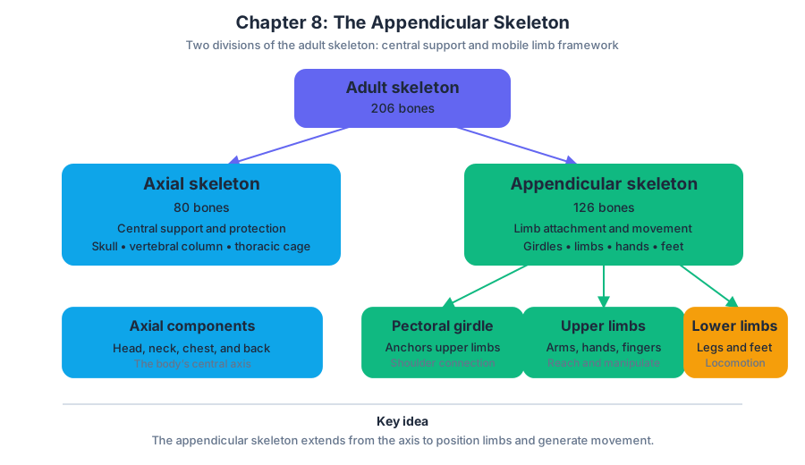
The skeleton has two organizational axes: the axial skeleton forms the body’s central supporting and protective axis, while the appendicular skeleton attaches to it and provides the framework for movement. The axial skeleton contains $80$ bones of the head, neck, chest, and back; the appendicular skeleton contains the bones of the upper and lower limbs plus the girdles that attach those limbs to the trunk.
This distinction is functional rather than merely geographic. The axial skeleton protects the brain, spinal cord, heart, and lungs and provides attachment sites for muscles of the head, neck, back, shoulders, and hips. The appendicular skeleton turns that stable central framework into movable levers: the pectoral girdle links the upper limb to the thoracic cage, and the pelvic girdle links the lower limb to the vertebral column.
The skull: axial protection at the top of the body
Although the limbs belong to the appendicular skeleton, the skull belongs to the axial skeleton. It is not a single solid bone but a carefully interlocked collection of bones that performs several jobs at once: it encloses the brain, forms the nasal cavity, surrounds the orbits, houses the structures of hearing and equilibrium, and supports the teeth of the upper and lower jaws.
An adult skull contains $22$ bones. Twenty-one are immobile and joined into a single structural unit by immovable joints called sutures. The twenty-second, the mandible, or lower jaw, is the only movable bone of the skull. This mobility is essential for chewing and speech, but it also makes the mandible distinct from the fixed bones of the braincase and face.
The neurocranium, or braincase
The neurocranium—also called the braincase—is the rounded portion of the skull that surrounds and protects the brain. Its bones form the walls and floor of the cranial cavity, the enclosed space in which the brain rests. The braincase also houses the middle and inner ear structures, although the external ear is formed largely by soft tissue and cartilage outside the skull.
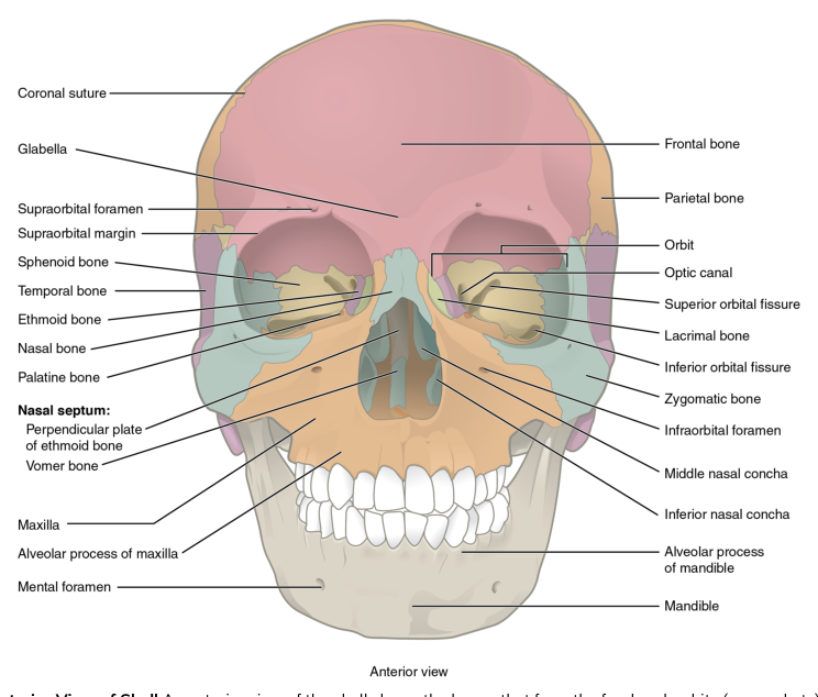
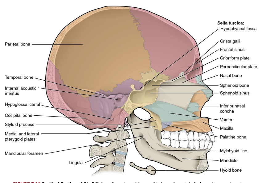
The bones of the neurocranium are the frontal bone, two parietal bones, two temporal bones, the occipital bone, the sphenoid bone, and the ethmoid bone.
- Frontal bone: forms the forehead, the roof of the orbits, and much of the anterior cranial floor.
- Parietal bones: paired bones forming most of the superior and lateral walls of the braincase.
- Temporal bones: paired bones forming the lateral skull and part of its floor; they contain or surround the major structures of the ear.
- Occipital bone: forms the posterior and inferior skull and contains the foramen magnum, the large opening through which the spinal cord passes.
- Sphenoid bone: a centrally placed, complex bone that contributes to the cranial floor, orbit, and skull base.
- Ethmoid bone: a lightweight bone between the nasal cavity and the orbits that contributes to the cranial floor and contains important olfactory structures.
Cranial cavity and cranial fossae
The cranial cavity is the space enclosed by the neurocranium. Its floor is not flat. Instead, it is shaped into three step-like depressions called cranial fossae. A fossa is a shallow depression in a bone; together, the fossae support different regions of the brain while allowing nerves and blood vessels to pass through openings in the skull.
- The anterior cranial fossa is the shallowest and most anterior depression. It supports the frontal lobes of the cerebrum. The frontal bone forms much of this region, with contributions from the ethmoid and sphenoid bones.
- The middle cranial fossa lies behind the anterior fossa and is deeper. It supports the temporal lobes of the cerebrum. The sphenoid and temporal bones form much of its floor.
- The posterior cranial fossa is the deepest and largest fossa. It supports the cerebellum and the brainstem. It is formed primarily by the occipital bone, with contributions from the temporal bones.
The fossae illustrate an important design principle: the skull is both a protective shell and a passageway. Its dense bone shields delicate neural tissue, while foramina—openings in bone—permit cranial nerves, blood vessels, and the spinal cord to connect the brain with the rest of the body.
Frontal bone and the orbital region
The frontal bone forms the forehead and the superior portion of the orbit, the bony socket that surrounds each eyeball. Its lower edge forms the supraorbital margin, the prominent ridge above each eye. You can feel this ridge by tracing a finger along the upper border of the orbit.
Near the supraorbital margin is the supraorbital notch or supraorbital foramen, an opening that allows the supraorbital nerve and blood vessels to pass from the orbit onto the forehead and scalp. The frontal bone also contains the frontal sinuses, air-filled spaces located above the eyebrows. These sinuses are part of the paranasal sinus system and communicate with the nasal cavity.
Temporal bone and the ear
The temporal bone forms much of the lateral wall and floor of the skull. Its position makes it a structural bridge between the braincase and the sensory apparatus of the ear. The external acoustic meatus is the canal that carries sound inward from the outside, while deeper portions of the temporal bone surround the middle and inner ear.
Important temporal-bone features include the following:
- The external acoustic meatus is the opening of the ear canal.
- The mastoid process is a rough projection posterior and inferior to the external acoustic meatus. It provides attachment for muscles of the neck.
- The styloid process is a slender projection that provides attachment for muscles and ligaments associated with the tongue and hyoid region.
- The mandibular fossa is a depression that receives the condyle of the mandible, forming the temporomandibular joint.
- The zygomatic process projects anteriorly to meet the zygomatic bone and help form the zygomatic arch.
The external ear collects sound, but the temporal bone is especially important for the deeper parts of hearing and balance. The middle ear contains the auditory ossicles, while the inner ear contains the cochlea for hearing and the vestibular structures for equilibrium. These structures lie within the protected temporal region rather than being exposed on the skull’s surface.
Ethmoid bone and olfaction
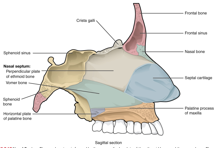
The ethmoid bone is a delicate, irregular bone located between the nasal cavity and the medial walls of the orbits. It contributes to the anterior cranial fossa, the nasal septum, and the walls of the nasal cavity. Its light, porous structure reflects its location and its role in separating the brain from the nasal passages.
The ethmoid’s cribriform plate is a thin, perforated portion of bone forming part of the roof of the nasal cavity. Tiny openings in the plate allow olfactory nerve fibers to pass from sensory receptors in the nasal mucosa to the brain. Rising from the cribriform plate is the crista galli, a sharp projection that provides attachment for a membrane supporting the brain.
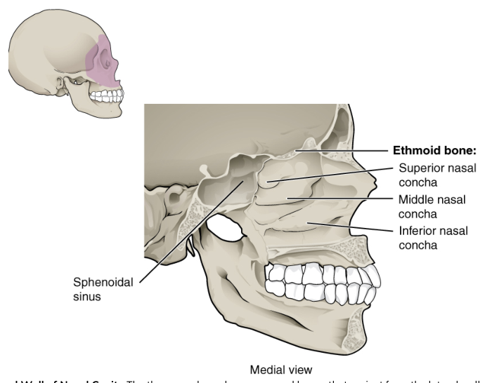
The ethmoid also forms the superior and middle nasal conchae. These curled shelves increase the surface area and turbulence of the nasal cavity, helping incoming air contact mucous membranes before it reaches the lower respiratory tract. The ethmoid air cells are small air-filled spaces located between the upper nasal cavity and medial orbit.
The facial skeleton
The facial skeleton, or viscerocranium, forms the framework of the face. It shapes the nose and orbits, forms the upper and lower jaws, supports the teeth, and contributes to the walls of the nasal and oral cavities. Its bones are smaller and more varied in shape than the braincase bones, but they are equally important for breathing, eating, speech, and facial expression.
The facial bones include paired maxillae, palatine bones, zygomatic bones, nasal bones, lacrimal bones, and inferior nasal conchae, plus the unpaired vomer and mandible.
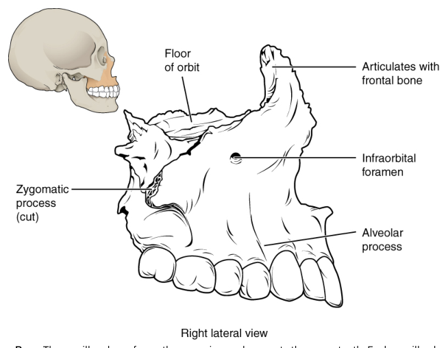
- Maxillae: paired bones forming the upper jaw, the anterior hard palate, part of the nasal cavity, and the floors of the orbits. They contain the sockets for the upper teeth.
- Mandible: the lower jaw and the only movable skull bone. It supports the lower teeth and moves at the temporomandibular joints.
- Zygomatic bones: the cheekbones. They form the lateral walls and floors of the orbits and join the temporal bones to form the zygomatic arches.
- Nasal bones: paired bones forming the bridge of the nose.
- Lacrimal bones: small bones on the medial orbital walls. They contribute to the passage through which tears drain toward the nasal cavity.
- Palatine bones: paired bones forming the posterior portion of the hard palate and contributing to the nasal cavity and orbit.
- Vomer: an unpaired bone forming the inferior portion of the nasal septum, which divides the nasal cavity into right and left passages.
- Inferior nasal conchae: paired, curved bones projecting from the lateral walls of the nasal cavity. Like the ethmoid conchae, they help warm, humidify, and filter inhaled air.
A common misconception is that the skull is simply a rigid helmet. In reality, its openings, cavities, projections, and articulations reveal several simultaneous systems: protection for the brain, passages for nerves and vessels, chambers for hearing and balance, air-filled spaces connected to the nose, and movable structures for chewing. The appendicular skeleton extends movement away from this protected axial core; the skull demonstrates how precisely the axial core is built to support and safeguard the organs that make that movement possible.
Source Materials
Study Anatomy and Physiology on Lykke
This course page is free to open: signed out, you can read its first two sections, and signing in, on any plan, opens the rest. The free Lykke Chrome extension syncs your own Canvas courses, so ChatGPT, Claude and Muse can answer from your classes and deadlines. It only reads Canvas.
Add the free extensionSign in to open every sectionAll sections of Anatomy and Physiology · Browse more course pages