From the free course Anatomy and Physiology on Lykke
Chapter 25: The Urinary System
Key concepts: Urinary system functions in blood filtration, urine formation, waste excretion, and homeostasis · Glomeruli are specialized capillary tufts that filter blood and help create the final urine composition · The initial filtrate is similar to plasma and contains ions, amino acids, vitamins, and wastes · Normal urine characteristics include a pH of 4.5–8.0 and a specific gravity of 1.003–1.032 · Urine volume and concentration vary with water intake and activity · Parathyroid hormone stimulates kidney tubule cells to reabsorb calcium from urine and release calcitriol · The kidneys remove circulating bilirubin and related metabolic products, such as urobilins, and secrete them into urine · The urethra transports urine from the bladder to the outside of the body · The male urethra passes through the prostate gland, whereas the female urethra is shorter · Loss of urinary-system function can cause weakness, lethargy, shortness of breath, anemia, edema, metabolic acidosis, elevated potassium, and cardiac arrhythmias
By the end of this chapter the student can: Describe the composition of urine; Label structures of the urinary system; Characterize the roles of each of the parts of the urinary system; Illustrate the macroscopic and microscopic structures of the kidney; Trace the flow of blood through the kidney; Outline how blood is filtered in the kidney nephron; Provide symptoms of kidney failure; List some of the solutes filtered, secreted, and reabsorbed in different parts of the nephron
Chapter 25: The Urinary System
The urinary system is a continuously operating chemical-control system: it filters blood, removes metabolic wastes, adjusts water and ion concentrations, and sends the resulting urine out of the body. Its central challenge is selective removal. The kidneys must eliminate substances such as urea and excess ions while retaining blood cells, proteins, nutrients, and the water the body still needs.
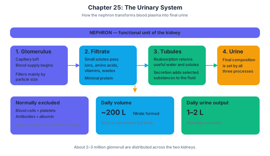
The two kidneys receive a large blood supply and contain approximately $2$ to $3$ million glomeruli, specialized tufts of capillaries that begin the process of urine formation. Together, the kidneys produce about $200$ liters of filtrate each day, yet most of that filtrate is recovered, leaving usually less than $2$ liters of urine for excretion.
The urinary system includes the kidneys, which form urine; the ureters, which transport urine; the urinary bladder, which stores urine; and the urethra, which conducts urine out of the body. The kidneys also function as endocrine organs, releasing hormones and producing substances that influence blood pressure, red blood cell production, calcium balance, and bone health.
A useful way to visualize the system is as a water-treatment plant with adjustable recovery pipes. Filtration creates a large initial volume, but tubular cells decide what returns to the blood and what remains in the tubular fluid. The analogy has a limit, however: renal tubules are living tissues whose transport changes in response to hormones and the body's internal conditions.
25.1 Physical Characteristics of Urine
Urine is the final fluid produced after the kidneys filter blood and modify the filtrate through tubular reabsorption and tubular secretion. Its composition reflects both waste removal and the body's current hydration, diet, activity, temperature, and hormonal state.
Normal urine is mostly water, but it also contains dissolved metabolic wastes and variable amounts of ions. Important waste products include urea, produced during amino acid metabolism; uric acid, produced during nucleic acid metabolism; and creatinine, produced by normal muscle metabolism. Urine may also contain ions such as sodium, potassium, chloride, phosphate, sulfate, and ammonium.
Fresh urine is normally transparent or slightly cloudy and ranges from pale yellow to deep amber. The yellow color is largely caused by urochrome, a pigment derived from hemoglobin breakdown. More concentrated urine generally appears darker, whereas dilute urine is paler. Color can also change with foods, medications, or disease, so color alone is not a complete diagnostic test.
Normal urine has a characteristic odor that becomes stronger as bacterial activity converts urea into ammonia. A sweet or fruity odor may indicate abnormal chemical conditions, while a strong foul odor may accompany infection. These observations are clues, not definitive diagnoses.
The normal urine pH ranges from $4.5$ to $8.0$. Its pH varies with diet and metabolism. For example, metabolism of proteins tends to generate acids, whereas other dietary patterns can produce a more alkaline urine. The kidneys help stabilize blood pH by adjusting the excretion of hydrogen ions and the conservation or production of bicarbonate.
Specific gravity compares the density of urine with the density of pure water. Normal values range from approximately $1.003$ to $1.032$. A higher value generally indicates more concentrated urine; a lower value indicates more dilute urine. Specific gravity depends on both the amount of water and the amount of dissolved material.
Urine volume varies substantially. Water intake, sweating, exercise, environmental temperature, nutrient intake, and hormone levels all influence output. During strenuous exercise on a hot day, substantial sweating reduces the water available for urine formation. The kidneys conserve water by producing a smaller volume of more concentrated urine.
A common misconception is that the kidneys simply “filter out toxins.” In reality, filtration is only the first step. The initial filtrate contains many useful substances, including water, ions, amino acids, and vitamins. Tubular reabsorption normally returns most of these substances to the blood, while secretion adds selected wastes and excess ions to the tubular fluid.
Concrete example: If a person drinks several liters of water, blood osmolarity tends to fall and the kidneys can excrete a larger volume of pale, dilute urine. If that same person exercises outdoors in heat, sweating and antidiuretic hormone release promote water conservation, producing a smaller volume of darker, more concentrated urine.
25.2 Gross Anatomy of Urine Transport
Urine moves through a one-way conducting pathway: collecting ducts drain into the renal papillae, urine enters the minor and major calyces, then flows into the renal pelvis, ureters, urinary bladder, and urethra.
The renal papilla is the apex of a renal pyramid. Openings from collecting ducts allow urine to pass from the pyramid into a minor calyx, a cup-shaped chamber. Several minor calyces unite to form a major calyx, and the major calyces merge into the funnel-shaped renal pelvis.
The renal pelvis narrows as it leaves the kidney and becomes the ureter. Each ureter is a muscular tube lined by transitional epithelium, an epithelium capable of stretching as urine volume changes. The ureter wall contains smooth muscle whose rhythmic contractions, called peristalsis, propel urine toward the bladder. Gravity can assist movement, but it is not required.
The urinary bladder is a muscular storage organ. Its wall contains the detrusor muscle, a smooth muscle layer that contracts during urination. The bladder lining also uses transitional epithelium, allowing it to expand while limiting the movement of urine across the wall.
The bladder's triangular region between the two ureteral openings and the urethral opening is the trigone. The trigone is relatively fixed compared with the rest of the bladder and helps define the routes by which urine enters and leaves.
The urethra conducts urine from the bladder to the exterior. Internal and external sphincters help control release. The internal urethral sphincter is composed of smooth muscle and is controlled involuntarily. The external urethral sphincter is skeletal muscle and can be controlled voluntarily after nervous-system development permits conscious regulation.
Urination, or micturition, begins when bladder stretch receptors detect increasing volume. Sensory signals reach the spinal cord, where a reflex pathway stimulates detrusor contraction and relaxation of the internal sphincter. Higher brain centers can facilitate or inhibit the reflex, while voluntary control of the external sphincter helps determine when urination occurs.
Concrete example: When urine enters the bladder, it does not normally flow backward into the ureters because the ureters pass obliquely through the bladder wall. As bladder pressure rises, this arrangement compresses the ureteral passages, helping prevent reflux.
25.3 Gross Anatomy of the Kidney
Each kidney is a retroperitoneal organ shaped somewhat like a bean. Its medial indentation, the hilum, provides passage for renal vessels, nerves, lymphatic vessels, and the ureter.
The kidney is protected by several layers. The tough fibrous capsule surrounds the organ directly. Outside it lies a layer of perirenal fat, which cushions the kidney. A connective-tissue renal fascia helps anchor the kidney to surrounding structures, while additional fat can provide further protection.
A frontal section reveals an outer renal cortex and an inner renal medulla. The medulla contains cone-shaped renal pyramids. Their bases face the cortex, and their tips, the renal papillae, point toward the renal sinus. Extensions of cortical tissue called renal columns separate adjacent pyramids.
The cortex contains renal corpuscles and much of the convoluted tubular system. The medulla contains loops of nephrons and collecting ducts arranged in a way that supports the concentration of urine. Together, the cortex and medulla provide the structural environment for filtration, reabsorption, secretion, and urine concentration.
The kidney receives blood through the renal artery, which branches into progressively smaller vessels: segmental arteries, interlobar arteries, arcuate arteries, and cortical radiate arteries. Blood then enters arterioles that supply the glomeruli. Venous blood returns through cortical radiate veins, arcuate veins, interlobar veins, and the renal vein.
The kidneys have an unusual vascular arrangement because blood passes through two capillary beds in sequence. An afferent arteriole carries blood into a glomerulus. The efferent arteriole carries blood away and forms the peritubular capillaries around cortical tubules or the vasa recta associated with long nephron loops. This arrangement allows filtration first and exchange with tubular fluid second.
The nephron is the functional unit of the kidney. Each nephron consists of a renal corpuscle and a renal tubule. The renal corpuscle begins with the glomerulus and the surrounding glomerular capsule. The tubule includes the proximal convoluted tubule, the loop of the nephron, and the distal convoluted tubule. The distal tubule drains into a collecting duct, although the collecting duct is not technically part of the nephron.
There are cortical nephrons, whose renal corpuscles lie in the outer cortex and whose loops are relatively short, and juxtamedullary nephrons, whose renal corpuscles lie near the cortex-medulla boundary and whose long loops extend deeply into the medulla. Juxtamedullary nephrons are especially important for producing concentrated urine.
Concrete example: Blood arriving at a cortical nephron travels through the renal artery and its branches, enters the afferent arteriole, passes through the glomerular capillaries, leaves through the efferent arteriole, and then flows through peritubular capillaries that surround the nephron tubule. The arrangement places filtration and tubular exchange in immediate sequence.
25.4 Microscopic Anatomy of the Kidney
The nephron's microscopic structure explains how the kidney can filter plasma while retaining cells and most proteins. Its key components are the renal corpuscle, the filtration barrier, the tubular epithelium, the peritubular capillaries, and the juxtaglomerular apparatus.
The glomerulus is a tuft of fenestrated capillaries enclosed by the glomerular capsule, also called Bowman's capsule. Blood pressure within these capillaries drives water and small solutes out of the blood and into the capsular space. Blood cells, platelets, antibodies, and albumin are normally excluded primarily because of their size and the properties of the filtration barrier.
The glomerular capsule has a parietal layer and a visceral layer. The visceral layer consists of specialized epithelial cells called podocytes. Podocytes extend branching processes that end in interlocking pedicels. Narrow filtration slits lie between the pedicels, contributing to the selective passage of fluid and small solutes.
The glomerular filtration barrier has three principal elements: the fenestrated capillary endothelium, a shared basement membrane, and the filtration slits between podocyte pedicels. The barrier permits water and small dissolved substances to pass but restricts blood cells and most plasma proteins.
The proximal convoluted tubule, or PCT, is lined by cuboidal cells with abundant microvilli. These microvilli form a brush border that greatly increases apical surface area. The PCT performs extensive reabsorption of water, ions, glucose, amino acids, and other useful materials, and it also secretes selected substances into the tubular fluid.
The loop of the nephron, often called the loop of Henle, has descending and ascending limbs. Their different permeabilities and transport properties help establish the medullary osmotic gradient. The descending limb is particularly permeable to water, whereas the thick ascending limb actively transports ions and is relatively impermeable to water.
The distal convoluted tubule, or DCT, has less prominent microvilli than the PCT and contributes to precise regulation of ions and acid-base balance. The DCT and collecting duct respond to hormones that alter sodium, potassium, calcium, and water handling.
The collecting ducts receive fluid from multiple nephrons and carry it through the medulla toward the renal papilla. Their permeability to water changes under the influence of antidiuretic hormone, allowing the body to adjust final urine concentration.
At the vascular pole of the renal corpuscle, the DCT passes between the afferent and efferent arterioles. This region forms the juxtaglomerular apparatus. The macula densa consists of specialized DCT cells that monitor tubular fluid, particularly sodium chloride concentration and flow. Juxtaglomerular cells in the afferent arteriole release renin when stimulated by low pressure, sympathetic input, or signals associated with low sodium chloride delivery.
Concrete example: If the macula densa detects reduced sodium chloride delivery, it can signal nearby juxtaglomerular cells to release renin. This microscopic sensor-effector arrangement links tubular composition to renal blood-flow regulation and systemic blood-pressure control.
25.5 Physiology of Urine Formation
Urine formation depends on four linked processes: glomerular filtration, tubular reabsorption, tubular secretion, and excretion. The overall relationship can be written as
$$ \text{Excretion}=\text{Filtration}-\text{Reabsorption}+\text{Secretion}. $$
Glomerular filtration is the movement of water and small solutes from glomerular blood into the capsular space. It is driven mainly by hydrostatic pressure in the glomerular capillaries. The resulting filtrate resembles plasma in its small dissolved substances but normally contains little or no blood cells and very little protein.
The filtration rate depends on the net pressure across the filtration barrier and the filtration surface available. Glomerular capillary pressure favors filtration. Capsular hydrostatic pressure opposes it, as does the osmotic pressure created by plasma proteins that remain in the capillary. A simplified expression is
$$ \text{Net filtration pressure}
P_{\text{glomerular}}
P_{\text{capsular}}
\pi_{\text{plasma}}. $$
The kidneys filter approximately $200$ liters of fluid per day. This enormous volume is not the daily urine output because the tubules recover most of it. Filtration therefore creates an abundant starting fluid, while selective tubular processing determines what the body actually loses.
Tubular reabsorption returns substances from the tubular fluid to the blood. It may occur through the tubular cells, between cells, or through both routes. Transport can be passive, driven by concentration or electrical gradients, or active, requiring cellular energy. Reabsorbed substances enter the interstitial fluid and then the peritubular capillaries or vasa recta.
Tubular secretion moves substances from peritubular blood into the tubular fluid. It helps eliminate hydrogen ions, potassium, ammonium, creatinine, drugs, and other substances. Secretion also supports acid-base regulation and allows the kidney to remove compounds that were not filtered efficiently.
The nephron processes fluid in a defined sequence:
- The glomerulus filters plasma into the glomerular capsule.
- The PCT reabsorbs most filtered water and many valuable solutes.
- The loop of the nephron establishes conditions that permit urine concentration.
- The DCT adjusts ion composition and acid-base status.
- The collecting duct determines final water recovery and delivers urine toward the renal papilla.
A major misconception is that filtration itself determines the final composition of urine. Filtration creates a preliminary fluid; the tubules perform the detailed editing. Glucose, for example, may be freely filtered but is normally almost completely reabsorbed, so it should not appear in substantial amounts in final urine.
Concrete example: Urea is filtered at the glomerulus, and some urea is reabsorbed as tubular fluid moves through the nephron. The amount ultimately excreted depends on the balance among filtration, reabsorption, and secretion rather than on filtration alone.
25.6 Tubular Reabsorption
Tubular reabsorption is the selective recovery of water and solutes from tubular fluid. It prevents the loss of substances needed for metabolism while allowing the kidneys to retain or discard water according to current conditions.
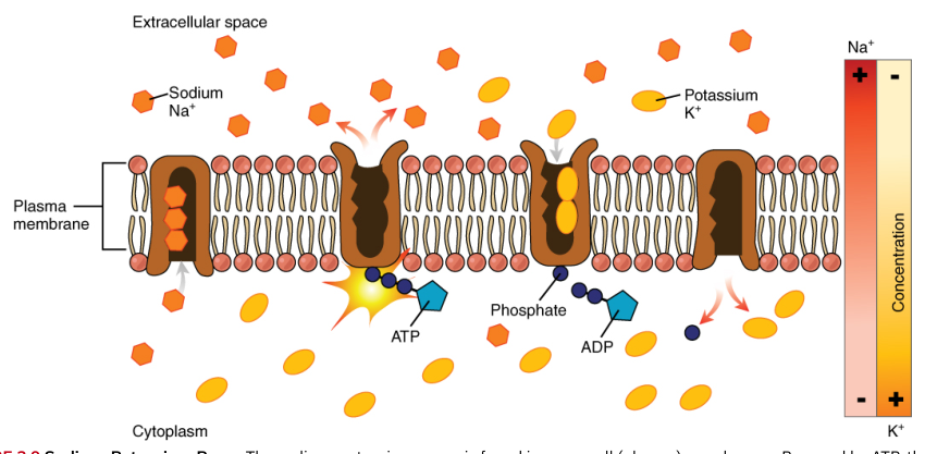
The PCT reabsorbs the largest share of filtered material. Sodium transport is central: active transport of sodium out of tubular cells helps create gradients that drive the reabsorption of chloride, bicarbonate, glucose, amino acids, and other solutes. Water follows osmotically, so the PCT recovers a large volume of fluid along with dissolved substances.
Filtered glucose and amino acids are normally reclaimed in the PCT through carrier-mediated transport. These transport systems have finite capacities. If the filtered load becomes too high, the carriers can reach their transport maximum, allowing glucose or another solute to remain in the urine.
The descending limb of the loop of the nephron permits substantial water movement but has relatively limited solute permeability. As tubular fluid descends into the increasingly concentrated medulla, water leaves the tubule and enters the interstitial fluid.
The ascending limb removes ions from tubular fluid. The thick ascending limb actively transports sodium, potassium, and chloride and is relatively impermeable to water. This separation of salt movement from water movement contributes to the medullary osmotic gradient required for concentrated urine formation.
The DCT and collecting duct perform regulated, fine-scale adjustments. Sodium reabsorption, potassium secretion, calcium reabsorption, hydrogen-ion secretion, and water permeability can all change in response to hormones and the body's needs.
Antidiuretic hormone, or ADH, increases water permeability in the late distal tubule and collecting ducts by promoting insertion of water channels into their membranes. When ADH is present, more water leaves the tubular fluid and returns to the blood. When ADH is absent, less water is reabsorbed and a larger volume of dilute urine is excreted.
The renin-angiotensin-aldosterone system, or RAAS, also changes tubular reabsorption. Angiotensin II promotes sodium reabsorption directly and stimulates aldosterone release. Aldosterone increases sodium reabsorption and potassium secretion, especially in the distal nephron. Because water follows retained sodium, RAAS activation can increase extracellular fluid volume and blood pressure.
Parathyroid hormone provides another example of regulated tubular reabsorption. When blood calcium falls, parathyroid hormone, or PTH, stimulates kidney tubule cells to reabsorb calcium from the tubular fluid, reducing urinary calcium loss. PTH also stimulates the kidneys to release or produce calcitriol, the active form of vitamin D, which increases intestinal calcium absorption.
Concrete example: After calcium levels fall, PTH acts on the kidney in two coordinated ways: calcium recovery from urine increases, and calcitriol production increases. Calcitriol then acts on the intestine to improve calcium absorption from digested food, helping restore blood calcium toward its normal range.
25.7 Regulation of Renal Blood Flow
The kidneys must receive enough blood to filter plasma, but excessive filtration would disrupt fluid balance. Renal autoregulation keeps renal blood flow and glomerular filtration relatively stable across a range of arterial pressures.
The first autoregulatory mechanism is the myogenic mechanism. When arterial pressure rises, the afferent arteriole wall stretches. Smooth muscle responds by contracting, increasing resistance and limiting the rise in glomerular blood flow. When pressure falls, the afferent arteriole relaxes, helping preserve flow into the glomerulus.
The second mechanism is tubuloglomerular feedback. The macula densa monitors sodium chloride concentration and flow in the distal tubule. High delivery can signal that filtration is excessive and promote afferent arteriolar constriction. Low delivery can promote signals that support renin release and help restore filtration and systemic pressure.
The juxtaglomerular apparatus therefore links local tubular information to vascular control. It does not merely detect pressure; it integrates information about blood flow, tubular sodium chloride, and sympathetic stimulation.
Systemic nervous and hormonal influences can override local autoregulation. Sympathetic stimulation constricts renal blood vessels, particularly during severe stress, exercise, or blood loss. This reduces renal blood flow while redirecting blood toward organs more immediately essential for maintaining circulation.
The kidney also uses local chemical signals. Some substances, such as prostaglandins and nitric oxide, promote vasodilation in appropriate contexts, whereas other signals promote vasoconstriction. The balance among these influences affects glomerular filtration and the delivery of blood to cortical and medullary tissues.
The RAAS begins when reduced renal perfusion, reduced sodium chloride delivery to the macula densa, or sympathetic stimulation causes juxtaglomerular cells to release renin. Renin cleaves liver-produced angiotensinogen to form angiotensin I. Angiotensin-converting enzyme, or ACE, especially in pulmonary blood vessels, converts angiotensin I into angiotensin II.
Angiotensin II is a potent vasoconstrictor. It increases systemic vascular resistance, supports blood pressure, promotes aldosterone release from the adrenal cortex, stimulates ADH release, and increases thirst. These actions raise blood pressure and conserve sodium and water.
Concrete example: During severe blood loss, renal perfusion falls. Renin release activates RAAS, angiotensin II constricts blood vessels, aldosterone promotes sodium retention, and ADH promotes water retention. The combined response helps preserve circulating volume, although prolonged or excessive activation can contribute to hypertension.
25.8 Endocrine Regulation of Kidney Function
The kidneys both respond to hormones and act as endocrine organs. Hormones regulate filtration, tubular transport, water balance, electrolyte composition, blood pressure, red blood cell production, and calcium metabolism.
Renin is released by juxtaglomerular cells when renal perfusion or sodium chloride delivery is low. It initiates RAAS rather than directly acting as the final blood-pressure hormone. The sequence is
$$ \text{Renin} \rightarrow \text{angiotensinogen} \rightarrow \text{angiotensin I} \rightarrow \text{angiotensin II}. $$
Angiotensin II acts on several targets. It constricts blood vessels, stimulates aldosterone release, promotes ADH secretion, and increases thirst. It also enhances sodium reabsorption in the nephron. These effects reinforce one another: retaining sodium favors water retention, while vasoconstriction raises vascular resistance.
Aldosterone is a steroid hormone released by the adrenal cortex. It acts mainly on cells in the late distal tubule and collecting duct. Aldosterone increases sodium reabsorption and promotes potassium secretion. Hydrogen-ion secretion can also increase, linking aldosterone activity with acid-base balance.
ADH, released from the posterior pituitary, responds primarily to increased plasma osmolarity or decreased blood volume. It increases water permeability in the late distal tubule and collecting ducts. In the presence of ADH, the collecting ducts allow more water to return to the hyperosmotic medullary interstitium, concentrating the urine.
The heart contributes to fluid regulation through atrial natriuretic peptide, or ANP, and related natriuretic signals. These hormones are released when cardiac chambers stretch because of increased blood volume. They promote sodium and water excretion and oppose the volume-retaining effects of RAAS and ADH.
The kidneys release erythropoietin, a hormone that stimulates red bone marrow to increase red blood cell production when renal tissues detect inadequate oxygen. Chronic kidney disease can reduce erythropoietin production and contribute to anemia.
The kidneys also convert vitamin D into calcitriol. PTH stimulates this renal activation when blood calcium is low. Calcitriol increases intestinal calcium absorption, supporting calcium homeostasis and normal bone physiology.
PTH also acts directly on kidney tubules. It increases calcium reabsorption, reducing calcium loss in urine, and decreases phosphate reabsorption in a way that promotes phosphate excretion. The coordinated result is support for an appropriate concentration of ionized calcium in the blood.
Concrete example: A fall in blood calcium stimulates PTH release. PTH increases renal calcium reabsorption and calcitriol production; calcitriol then increases intestinal calcium absorption. If blood calcium becomes high, thyroid parafollicular cells release calcitonin, which helps oppose the rise by supporting calcium deposition and reducing calcium elevation.
25.9 Regulation of Fluid Volume and Composition
The kidneys maintain the volume and chemical composition of body fluids by changing the amounts of water and solutes excreted. The controlled variables include osmolarity, sodium, potassium, calcium, phosphate, hydrogen ions, bicarbonate, and blood volume.
Osmolarity describes the concentration of dissolved particles in a solution. Blood osmolarity must remain within a narrow range because water moves across cell membranes toward the compartment with greater solute concentration. Severe hypo-osmolarity can cause cells to swell and rupture, while severe hyperosmolarity draws water out of cells.
The kidneys regulate osmolarity by adjusting water excretion relative to solute excretion. When plasma osmolarity rises, osmoreceptors promote ADH release and thirst. ADH increases water reabsorption, and drinking supplies additional water. When plasma osmolarity falls, ADH release decreases, making the collecting ducts less permeable to water.
Sodium is the principal extracellular cation and a major determinant of extracellular fluid volume. RAAS activation increases sodium reabsorption through aldosterone and angiotensin II. Conversely, natriuretic peptides promote sodium excretion. Because water follows sodium, these mechanisms strongly influence blood volume and pressure.
Potassium balance is especially important because small changes in extracellular potassium can alter membrane potentials and cardiac rhythm. Aldosterone increases potassium secretion by the distal nephron while promoting sodium reabsorption. Thus, a hormone that conserves volume also affects electrical physiology.
The kidneys help regulate acid-base balance by secreting hydrogen ions, reabsorbing filtered bicarbonate, and generating or conserving additional bicarbonate when necessary. These processes allow the kidneys to compensate for persistent metabolic acid production more slowly but more powerfully than the respiratory system.
Protein normally remains in the blood because the glomerular filtration barrier restricts large molecules and negatively charged plasma proteins. If glomerular damage allows substantial protein loss, plasma osmolarity can fall. Water then shifts toward interstitial spaces, producing edema. Severe edema can include dangerous swelling of the brain.
The kidneys also regulate calcium and phosphate. PTH increases calcium reabsorption and stimulates calcitriol formation, while influencing phosphate handling. These actions coordinate renal excretion with intestinal absorption and bone storage.
Urine concentration depends on the medullary osmotic gradient, the permeability of different nephron segments, and ADH. The loop of the nephron establishes the gradient through countercurrent multiplication: the ascending limb removes solute without equivalent water movement, while the descending limb allows water to leave. The vasa recta help preserve the gradient as blood circulates through the medulla.
Concrete example: After prolonged sweating, plasma osmolarity and blood volume may both change. Increased ADH conserves water, while RAAS activation conserves sodium and stimulates thirst. The resulting urine is typically lower in volume and higher in concentration than urine produced after abundant water intake.
25.10 The Urinary System and Homeostasis
Homeostasis depends on the urinary system's ability to adjust output rather than merely remove waste. By modifying filtration, reabsorption, secretion, and endocrine signaling, the kidneys stabilize the internal environment over changing conditions.
The urinary system contributes to homeostasis in several connected ways:
- It removes nitrogenous wastes and other metabolic products.
- It regulates blood volume and blood pressure.
- It controls plasma osmolarity.
- It maintains electrolyte concentrations.
- It contributes to acid-base balance.
- It supports calcium and phosphate balance.
- It stimulates red blood cell production through erythropoietin.
- It activates vitamin D through calcitriol production.
The system's logic is selective rather than wasteful. The glomeruli filter a plasma-like fluid, excluding most cells and proteins. The tubules then reclaim valuable molecules and water, secrete additional unwanted substances, and adjust the final composition under hormonal control.
A useful summary is:
$$ \text{Blood} \xrightarrow{\text{glomerular filtration}} \text{initial filtrate} \xrightarrow{\text{reabsorption and secretion}} \text{urine} \xrightarrow{\text{ureters}} \text{bladder} \xrightarrow{\text{urethra}} \text{outside the body}. $$
The first arrow is highly permissive for small solutes but restrictive for cells and most proteins. The second arrow is highly selective and regulated. The final arrows transport and store the product rather than chemically modify it.
Kidney failure demonstrates why these processes matter. If filtration declines severely, wastes accumulate in the blood. If tubular regulation fails, water, potassium, sodium, calcium, phosphate, and acid-base balance can become abnormal. If endocrine functions fail, anemia and disordered calcium metabolism may develop even when urine formation is considered separately.
The urinary system also works with other organ systems. The cardiovascular system supplies the pressure and blood flow required for filtration. The endocrine system provides ADH, aldosterone, PTH, and natriuretic peptides. The respiratory system helps regulate carbon dioxide and acid-base status. Bone and intestine participate in calcium and phosphate balance, while the nervous system influences thirst, sympathetic renal control, and micturition.
Concrete example: Consider a person who loses water through sweating. Reduced circulating volume decreases renal perfusion, encouraging renin release. RAAS raises vascular resistance and promotes sodium retention; ADH increases water reabsorption; thirst encourages water intake. At the nephron, the loop and collecting duct concentrate the remaining urine. The response illustrates how filtration, tubular transport, hormones, circulation, behavior, and excretion operate as one homeostatic system.
The most important distinction is between filtrate and urine. Filtrate is the initial, plasma-like fluid entering the nephron. Urine is the final fluid remaining after the nephron has recovered useful substances, secreted selected wastes, adjusted water content, and responded to the body's regulatory signals. Understanding that distinction explains how the kidneys can process approximately $200$ liters of filtrate daily while excreting less than $2$ liters of urine.
Content adapted from OpenStax Anatomy and Physiology 2e by J. Gordon Betts et al., licensed CC BY 4.0.
Source Materials
Study Anatomy and Physiology on Lykke
This course page is free to open: signed out, you can read its first two sections, and signing in, on any plan, opens the rest. The free Lykke Chrome extension syncs your own Canvas courses, so ChatGPT, Claude and Muse can answer from your classes and deadlines. It only reads Canvas.
Add the free extensionSign in to open every sectionAll sections of Anatomy and Physiology · Browse more course pages