From the free course Anatomy and Physiology on Lykke
Chapter 9: Joints
Key concepts: Joints (articulations) and their role in connecting bones or bone and cartilage · Structural classification of joints: fibrous, cartilaginous, and synovial · Functional classification: synarthrosis, amphiarthrosis, and diarthrosis · Relationship between joint mobility and stability · Fibrous joints and their strong connective-tissue connections · Cartilaginous joints, including symphyses · Synovial joints and their fluid-filled joint cavities · Movement axes and planes in diarthroses · Intervertebral joints and discs · Examples of joints in the skull, forearm, leg, pelvis, and vertebral column
By the end of this chapter the student can: Discuss both functional and structural classifications for body joints; Describe the characteristic features for fibrous, cartilaginous, and synovial joints and give examples of each; Define and identify the different body movements; Discuss the structure of specific body joints and the movements allowed by each; Explain the development of body joints.
Chapter 9: Joints
A kayak stroke, a handshake, and the protection of the brain all depend on joints, places where skeletal elements meet. A joint may connect two adjacent bones or bring a bone and cartilage together. Without these articulations, muscles could pull on bones but could not change the position of one body region relative to another.
Joints are also called articulations. They do more than permit movement: they connect the skeleton, transmit forces, provide stability, and sometimes protect internal organs. The skull, for example, is built mainly from immobile joints that help protect the brain, whereas joints of the limbs permit the extensive movement needed to walk, dance, grasp, or kick.
Two Ways to Classify Joints
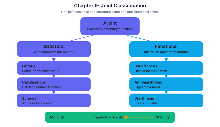
Joints are classified in two complementary ways. Structural classification asks what physically connects the bones and whether a joint cavity is present. Functional classification asks how much movement occurs between the bones. These systems describe different properties, so a single joint can receive one structural name and one functional name.
| Classification | Main question | Categories |
|---|---|---|
| Structural | What connects the bones? Is a joint cavity present? | Fibrous, cartilaginous, or synovial |
| Functional | How much movement is possible? | Synarthrosis, amphiarthrosis, or diarthrosis |
The two classification systems are related but not identical. Fibrous and cartilaginous joints are generally immobile or only slightly movable, while synovial joints are freely movable. However, the exact movement of a fibrous or cartilaginous joint depends on its location and the strength and arrangement of the tissues connecting it.
A central principle is the relationship between mobility and stability. In general, increasing mobility means decreasing stability. Joints that allow little or no movement can strongly unite bones, support body weight, or protect organs. Joints that allow extensive movement must accept greater mechanical instability and therefore depend on surrounding ligaments, muscles, and joint shape for control.
Functional Classification: How Much Movement?
The functional classification describes joint mobility using three terms. A synarthrosis is an immobile joint. An amphiarthrosis is a slightly movable joint. A diarthrosis is a freely movable joint. The word arthron refers to a joint, while the prefixes help indicate the amount of movement.
Synarthroses are designed primarily for firm connection and protection. The joints between many skull bones are examples: their limited movement helps form a strong enclosure for the brain. Amphiarthroses permit small movements that can absorb forces or provide flexibility without sacrificing substantial stability. Diarthroses provide the broad movement needed for most limb actions.
Freely movable diarthroses can be further described according to the number of movement axes they permit. A uniaxial joint allows movement primarily in one plane or around one axis. A biaxial joint permits movement in two planes. A multiaxial joint permits movement in all three planes. The movement available at a particular joint is determined not only by its classification but also by neighboring bones, ligaments, and the shape of its articulating surfaces.
Structural Classification: What Connects the Bones?
Structural classification divides joints into fibrous, cartilaginous, and synovial joints. The first two categories connect bones directly through strong connective tissue or cartilage. Synovial joints differ because the bones are not directly joined to one another; instead, their articulating surfaces face each other across a fluid-filled joint cavity.
Fibrous Joints
Fibrous joints are united by dense connective tissue, especially strong collagen fibers. They do not have a joint cavity. Their connective-tissue connections bind adjacent bones firmly, so fibrous joints are functionally classified as either synarthroses or amphiarthroses, depending on their location and the amount of movement permitted.
Sutures
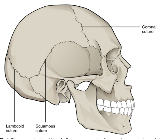
A suture is a fibrous joint between the bones of the skull. Interlocking edges and dense connective tissue create a strong connection with essentially no movement in the adult skull. This arrangement illustrates how immobility can be advantageous: the joint contributes to a protective case around the brain rather than providing a route for limb-like motion.
Syndesmoses
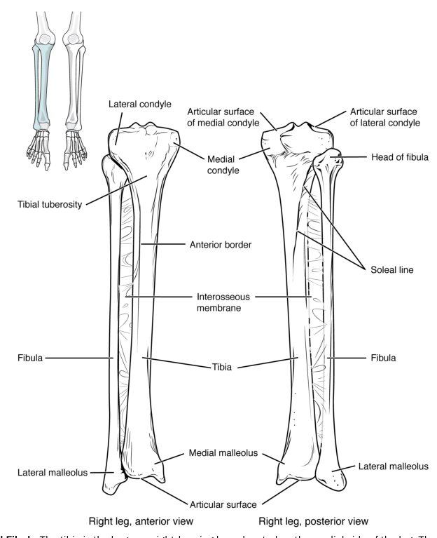
A syndesmosis is a fibrous joint in which bones are connected by ligaments or by a sheet of connective tissue called an interosseous membrane. The interosseous membrane between the radius and ulna forms a syndesmosis. It binds the forearm bones together while allowing limited movement between them, making this joint an amphiarthrosis. Syndesmoses are also found between the tibia and fibula of the leg.
The radius–ulna interosseous membrane demonstrates that a strong connection does not always mean absolute immobility. The tissue can stabilize the paired bones and help transmit forces while still permitting the small relative movements required during forearm function.
Gomphoses
A gomphosis is a fibrous joint that anchors a tooth in its bony socket. The tooth is held in place by dense connective tissue, forming a strong connection specialized for resisting the forces of biting and chewing. A gomphosis is functionally a synarthrosis because movement is extremely limited.
Cartilaginous Joints
Cartilaginous joints connect bones through cartilage and contain no joint cavity. Cartilage provides firm support while allowing more flexibility than a direct bony connection. Like fibrous joints, cartilaginous joints are functionally classified as synarthroses or amphiarthroses according to how much movement they permit.
Symphyses
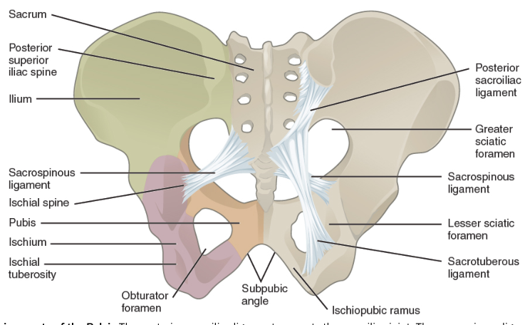
A symphysis is a cartilaginous joint in which bones are united by fibrocartilage. Fibrocartilage is well suited to locations that must resist compression while allowing a small amount of movement. The pubic symphysis is a cartilaginous amphiarthrosis: it firmly connects the left and right pubic bones but permits limited movement.
The joints between the vertebrae provide another important example. Adjacent vertebrae are connected by intervertebral discs, which are made of fibrocartilage and form symphyses. Each individual intervertebral joint permits only a small amount of movement, but the vertebrae work together as a column. The small movements of many joints can add together to produce a large overall range of bending and rotation of the vertebral column.
This is a crucial distinction between local and whole-body movement: a joint does not need to move extensively by itself to contribute to substantial movement of a body region. The vertebral column gains flexibility through the combined effect of many slightly movable joints, while the discs and surrounding connective tissues help maintain support and distribute forces.
Synovial Joints
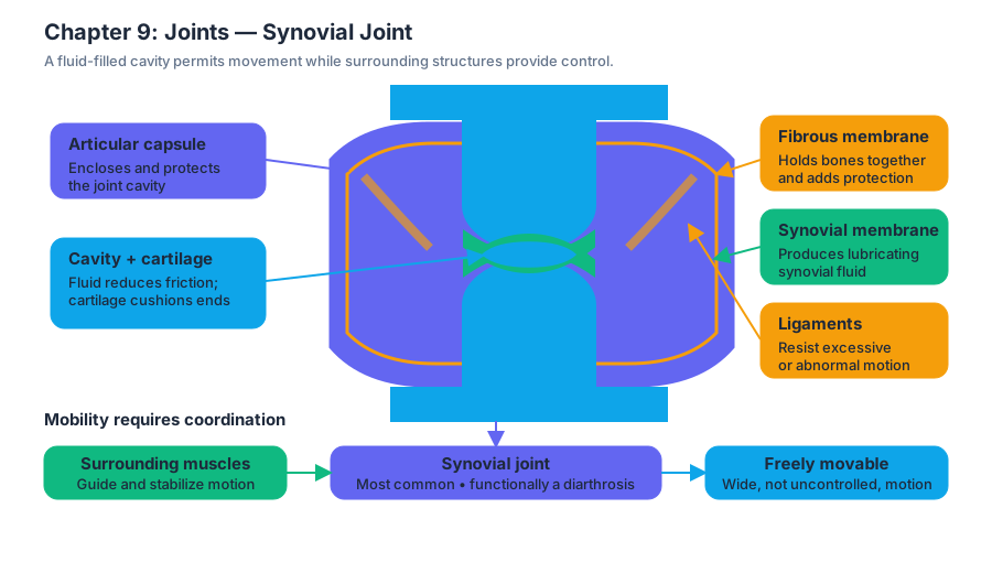
Synovial joints are the most common joints of the body and are functionally classified as diarthroses. Unlike fibrous and cartilaginous joints, the articulating bones are not directly connected. Their bone ends come into contact within a joint cavity, a space filled with lubricating synovial fluid. This arrangement permits free movement between the bones.
The joint cavity is enclosed by an articular capsule. Its outer fibrous membrane helps hold the bones together and provides structural protection. Its inner synovial membrane produces synovial fluid. The articulating bone surfaces are covered by a thin layer of articular cartilage, which cushions the bone ends and reduces friction as the joint moves. Ligaments reinforce the joint by resisting excessive or abnormal movement.
The phrase “freely movable” does not mean “uncontrolled.” A synovial joint can move widely only because several structures coordinate mobility with stability. The cavity and fluid reduce friction, articular cartilage protects the contacting surfaces, the capsule encloses the joint, ligaments limit excessive motion, and surrounding muscles actively guide and stabilize the joint.
Joint Shape, Stability, and Movement Axes
The shape of a synovial joint helps determine which movements are possible. A hinge-like arrangement favors movement primarily in one plane and is therefore uniaxial. A joint with surfaces shaped to permit movement in two planes is biaxial. A multiaxial joint permits movement in all three planes, although surrounding ligaments and neighboring bones may restrict the theoretical range.
A saddle joint has articulating surfaces that are each concave in one direction and convex in the other. The surfaces fit together like a rider sitting on a saddle, allowing biaxial movement. The first carpometacarpal joint, between the trapezium and the first metacarpal bone at the base of the thumb, is the primary example. Its structure allows the thumb to move away from the palm and across the palm, supporting the thumb’s broad range of functional movement.
A plane joint has relatively flat or slightly curved articulating surfaces. Its surfaces may allow several kinds of movement, including rotation, so a plane joint can be considered multiaxial based on shape. In practice, however, the movement of an individual plane joint is often limited by tight ligaments and neighboring bones. Plane joints occur between the carpal bones of the wrist, between the tarsal bones of the foot, and between the clavicle and acromion of the scapula.
This explains why structural possibility and actual movement are not identical. A joint’s surfaces may theoretically permit several directions of movement, but ligaments, surrounding bones, and muscle control determine how much movement is actually available. Stability is therefore produced by the combined design of the joint rather than by one structure alone.
The Mobility–Stability Trade-Off in the Body
The skeleton must solve two competing mechanical problems: it must be stable enough to bear loads and protect tissues, yet mobile enough to position the body. Fibrous joints such as skull sutures prioritize protection. Syndesmoses stabilize paired bones such as the radius and ulna or tibia and fibula. Cartilaginous symphyses support and absorb forces while permitting limited flexibility. Synovial joints prioritize movement but require capsules, ligaments, cartilage, synovial fluid, and muscle activity to prevent harmful motion.
The best example of stability emerging from many small movements is the vertebral column. Each intervertebral symphysis moves only slightly, but the combined movements of the vertebrae produce substantial bending and rotation. Conversely, the extensive movement of limb joints is possible because their surrounding connective tissues and muscles continually limit and guide that movement. Joint function is therefore not simply a choice between “moving” and “not moving”; it is a carefully balanced interaction between connection, permitted axes, friction reduction, and resistance to excessive force.
A common misconception is that a freely movable joint is inherently weak or that an immobile joint is always mechanically superior. In reality, each arrangement is specialized. A skull suture is stable because protection is its priority, while a thumb saddle joint is valuable because controlled mobility is its priority. The functional success of a joint depends on matching its mobility and stability to the demands placed on that body region.
Content adapted from OpenStax Anatomy and Physiology 2e by J. Gordon Betts et al., licensed CC BY 4.0.

Source Materials
Study Anatomy and Physiology on Lykke
This course page is free to open: signed out, you can read its first two sections, and signing in, on any plan, opens the rest. The free Lykke Chrome extension syncs your own Canvas courses, so ChatGPT, Claude and Muse can answer from your classes and deadlines. It only reads Canvas.
Add the free extensionSign in to open every sectionAll sections of Anatomy and Physiology · Browse more course pages