From the free course Anatomy and Physiology on Lykke
Chapter 13: Anatomy of the Nervous System
Key concepts: Gross organization of the human nervous system into central and peripheral divisions · Embryonic development of the nervous system from the neural tube · Neural crest development and differentiation · Primary brain divisions: forebrain, midbrain, and hindbrain · Developmental relationship between the prosencephalon, telencephalon, and cerebrum · Developmental relationship between the rhombencephalon, metencephalon, and pons · Diencephalon anatomy and its position between the cerebrum and the rest of the nervous system · Neuraxis flexures associated with upright, bipedal posture · Spinal cord organization in cross-section and the central canal · Functional brain imaging and visual-cortex activation shown by fMRI
By the end of this chapter the student can: Relate the developmental processes of the embryonic nervous system to the adult structures; Name the major regions of the adult nervous system; Locate regions of the cerebral cortex on the basis of anatomical landmarks common to all human brains; Describe the regions of the spinal cord in cross-section; List the cranial nerves in order of anatomical location and provide the central and peripheral connections; List the spinal nerves by vertebral region and by which nerve plexus each supplies.
Chapter 13: Anatomy of the Nervous System
A person can balance on one foot because the nervous system continuously combines sensory information, generates motor commands, and adjusts those commands as the body moves. The same system also regulates breathing, heart rate, digestion, posture, sensation, and conscious thought. Its organization is therefore both anatomical and functional: the location of a structure helps predict what it does, and damage to that structure can produce a characteristic loss of function.
A central principle is localization of function, the idea that particular nervous-system structures are specialized for particular functions. The cerebral cortex contains regions associated with sensation, voluntary movement, language, vision, hearing, and other activities. However, localization does not mean that any function is performed by one isolated structure. Most behavior results from communication among multiple regions connected by neural pathways.
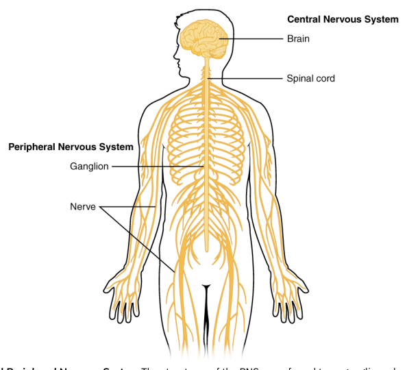
The nervous system is divided broadly into the central nervous system (CNS) and peripheral nervous system (PNS). The CNS consists of the brain and spinal cord. The PNS consists of nervous tissue outside the brain and spinal cord, including cranial nerves, spinal nerves, peripheral nerves, sensory receptors, and ganglia, which are collections of neuronal cell bodies in the periphery. Within the PNS, the somatic nervous system is associated primarily with conscious sensation and voluntary skeletal-muscle control, whereas the autonomic nervous system regulates involuntary targets such as cardiac muscle, smooth muscle, and glands.
A useful distinction is between gray matter and white matter. Gray matter contains many neuronal cell bodies, dendrites, unmyelinated axons, and supporting neuroglia. White matter is dominated by bundles of myelinated axons; myelin gives it a lighter appearance in fresh tissue. In the brain, gray matter forms the outer cerebral and cerebellar cortices and also occurs in deeper nuclei. In the spinal cord, gray matter is central and white matter surrounds it.
13.1 The Embryologic Perspective
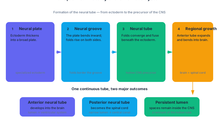
The adult nervous system begins as a relatively simple sheet of embryonic tissue. During early development, the neural plate, a thickened region of ectoderm, bends inward to form the neural groove. The raised edges of this groove are the neural folds. As the folds move toward one another and fuse, they create the neural tube, the embryonic structure that gives rise to the brain and spinal cord.
The transformation can be followed as a sequence:
- Neuroectoderm thickens to form the neural plate.
- The plate bends inward, producing the neural groove.
- The neural folds elevate along the sides of the groove.
- The folds fuse dorsally, closing the neural tube.
- The tube separates from the surface ectoderm.
- The tube’s rostral end expands into the developing brain, while its caudal portion becomes the spinal cord.
- Cells at the junction between the neural tube and surface ectoderm detach and migrate as neural crest cells.
The neural tube is the embryologic foundation of the CNS. Its lumen remains as the ventricular system of the brain and the central canal of the spinal cord. This continuity explains why the adult brain’s ventricles and the spinal cord’s central canal are not unrelated spaces: both are remnants of the internal cavity of the embryonic neural tube.
The neural crest is equally important, but it does not become the main body of the CNS. Neural crest cells migrate widely and differentiate into several peripheral structures, including many sensory and autonomic neurons, supporting cells of peripheral nerves, and other specialized tissues. Their migration is an example of how a temporary embryonic population can generate anatomically distant adult structures.
A concrete example is the formation of a spinal nerve pathway. Neurons that will contribute to a sensory ganglion arise from migrating neural crest cells. Their cell bodies remain outside the spinal cord, while their processes extend toward the body and into the CNS. The resulting ganglion belongs to the PNS, even though its axons connect directly with the spinal cord.
Primary brain vesicles
At approximately the three-to-four-week stage, the rostral neural tube enlarges into three primary brain vesicles. From anterior to posterior, these are the prosencephalon, or forebrain; the mesencephalon, or midbrain; and the rhombencephalon, or hindbrain.
The primary vesicles then produce five secondary brain vesicles:
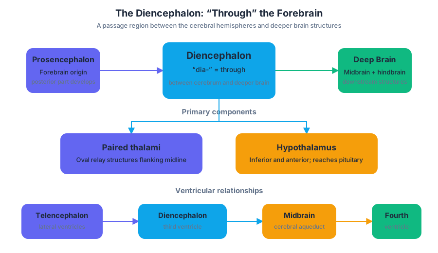
- The prosencephalon divides into the telencephalon and diencephalon.
- The mesencephalon remains undivided as the midbrain.
- The rhombencephalon divides into the metencephalon and myelencephalon.
These names describe developmental relationships rather than five unrelated adult organs. The telencephalon expands greatly and forms the cerebral hemispheres, including the cerebral cortex and underlying structures. The diencephalon forms deep forebrain structures such as the thalamus and hypothalamus. The mesencephalon becomes the adult midbrain. The metencephalon gives rise to the pons and cerebellum, while the myelencephalon becomes the medulla oblongata.
The developmental sequence is especially useful when learning adult anatomy because it links structures that occupy different positions in the mature brain. For example, the prosencephalon is the embryonic forebrain division, the telencephalon is one of its secondary vesicles, and the cerebrum is the major adult structure derived from the telencephalon. Similarly, the rhombencephalon is the embryonic hindbrain, the metencephalon is one of its secondary vesicles, and the pons is one of the adult structures derived from the metencephalon.
A common misconception is that the cerebrum develops directly from the entire forebrain as a single undivided mass. More precisely, the prosencephalon first divides, and the telencephalon then expands to form the cerebral hemispheres. The diencephalon remains deep and contributes the thalamus, hypothalamus, and related structures.
13.2 The Central Nervous System
The CNS consists of the brain and spinal cord, enclosed and protected by bone, connective-tissue coverings, and fluid. The brain occupies the cranial cavity, and the spinal cord extends through the vertebral canal. Together they receive sensory information, integrate it, and produce motor and regulatory responses.
The brain
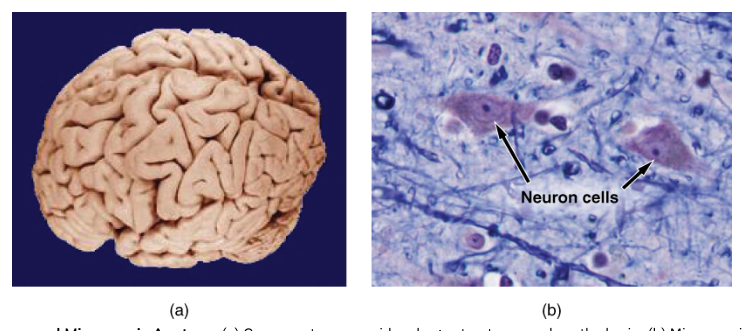
The adult brain is organized into the cerebrum, diencephalon, brain stem, and cerebellum. The cerebrum is the largest region and is divided into right and left cerebral hemispheres. A deep longitudinal fissure separates the hemispheres, while the corpus callosum, a large commissural tract, connects them. Each hemisphere is covered by a folded layer of gray matter called the cerebral cortex.
The folds of the cortex increase its surface area. Elevated ridges are gyri, shallow grooves are sulci, and deeper grooves are fissures. Major anatomical landmarks divide each hemisphere into lobes. The frontal lobe lies anterior to the central sulcus, the parietal lobe lies posterior to it, the temporal lobe lies inferior to the lateral sulcus, and the occipital lobe forms the posterior region. The insula is located deep to the lateral sulcus and is covered by portions of the frontal, parietal, and temporal lobes.
The lobes provide useful anatomical landmarks, but they should not be treated as sealed compartments. The frontal lobe contains important motor and executive regions; the parietal lobe processes much somatic sensory information; the temporal lobe includes auditory and memory-related regions; and the occipital lobe is strongly associated with visual processing. These functions depend on networks that cross boundaries within and between lobes.
Deep to the cortex is cerebral white matter, formed by myelinated axons connecting cortical areas with one another and with lower CNS regions. Deeper gray-matter masses are called basal nuclei. These nuclei participate in the regulation and coordination of movement. A simple voluntary action, such as reaching for a cup, depends not only on the motor cortex but also on sensory feedback, basal-nuclei circuits, the cerebellum, brain stem pathways, and spinal motor neurons.
The diencephalon
The diencephalon lies deep within the cerebral hemispheres and surrounds the third ventricle. Its major components include the thalamus, hypothalamus, and epithalamus. The thalamus consists of paired, ovoid masses on either side of the third ventricle. It acts as a major relay for sensory information traveling to the cerebral cortex, although olfactory information is a notable exception to the general pattern.
The hypothalamus lies inferior and anterior to the thalamus. It helps maintain homeostasis by influencing autonomic functions, endocrine activity, body temperature, hunger, thirst, and circadian rhythms. The pituitary gland attaches to the hypothalamus by a stalk-like connection. The epithalamus forms part of the roof of the third ventricle and includes the pineal gland.
For example, when body temperature rises, the hypothalamus can coordinate several responses: it can alter autonomic activity to increase heat loss, influence behavior such as seeking a cooler environment, and regulate endocrine signals. The response is distributed across systems, but the hypothalamus provides a major integrating center.
The brain stem
The brain stem is a tapering connection between the forebrain and the spinal cord. It consists of the midbrain, pons, and medulla oblongata. It contains ascending sensory tracts, descending motor tracts, and nuclei associated with several cranial nerves. It also contributes to vital functions such as cardiovascular regulation, respiratory control, consciousness, and sleep-wake states.
The midbrain is the superior part of the brain stem and is derived from the embryonic mesencephalon. It contains pathways between higher and lower regions and nuclei involved in visual and auditory reflexes. The pons forms a prominent bulge inferior to the midbrain. Derived from the metencephalon, it contains pathways linking the cerebrum, cerebellum, and spinal cord. The medulla oblongata is the inferior part of the brain stem and continues into the spinal cord. It contains centers that influence heart rate, blood pressure, and respiration.
The brain stem’s importance becomes clear in a simple example: breathing continues during sleep because respiratory control does not depend entirely on conscious cortical activity. Brain-stem networks receive chemical and sensory information and adjust respiratory output through descending pathways to spinal motor neurons.
The cerebellum
The cerebellum lies posterior to the brain stem and inferior to the posterior cerebral hemispheres. It has two hemispheres connected by a narrow central region and a highly folded cortex. Deep cerebellar white matter forms a branching pattern called the arbor vitae.
The cerebellum compares intended movement with sensory information about the movement actually occurring. It contributes to coordination, precision, balance, posture, and motor learning. It does not initiate voluntary movement in the same way the primary motor cortex does; instead, it helps make movement smooth and accurately timed.
When a person reaches for a moving object, the cerebellum receives information from the eyes, muscles, joints, vestibular system, and cerebral motor areas. It compares the expected trajectory with the actual trajectory and adjusts motor commands through connections with the brain stem and cerebral regions. Damage can produce inaccurate, poorly coordinated movement even when muscle strength remains relatively preserved.
The spinal cord
The spinal cord is a long cylindrical CNS structure that begins at the medulla oblongata and descends through the vertebral canal. It carries sensory information toward the brain and motor commands away from the brain. It also contains circuits capable of producing spinal reflexes without waiting for conscious brain processing.
In cross-section, spinal gray matter forms a roughly butterfly-shaped central region with posterior, anterior, and, in some regions, lateral horns. The posterior horns receive much sensory information. The anterior horns contain somatic motor neurons whose axons leave the spinal cord to control skeletal muscle. Lateral horns, prominent in particular spinal regions, contain autonomic motor neurons.
Surrounding the gray matter are columns of white matter: posterior, lateral, and anterior columns. These columns contain ascending and descending tracts. A withdrawal from a hot surface illustrates the arrangement: sensory neurons carry information into the posterior spinal cord, interneurons organize the reflex, and motor neurons in the anterior horn send commands to skeletal muscle. At the same time, ascending pathways notify the brain, allowing conscious awareness of pain and heat.
13.3 Circulation and the Central Nervous System
Neural tissue requires a continuous supply of oxygen and glucose, yet it is especially vulnerable to changes in blood flow. The CNS is therefore supported by specialized circulation, protective membranes, and cerebrospinal fluid. A brief interruption of blood supply can injure neurons because they have high metabolic demands and limited energy reserves.
Blood supply to the brain
The brain receives arterial blood through two paired routes: the internal carotid arteries and the vertebral arteries. The internal carotid arteries supply much of the anterior brain. The vertebral arteries ascend through the neck, unite to form the basilar artery, and supply posterior brain regions and the brain stem. Branches of these vessels form an arterial connection called the cerebral arterial circle, or circle of Willis, at the base of the brain.
The circle of Willis provides potential alternative routes for blood flow if one arterial pathway becomes narrowed. It does not guarantee that a stroke cannot occur; severe blockage or rupture can still deprive tissue of oxygen. Its importance lies in the possibility of collateral circulation among branches serving different brain regions.
Venous blood leaves the brain through cerebral veins that drain into dural venous sinuses. These channels ultimately connect with the internal jugular veins. The arrangement differs from ordinary peripheral veins because the dural sinuses are spaces formed between layers of the cranial dura mater rather than typical collapsible veins.
The blood-brain barrier
The blood-brain barrier (BBB) is a selective barrier between the blood and neural tissue. It depends principally on tight junctions between capillary endothelial cells, a continuous basement membrane, and the close association of astrocyte processes. The BBB helps stabilize the chemical environment required for neuronal signaling.
The barrier is selective rather than absolute. Oxygen, carbon dioxide, and many lipid-soluble molecules cross relatively easily. Some nutrients require transport proteins, while many toxins, pathogens, and large or charged molecules are restricted. Regions involved in monitoring the chemical composition of blood have weaker barrier properties, allowing them to detect circulating signals.
A practical example is medication delivery. A drug circulating in the blood may fail to reach effective concentrations in the brain if it cannot cross the BBB. This protection is beneficial because it limits harmful substances, but it also makes treatment of CNS disorders difficult.
Meninges and cerebrospinal fluid
The brain and spinal cord are surrounded by three connective-tissue membranes called the meninges. The outer dura mater is tough and protective. The middle arachnoid mater has a web-like appearance. The delicate inner pia mater closely follows the surface of the brain and spinal cord. The subarachnoid space between the arachnoid mater and pia mater contains cerebrospinal fluid and major blood vessels.
Cerebrospinal fluid (CSF) is a clear fluid that cushions the CNS, provides buoyancy, helps maintain a stable chemical environment, and assists in the movement of nutrients and wastes. It is produced mainly by the choroid plexuses, specialized vascular tissue within the brain’s ventricles.
CSF circulates through a defined pathway:
- It is secreted into the lateral ventricles.
- It passes through interventricular foramina into the third ventricle.
- It flows through the cerebral aqueduct into the fourth ventricle.
- It exits through openings of the fourth ventricle.
- It enters the subarachnoid space around the brain and spinal cord.
- It is reabsorbed into the dural venous sinuses through arachnoid granulations.
This circulation explains why obstruction can be dangerous. If a narrow channel such as the cerebral aqueduct becomes blocked, CSF can accumulate upstream, enlarging the ventricles and increasing pressure on brain tissue. The condition is called hydrocephalus. Thus, CSF is not merely a passive cushion; its production, movement, and reabsorption must remain balanced.
13.4 The Peripheral Nervous System
The PNS connects the CNS with the rest of the body. Its nerves carry information toward the CNS through afferent pathways and carry commands away from the CNS through efferent pathways. Afferent signals report conditions such as touch, temperature, pain, stretch, and body position. Efferent signals activate muscles and glands.
Nerves and ganglia
A nerve is a bundle of axons in the PNS. The connective-tissue layers surrounding a peripheral nerve are the endoneurium, which surrounds individual axons; the perineurium, which surrounds bundles called fascicles; and the epineurium, which surrounds the entire nerve. A ganglion is a collection of neuronal cell bodies outside the CNS.
Cranial nerves arise from the brain, whereas spinal nerves arise from the spinal cord. Each spinal nerve is formed by the union of a posterior sensory root and an anterior motor root. The posterior root contains a posterior root ganglion, which houses the cell bodies of sensory neurons. Once the roots unite, the mixed spinal nerve contains both sensory and motor fibers.
The root arrangement can be demonstrated by a pain signal from the skin. A sensory receptor detects the stimulus, and the sensory neuron carries the signal through a peripheral nerve. Its cell body lies in a posterior root ganglion. The axon enters the spinal cord through the posterior root, where it may participate in a reflex circuit and also send information toward the brain.
Spinal nerves and plexuses
There are 31 pairs of spinal nerves, organized into cervical, thoracic, lumbar, sacral, and coccygeal groups. Most spinal nerves quickly divide into branches called rami. The posterior rami supply structures of the posterior trunk, while the anterior rami supply the anterolateral trunk and limbs.
Anterior rami in several body regions interweave to form nerve plexuses. The major plexuses are the cervical plexus, brachial plexus, lumbar plexus, and sacral plexus. Thoracic anterior rami generally remain separate as intercostal nerves rather than forming a large plexus.
The cervical plexus supplies the neck and contributes to the phrenic nerve, which controls the diaphragm. The brachial plexus supplies the upper limb. The lumbar plexus supplies portions of the abdominal wall and lower limb, and the sacral plexus supplies much of the pelvis and lower limb. Plexuses redistribute fibers from spinal nerves so that a peripheral nerve often contains axons from more than one spinal cord level.
For example, injury to one spinal nerve root may weaken several muscles but usually does not completely paralyze an entire limb, because peripheral nerves commonly contain fibers from multiple spinal levels. Conversely, damage to a named peripheral nerve can produce a characteristic pattern of sensory loss and muscle weakness based on that nerve’s distribution.
Cranial nerves
The cranial nerves are twelve pairs of nerves associated with the brain. They are numbered from anterior to posterior:
- Olfactory nerve, associated with smell.
- Optic nerve, associated with vision.
- Oculomotor nerve, which controls several eye muscles and functions of the pupil.
- Trochlear nerve, which controls the superior oblique eye muscle.
- Trigeminal nerve, carrying facial sensation and controlling muscles of mastication.
- Abducens nerve, which controls the lateral rectus eye muscle.
- Facial nerve, involved in facial expression, taste from part of the tongue, and glandular secretion.
- Vestibulocochlear nerve, carrying information for hearing and balance.
- Glossopharyngeal nerve, involved in taste and sensation from the posterior tongue and in swallowing-related functions.
- Vagus nerve, with extensive sensory and motor connections involving the pharynx, larynx, thoracic organs, and abdominal organs.
- Accessory nerve, which controls selected neck and shoulder muscles.
- Hypoglossal nerve, which controls muscles of the tongue.
Cranial nerves may be sensory, motor, or mixed. Their central connections are located in the brain, while their peripheral branches reach sensory receptors, skeletal muscles, smooth muscle, cardiac muscle, and glands. The vagus nerve illustrates the broad reach of the PNS: although it begins at the brain, it carries signals to and from organs in the thorax and abdomen.
Functional divisions of the PNS
The somatic nervous system includes sensory pathways from the skin, skeletal muscles, and joints and motor pathways to skeletal muscle. It supports conscious sensation and voluntary movement, but somatic motor responses can also occur reflexively. The autonomic nervous system regulates cardiac muscle, smooth muscle, glands, and adipose tissue. Its major divisions are the sympathetic, parasympathetic, and enteric divisions.
The sympathetic division is associated with mobilizing energy and coordinating responses to stress. The parasympathetic division supports maintenance, digestion, and energy conservation. The enteric division controls much of the intrinsic activity of the gastrointestinal tract, although it is influenced by sympathetic and parasympathetic pathways.
Consider touching a hot pan. Somatic sensory receptors detect heat and pain; afferent fibers carry the information into the spinal cord; interneurons organize a withdrawal reflex; and somatic motor neurons activate skeletal muscles. Autonomic pathways may simultaneously alter cardiovascular and glandular activity. One event therefore recruits both central processing and coordinated peripheral pathways.
The CNS and PNS should not be viewed as independent systems. The CNS integrates information and establishes responses, while the PNS provides the communication lines that connect those decisions to the body and bring new sensory information back. The embryonic neural tube establishes the CNS, neural crest cells contribute extensively to the PNS, and the mature nervous system preserves those developmental relationships in its anatomy.
Content adapted from OpenStax Anatomy and Physiology 2e by J. Gordon Betts et al., licensed CC BY 4.0.
Source Materials
Study Anatomy and Physiology on Lykke
This course page is free to open: signed out, you can read its first two sections, and signing in, on any plan, opens the rest. The free Lykke Chrome extension syncs your own Canvas courses, so ChatGPT, Claude and Muse can answer from your classes and deadlines. It only reads Canvas.
Add the free extensionSign in to open every sectionAll sections of Anatomy and Physiology · Browse more course pages