From the free course Anatomy and Physiology on Lykke
Chapter 19: The Cardiovascular System: The Heart
Key concepts: The heart as a muscular pump · Heart location within the thorax · Pathways of oxygenated and deoxygenated blood through the heart · Aorta and pulmonary trunk as major vessels receiving blood from the heart · Electrocardiogram (ECG): process and purpose · The cardiac cycle · Cardiac output and its calculation · Effects of exercise on cardiac output and heart rate · Brain centers that control heart rate · Cardiopulmonary resuscitation (CPR) and the urgency of restoring blood flow
By the end of this chapter the student can: Identify and describe the interior and exterior parts of the human heart; Describe the path of blood through the cardiac circuits; Describe the size, shape, and location of the heart; Compare cardiac muscle to skeletal and smooth muscle; Explain the cardiac conduction system; Describe the process and purpose of an electrocardiogram; Explain the cardiac cycle; Calculate cardiac output
Chapter 19: The Cardiovascular System: The Heart
At an average rate of about $75$ contractions per minute, the heart moves approximately $14{,}000\ \text{L}$ of blood each day. Over one year, that is roughly $10{,}000{,}000\ \text{L}$ pushed through nearly $60{,}000$ miles of blood vessels. The heart is therefore not merely an organ that “contains” blood; it is a muscular pump that repeatedly generates pressure, directs blood through two circuits, and keeps tissues supplied.
The heart as a muscular pump
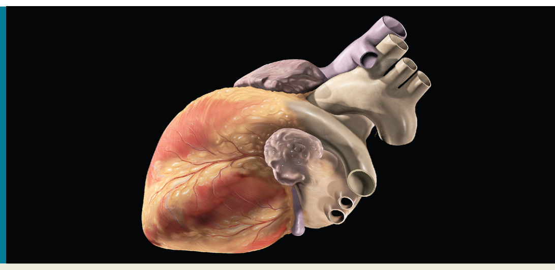
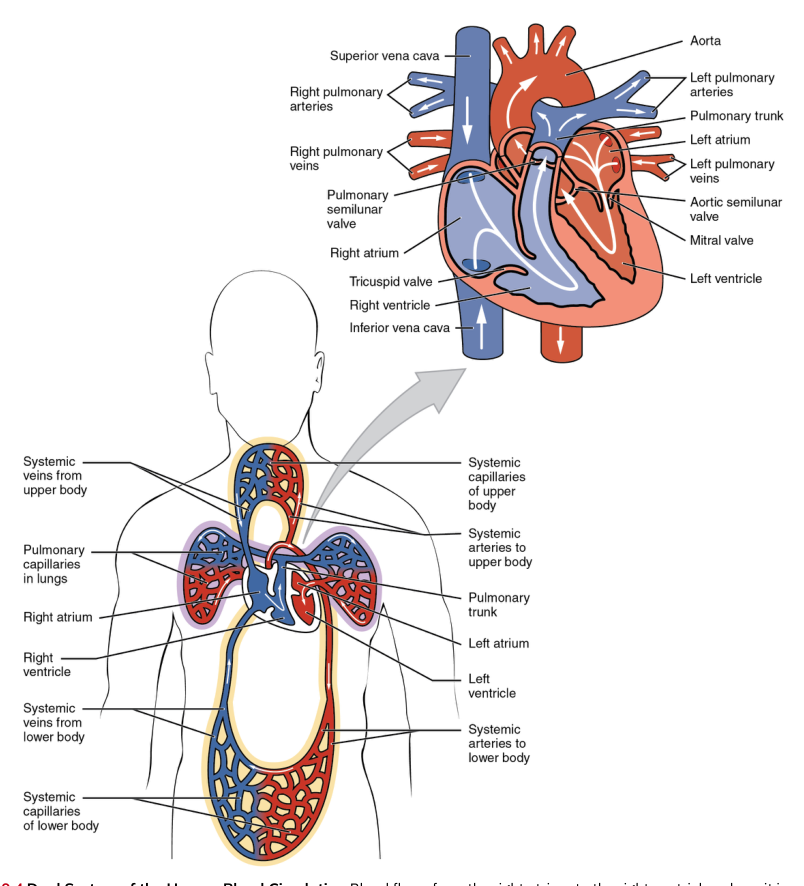
A useful mental picture is a two-sided pump arranged in series. The right side sends blood to the lungs, where gas exchange occurs; the left side sends blood to the rest of the body. Each side has an upper receiving chamber, an atrium, and a lower pumping chamber, a ventricle. The atria receive blood, while the ventricles develop the pressure needed to eject it.
The heart’s contractions are produced by cardiac muscle. When the ventricles contract, pressure rises inside them and blood is forced into the large vessels leaving the heart: the pulmonary trunk on the right and the aorta on the left. When the ventricles relax, their pressure falls and they can fill again. This alternating contraction and relaxation is the mechanical basis of the heartbeat.
Four valves keep the pump operating in one direction. The right and left atrioventricular valves lie between the atria and ventricles: the tricuspid valve is on the right, and the bicuspid, or mitral, valve is on the left. The semilunar valves lie at the exits of the ventricles: the pulmonary valve opens into the pulmonary trunk, and the aortic valve opens into the aorta. Valves do not actively push blood; pressure differences open and close them. They prevent backflow when pressure reverses.
Location and orientation in the thorax
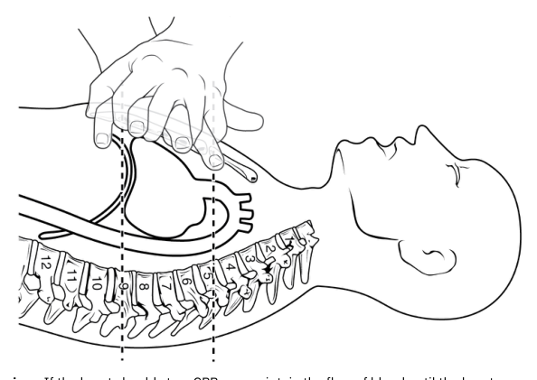
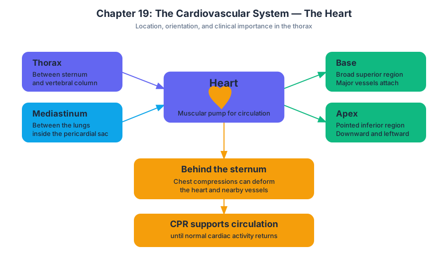
The heart lies in the thorax, between the vertebral column and the sternum. More precisely, it occupies the mediastinum between the lungs and is enclosed within the pericardial sac. Its broad superior region is the base, where the major vessels attach; its pointed inferior region is the apex, which points downward and toward the left side of the body.
This orientation matters clinically. The sternum lies anterior to the heart, and the heart’s position behind the sternum helps explain how chest compressions can deform the heart and nearby vessels. Effective cardiac pumping is essential for circulation; when pumping stops, CPR can temporarily support circulation until normal cardiac activity is restored, because the brain can sustain irreversible neuronal injury after only a few minutes without adequate blood flow.
The route of deoxygenated and oxygenated blood
The heart’s right and left sides are separated by internal partitions, so oxygen-poor and oxygen-rich blood normally do not mix. The complete pathway can be traced as a sequence of chambers, valves, and vessels:
- Deoxygenated blood enters the right atrium from the superior vena cava, inferior vena cava, and coronary sinus. The venae cavae return blood from the body, while the coronary sinus returns blood from the heart muscle itself.
- Blood moves through the tricuspid valve into the right ventricle.
- Contraction of the right ventricle opens the pulmonary semilunar valve, sending blood into the pulmonary trunk.
- The pulmonary trunk divides into pulmonary arteries that carry blood to the lungs. In the lungs, carbon dioxide leaves the blood and oxygen enters it.
- Oxygenated blood returns to the left atrium through the pulmonary veins.
- Blood passes through the bicuspid, or mitral, valve into the left ventricle.
- Contraction of the left ventricle opens the aortic semilunar valve, ejecting blood into the aorta.
- The aorta distributes blood through systemic arteries to the tissues of the body.
The terms “artery” and “vein” are defined by direction relative to the heart, not by oxygen content. Arteries carry blood away from the heart, and veins carry blood toward it. Thus, the pulmonary arteries carry deoxygenated blood, whereas the pulmonary veins carry oxygenated blood.
The cardiac cycle
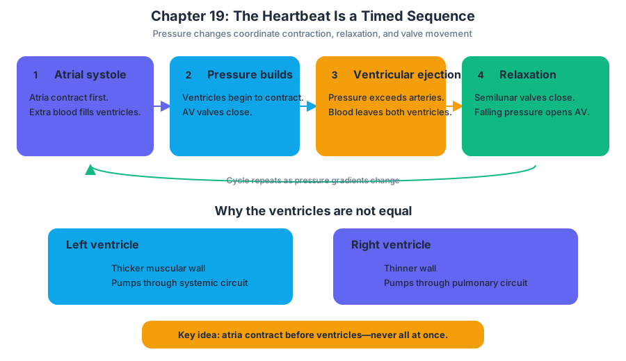
The cardiac cycle is one complete sequence of atrial contraction, ventricular contraction, and relaxation. The terms systole and diastole describe contraction and relaxation, respectively. During systole, a chamber develops pressure as its muscle contracts. During diastole, the chamber relaxes and fills.
The cycle begins with general relaxation, or ventricular diastole. Blood returning to the heart flows into the atria and passes through the open atrioventricular valves into the ventricles. The semilunar valves remain closed because pressure in the ventricles is lower than pressure in the pulmonary trunk and aorta.
Next, the atria contract. This atrial systole pushes additional blood into the ventricles, completing ventricular filling. The atrioventricular valves remain open because atrial pressure exceeds ventricular pressure.
Ventricular systole follows. As the ventricles begin to contract, ventricular pressure rises above atrial pressure, closing the atrioventricular valves. For a brief interval, all four valves are closed while the ventricles contract and pressure builds; this is called isovolumetric contraction because ventricular volume does not yet change.
When ventricular pressure exceeds pressure in the pulmonary trunk and aorta, the pulmonary and aortic semilunar valves open. Blood is ejected from the ventricles. The left ventricle has a thicker muscular wall than the right ventricle because it must generate enough pressure to propel blood through the systemic circuit, whereas the right ventricle pumps only through the pulmonary circuit.
Finally, the ventricles relax. Ventricular pressure falls below pressure in the great arteries, causing the semilunar valves to close. The ventricles then enter another brief period in which all valves are closed, followed by renewed filling as ventricular pressure falls below atrial pressure and the atrioventricular valves open.
A common misconception is that the heart “squeezes all at once.” In reality, the atria contract before the ventricles, and the valves coordinate the changing pressure gradients. The heart’s pumping action is therefore a timed sequence, not a single undifferentiated squeeze.
Cardiac output: turning each beat into a flow rate
Cardiac output is the volume of blood pumped by one ventricle in one minute. It depends on two quantities: heart rate, the number of beats per minute, and stroke volume, the amount of blood ejected by a ventricle with each beat.
$$\text{Cardiac output} = \text{heart rate} \times \text{stroke volume}$$
For example, if the heart rate is $75\ \text{beats/min}$ and the stroke volume is $70\ \text{mL/beat}$, then:
$$\text{Cardiac output} = \left(75\ \frac{\text{beats}}{\text{min}}\right)\left(70\ \frac{\text{mL}}{\text{beat}}\right)$$ $$\text{Cardiac output} = 5{,}250\ \text{mL/min}$$ $$\text{Cardiac output} = 5.25\ \text{L/min}$$
The unit “beats” cancels, leaving volume per unit time. This calculation describes the output of one ventricle; under normal conditions, the right and left ventricles pump approximately equal volumes over time. If their outputs differed substantially, blood would accumulate in one circuit.
ECG: recording the heart’s electrical activity
Mechanical contraction requires electrical coordination. An electrocardiogram, or ECG, records voltage changes at the skin produced by the heart’s electrical activity. It does not directly record the force of contraction or the volume of blood ejected. Instead, it provides an indirect but valuable record of the sequence that initiates contraction.
The normal ECG contains three major features. The P wave represents atrial depolarization, the electrical event that precedes atrial contraction. The QRS complex represents ventricular depolarization, which precedes ventricular contraction; atrial repolarization occurs at the same time but is hidden within the larger QRS signal. The T wave represents ventricular repolarization, which precedes ventricular relaxation.
Because the ECG displays timing, clinicians can evaluate heart rate and rhythm and identify conduction abnormalities. An abnormal rhythm may interfere with the coordinated opening and closing of valves or reduce ventricular filling and ejection. The ECG is therefore a recording of electrical events, while the cardiac cycle describes the corresponding mechanical events.
Exercise and cardiac output
During exercise, skeletal muscles demand more oxygen and produce more metabolic waste. The heart responds by increasing cardiac output, chiefly through an increase in heart rate and usually an increase in stroke volume. The result is greater blood flow through the pulmonary and systemic circuits, helping deliver oxygenated blood to active tissues and remove carbon dioxide.
For instance, if exercise raises heart rate to $150\ \text{beats/min}$ and stroke volume to $100\ \text{mL/beat}$:
$$ \text{Cardiac output} = \left(150\ \frac{\text{beats}}{\text{min}}\right)\left(100\ \frac{\text{mL}}{\text{beat}}\right) = 15{,}000\ \text{mL/min} = 15\ \text{L/min} $$
This is almost three times the resting output in the earlier example. The key idea is that exercise does not merely make the heart beat faster: cardiac output reflects the combined effects of heart rate and the volume ejected per beat. A faster rate with an unchanged or reduced stroke volume may not increase output as effectively, because very rapid beating shortens the time available for ventricular filling.
The heart’s extraordinary lifetime workload comes from repeating this cycle continuously: receiving blood, filling, generating pressure, ejecting blood into the pulmonary trunk and aorta, and relaxing in preparation for the next beat. Its location, valves, electrical system, muscle, and coordinated cycle are all parts of one integrated pump.
Content adapted from OpenStax Anatomy and Physiology 2e by J. Gordon Betts et al., licensed CC BY 4.0.
Source Materials
Study Anatomy and Physiology on Lykke
This course page is free to open: signed out, you can read its first two sections, and signing in, on any plan, opens the rest. The free Lykke Chrome extension syncs your own Canvas courses, so ChatGPT, Claude and Muse can answer from your classes and deadlines. It only reads Canvas.
Add the free extensionSign in to open every sectionAll sections of Anatomy and Physiology · Browse more course pages