From the free course Anatomy and Physiology on Lykke
Chapter 7: Axial Skeleton
Key concepts: Axial and appendicular skeletons · Bones and regions of the skull · Zygomatic arch and its processes · Temporal fossa and infratemporal fossa · Cranial fossae and their relationship to brain regions · Temporal bone landmarks · Mandibular fossa, articular tubercle, and temporomandibular joint · Orbit as a bony socket for the eye · Styloid process and hyoid-supporting attachments · Carotid canal and passage of a major brain-supplying artery
By the end of this chapter the student can: Describe the functions of the skeletal system and define its two major subdivisions; Identify the bones and bony structures of the skull, the cranial suture lines, the cranial fossae, and the openings in the skull; Discuss the vertebral column and regional variations in its bony components and curvatures; Describe the components of the thoracic cage; Discuss the embryonic development of the axial skeleton.
Chapter 7: Axial Skeleton
7.1 Divisions of the Skeletal System
The skeletal system is a living framework made of bones, cartilage, and ligaments. It supports the body, protects delicate organs, permits movement, stores minerals, and provides a site for blood-cell production. Bones form the rigid structure; cartilage supplies flexible support and cushioning; ligaments bind bones together at joints and restrict excessive movement.
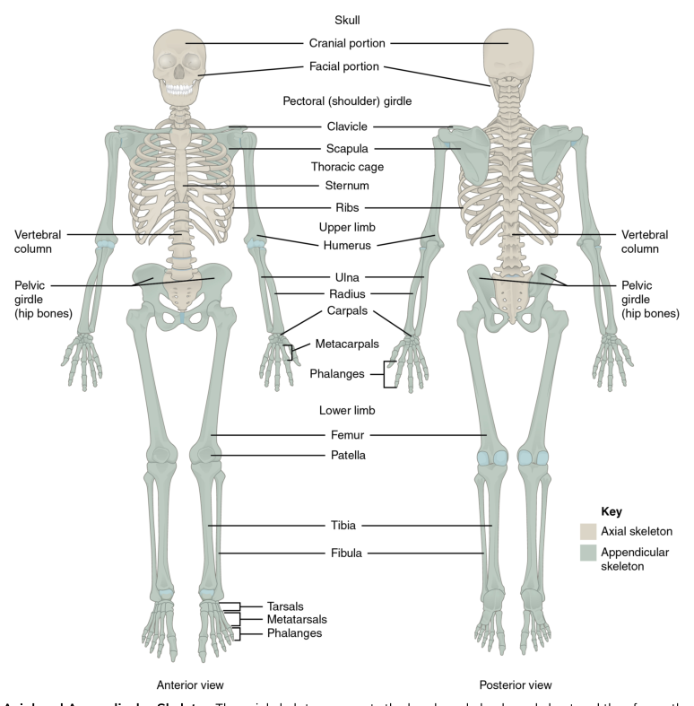
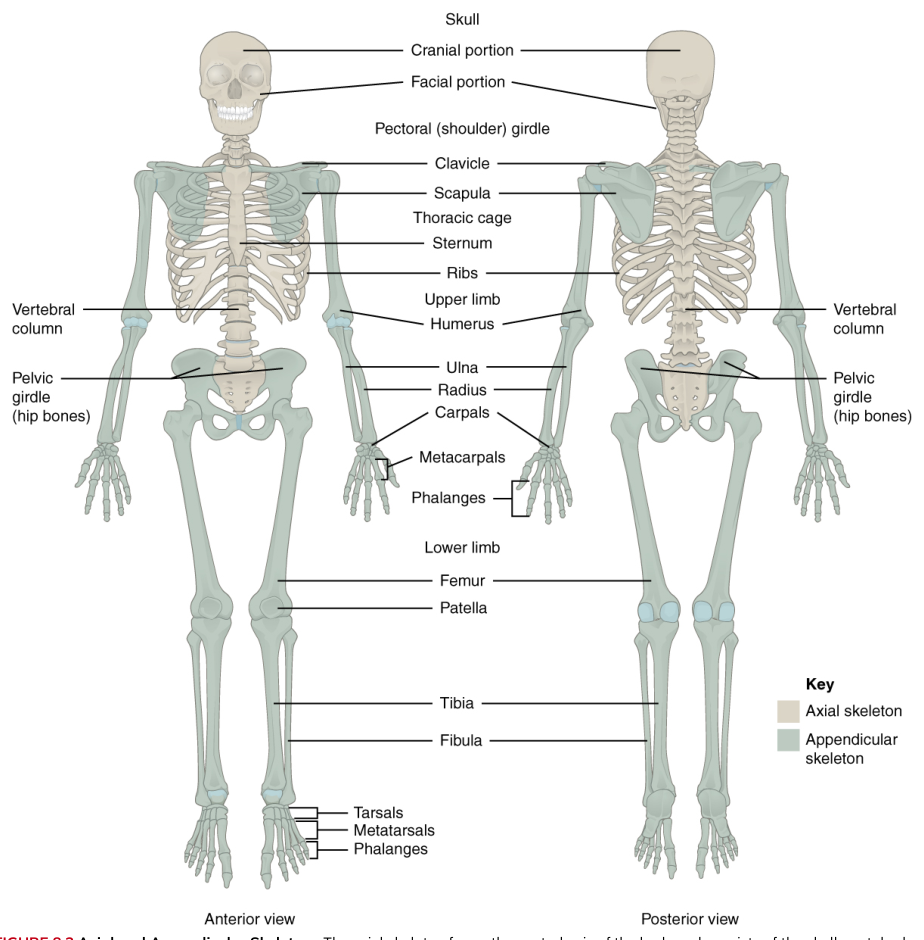
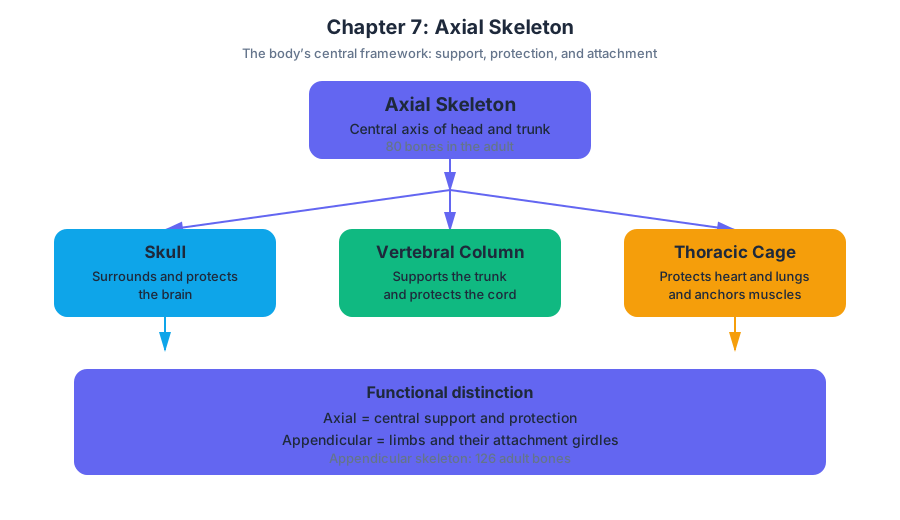
The skeleton has two major subdivisions: the axial skeleton and the appendicular skeleton. The axial skeleton forms the central axis of the body. The appendicular skeleton consists of the bones of the limbs and the bones that attach the limbs to the axial skeleton.
| Division | Principal components | Main structural role |
|---|---|---|
| Axial skeleton | Skull, vertebral column, and thoracic cage | Forms the central axis and protects the brain, spinal cord, and organs of the thorax |
| Appendicular skeleton | Pectoral girdles, upper limbs, pelvic girdle, and lower limbs | Attaches the limbs to the axial skeleton and enables movement |
The axial skeleton includes the skull, vertebral column, and thoracic cage. The skull protects the brain and supports the structures of the face. The vertebral column surrounds and protects the spinal cord while supporting the head and trunk. The thoracic cage protects the heart and lungs and provides attachment sites for muscles involved in breathing and movement.
The appendicular skeleton includes the pectoral girdle, formed by the clavicles and scapulae; the bones of the upper limbs; the pelvic girdle, formed by the hip bones; and the bones of the lower limbs. Its name comes from appendages, meaning structures attached to a central axis. The appendicular skeleton is therefore specialized for mobility, whereas the axial skeleton is more strongly associated with support and protection.
A useful way to distinguish the divisions is to trace a limb inward. The fingers, hand, forearm, arm, and shoulder girdle belong to the appendicular skeleton. The sternum and ribs to which some shoulder and chest muscles attach belong to the axial skeleton. The boundary is anatomical, not functional: muscles often cross from one division to the other to move the limbs or stabilize the trunk.
The skeleton does not act as a collection of isolated struts. Cartilage provides flexible support in the external ear, trachea, larynx, and thoracic cage. At joints, cartilage may unite adjacent bones or form a smooth, resilient surface that reduces friction and absorbs compression. Ligaments are strong bands of connective tissue that hold bones together at movable joints. They help guide normal movement and prevent movements large enough to damage the joint.
Bone also functions as a metabolic tissue. The skeleton stores minerals, especially calcium and phosphate, which can be released into the blood when needed. Within certain bones, red bone marrow produces blood cells. Thus, the skeleton is not an inert scaffolding: it is mechanically active, chemically responsive, and continuously remodeled.
Concrete example: When a person jumps, the femur, tibia, and foot bones transmit forces through the appendicular skeleton, while the vertebral column and pelvis stabilize the body’s central axis. The ribs and sternum remain part of the axial skeleton even though muscles attached to them help maintain posture and control breathing during the jump.
Key distinction: The axial skeleton is the body’s central protective and supporting axis; the appendicular skeleton consists of the limb bones and the girdles that attach those limbs to the axis.
7.2 The Skull
The skull is both a protective case for the brain and a structural platform for the face, sensory organs, muscles of facial expression, chewing muscles, and the openings through which nerves and blood vessels pass. It is made of cranial bones, which enclose the brain, and facial bones, which form the framework of the face, the nasal cavity, the orbit, and the jaws.
Cranial and facial bones
The cranium is the portion of the skull that surrounds the brain. Its bones include the unpaired frontal, occipital, sphenoid, and ethmoid bones, together with the paired parietal and temporal bones. The facial skeleton includes the paired maxillae, zygomatic bones, nasal bones, lacrimal bones, palatine bones, and inferior nasal conchae, as well as the unpaired mandible and vomer.
The frontal bone forms the forehead, the roof of the orbit, and much of the anterior cranial floor. The paired parietal bones form most of the superior and lateral walls of the cranium. The occipital bone forms the posterior and inferior skull and contains the foramen magnum, the large opening through which the spinal cord connects with the brain.
The temporal bones form the inferolateral walls of the cranium and contribute to the skull base. They surround structures of the ear and articulate with the mandible. The sphenoid bone lies centrally at the skull base and connects with many other cranial bones. The ethmoid bone contributes to the nasal septum, nasal cavity, and medial orbital walls.
The paired zygomatic bones form the prominence of the cheeks and part of the lateral walls and floors of the orbits. The maxillae form the upper jaw, the anterior hard palate, and portions of the nasal cavity and orbits. The mandible is the lower jaw and is the only freely movable bone of the skull, moving at the temporomandibular joints.
Sutures and surface landmarks
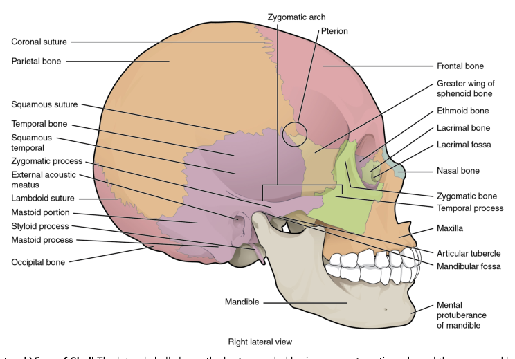
Most skull bones meet at immovable fibrous joints called sutures. The coronal suture lies between the frontal and parietal bones. The sagittal suture runs between the two parietal bones. The lambdoid suture separates the parietal bones from the occipital bone. On the lateral skull, the squamous suture joins the parietal and temporal bones.
The pterion is a region where the frontal, parietal, temporal, and sphenoid bones meet. It is clinically important because the bone is relatively thin in this area and lies near the middle meningeal artery. A blow to the side of the head can fracture the pterion and damage this vessel.
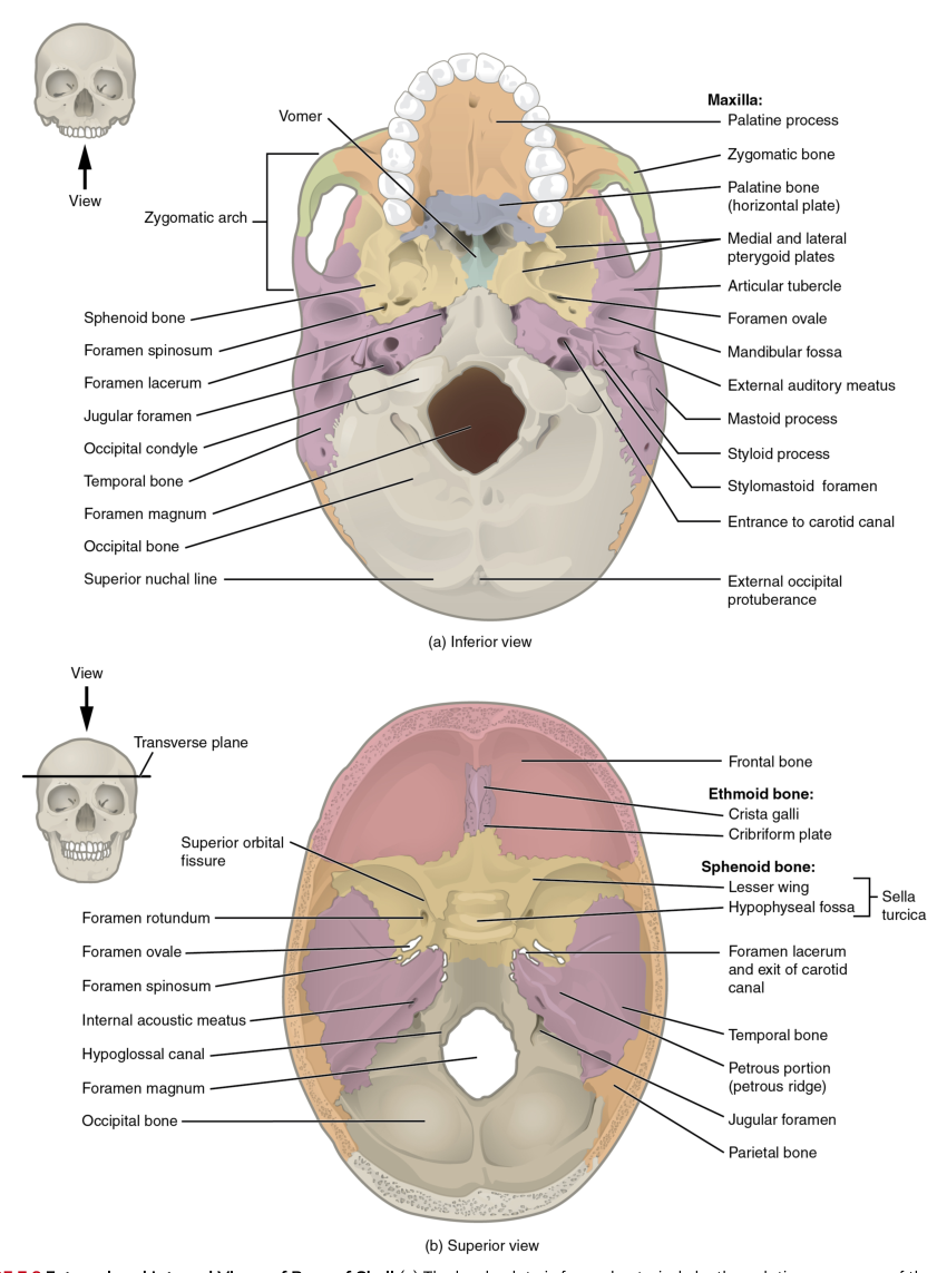
The skull also contains many openings and depressions. A foramen is an opening through a bone, usually for a nerve or blood vessel. A canal is a passageway through bone. A fossa is a shallow depression, often serving as a surface for a brain region, muscle, or gland. These landmarks are not decorative names: they describe the routes and spaces required by the nervous and circulatory systems.
The zygomatic arch and temporal fossae
The zygomatic arch is the bony bridge on each side of the skull that forms the cheek region. It is created by the union of the temporal process of the zygomatic bone with the zygomatic process of the temporal bone. This arch provides attachment for muscles and forms a boundary between the temporal region above and the infratemporal region below.
The temporal fossa is the broad depression on the lateral side of the skull, superior to the zygomatic arch. It is bounded in part by the temporal lines and contains the temporalis muscle, which elevates and retracts the mandible during chewing. The infratemporal fossa lies deep to the zygomatic arch and inferior to the temporal fossa. It contains muscles, vessels, and nerves associated with mastication.
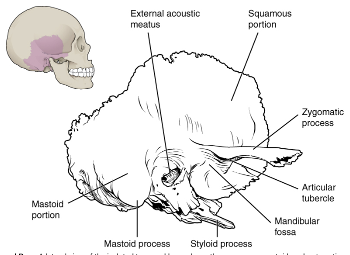
The temporal bone has several named regions and projections. Its squamous portion is the relatively flat, thin part that forms the lateral cranial wall. The zygomatic process projects anteriorly to meet the zygomatic bone. The mastoid portion lies posteriorly and includes the rounded mastoid process, which provides attachment for several neck muscles. The slender styloid process projects inferiorly and serves as an attachment site for muscles and ligaments of the tongue, pharynx, and hyoid region.
The external acoustic meatus is the ear canal opening in the temporal bone. Anterior to it are the mandibular fossa and articular tubercle. The mandibular condyle fits into the mandibular fossa to form the temporomandibular joint; the articular tubercle helps limit excessive forward displacement of the mandible.
Concrete example: During chewing, the temporalis muscle occupies the temporal fossa and pulls on the mandible. The muscle’s force is transmitted through the coronoid process of the mandible, while the mandibular condyle rotates and glides against the mandibular fossa and articular tubercle of the temporal bone.
Cranial fossae and skull openings
The internal floor of the cranium is divided into three cranial fossae: the anterior, middle, and posterior cranial fossae. These are not separate sealed chambers. They are progressively deeper regions of the skull base that support different parts of the brain.
The anterior cranial fossa is the shallowest and most anterior. It supports the frontal lobes of the cerebrum. Its floor is formed mainly by the frontal bone, with contributions from the ethmoid and sphenoid bones. The ethmoid’s cribriform plate contains numerous small openings through which the olfactory nerves pass from the nasal cavity to the brain.
The middle cranial fossa is deeper than the anterior fossa and supports the temporal lobes of the cerebrum. It is formed largely by the sphenoid and temporal bones. The sella turcica, a saddle-shaped region of the sphenoid bone, houses the pituitary gland. Other openings in this region transmit cranial nerves and blood vessels.
The posterior cranial fossa is the deepest and largest of the three. It supports the cerebellum and the brainstem. It is formed mainly by the occipital bone, with contributions from the temporal and sphenoid bones. The foramen magnum opens in this fossa and allows passage of the spinal cord, vertebral arteries, and associated coverings.
The orbit is a bony socket formed by seven bones: the frontal, sphenoid, zygomatic, maxilla, palatine, ethmoid, and lacrimal bones. At the posterior orbit, the optic canal transmits the optic nerve and the ophthalmic artery. The superior orbital fissure transmits nerves and vessels between the cranial cavity and orbit.
A reliable way to interpret a skull opening is to follow three steps: first, identify its location; second, identify the cavity or region it connects; third, determine whether it provides passage for a nerve, artery, vein, or other structure. This prevents the common misconception that all foramina serve the same purpose.
The mandible and hyoid bone
The mandible consists of a horizontal body and two vertical rami. Each ramus ends superiorly in a posterior condylar process, which participates in the temporomandibular joint, and an anterior coronoid process, which receives attachment from the temporalis muscle. The alveolar process contains the sockets for the lower teeth. The mental protuberance forms the chin.
On the medial surface of each ramus is the mandibular foramen, an opening for the inferior alveolar nerve and vessels. The lingula, a small tongue-shaped projection near the mandibular foramen, helps identify this region. An injection for an inferior alveolar nerve block is placed near this opening to numb the lower teeth.
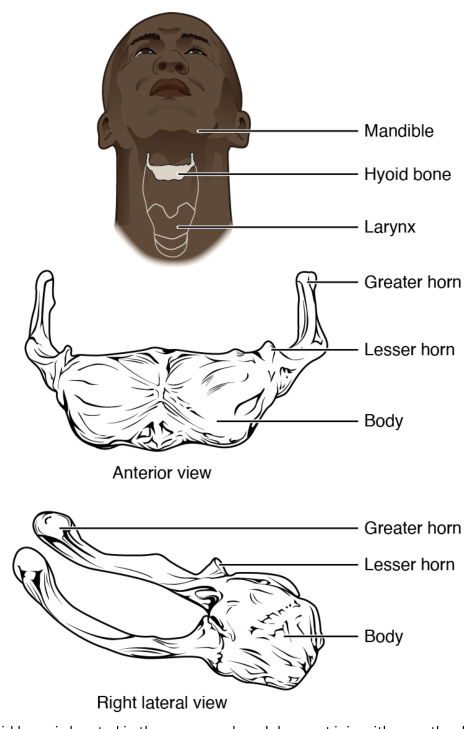
The hyoid bone is suspended in the neck by muscles and ligaments rather than articulating directly with another bone. It supports the tongue and provides attachment for muscles involved in swallowing and speech. Its position allows the larynx and tongue to move while remaining mechanically connected to the skull and mandible.
7.3 The Vertebral Column
The vertebral column is a flexible, segmented column that supports the head and trunk, protects the spinal cord, provides attachment for ribs and muscles, and permits bending and rotation. It normally contains $33$ vertebrae, although some fuse during development to form the sacrum and coccyx.
Regions and normal curvatures
The vertebral column is divided into five regions: $7$ cervical vertebrae in the neck, $12$ thoracic vertebrae in the upper and middle back, $5$ lumbar vertebrae in the lower back, $5$ fused sacral vertebrae, and usually $4$ fused coccygeal vertebrae.
The column’s curvatures distribute body weight and help absorb shock. The cervical and lumbar regions have lordotic curvatures, which are convex anteriorly. The thoracic and sacral regions have kyphotic curvatures, which are convex posteriorly. These curves are not abnormalities; they are normal features of the adult spine.
At birth, the vertebral column has a single primary curvature that is concave anteriorly. The cervical and lumbar curvatures develop later as secondary curvatures. The cervical curvature develops when an infant begins to hold up the head; the lumbar curvature develops as the infant begins to stand and walk.
Typical vertebral structure
A typical vertebra has an anterior body, or centrum, and a posterior vertebral arch. The body is the thick, weight-bearing portion. The vertebral arch surrounds the vertebral foramen, the opening through which the spinal cord passes. When vertebrae are stacked, their vertebral foramina form the vertebral canal.
The vertebral arch includes paired pedicles and laminae. It also gives rise to a median spinous process and paired transverse processes. These processes provide attachment sites for muscles and ligaments and act as levers that increase the mechanical advantage of spinal muscles.
Each vertebra has paired superior and inferior articular processes. Their articular facets form joints with neighboring vertebrae and help control the direction and range of movement. Between adjacent vertebrae are intervertebral foramina, openings through which spinal nerves leave the vertebral canal.
Between most vertebral bodies are intervertebral discs. Each disc has an outer annulus fibrosus, made of fibrocartilage, and an inner nucleus pulposus, a gel-like core. The discs bind vertebrae together, permit limited movement, and absorb compression.
Concrete example: When a person lands from a small jump, the intervertebral discs deform slightly and distribute compressive forces across the vertebral bodies. The vertebral curvatures and surrounding muscles then help prevent the force from concentrating at a single point.
Regional differences
Cervical vertebrae are relatively small and designed for mobility. Most have a transverse foramen in each transverse process, allowing passage of vertebral arteries and veins. The first two cervical vertebrae are specialized. The atlas, or C1, supports the skull and lacks a body. The axis, or C2, has the tooth-like dens, around which the atlas and head rotate.
The seventh cervical vertebra, called the vertebra prominens, has a long, easily palpable spinous process. This landmark can help identify the transition between the cervical and thoracic regions.
Thoracic vertebrae articulate with ribs. Their bodies and transverse processes contain facets for rib attachment. Thoracic spinous processes are generally long and angle inferiorly. Lumbar vertebrae have large, thick bodies because they bear substantial body weight. Their short, broad spinous processes provide attachment for powerful lower-back muscles.
The sacrum is a triangular bone formed by the fusion of five sacral vertebrae. It forms the posterior part of the pelvic girdle and articulates laterally with the hip bones at the sacroiliac joints. The coccyx is a small terminal bone formed by fused coccygeal vertebrae and serves as an attachment site for muscles and ligaments of the pelvic floor.
Movement and protection
The vertebral column protects the spinal cord through a sequence of structures: the vertebral bodies and arches form a canal, the articular processes stabilize one vertebra against the next, and the discs and ligaments hold the column together while preserving limited flexibility. The column therefore balances two apparently competing requirements—rigidity for support and mobility for movement.
A disc herniation illustrates what happens when this balance fails. If the annulus fibrosus weakens or tears, the nucleus pulposus may protrude. The protruding material can compress a spinal nerve near an intervertebral foramen, producing pain, numbness, or weakness along the nerve’s distribution.
7.4 The Thoracic Cage
The thoracic cage is formed by the thoracic vertebrae, ribs, sternum, and costal cartilages. It surrounds the heart and lungs, protects portions of the liver and other upper abdominal organs, supports the pectoral girdles and upper limbs through muscular attachments, and expands and contracts during breathing.
Ribs and costal cartilages
There are $12$ pairs of ribs. Each rib is a curved, flattened bone with a posterior attachment to a thoracic vertebra. The anterior ends of the ribs attach either directly or indirectly to the sternum through costal cartilage, which provides flexibility and allows the thoracic cage to change volume.
The first seven pairs are true ribs, because their costal cartilages attach directly to the sternum. Ribs $8$ through $10$ are false ribs, because their cartilages join the cartilage of the rib above rather than attaching independently to the sternum. Ribs $11$ and $12$ are floating ribs because they have no anterior attachment to the sternum.
A typical rib has a posterior head, neck, and tubercle. The head articulates with vertebral bodies, while the tubercle articulates with a transverse process. The shaft curves forward and contains an inferior costal groove, which protects intercostal nerves and blood vessels.
The first rib differs from typical ribs. It is shorter, broader, and more sharply curved. Its superior surface contains grooves for the subclavian vessels and a tubercle for attachment of the anterior scalene muscle. These differences reflect the first rib’s position at the junction between the neck and thorax.
The sternum
The sternum, or breastbone, is a flat bone in the anterior midline of the thoracic cage. It consists of the superior manubrium, the central body, and the inferior xiphoid process.
The manubrium articulates with the clavicles and the first pair of ribs. The junction between the manubrium and body forms the sternal angle, an important surface landmark. The second rib attaches at the sternal angle, making it useful for counting ribs during a physical examination. The body provides attachment for costal cartilages from the second through seventh ribs.
The xiphoid process begins as cartilage and usually becomes ossified in adulthood. It provides attachment for muscles and connective tissues of the thorax and abdominal wall. Because its position is relatively superficial, forceful pressure over the xiphoid process can injure underlying structures.
Mechanics and clinical meaning
The thoracic cage must be strong enough to protect organs but flexible enough to move during ventilation. During inhalation, contraction of the diaphragm and external intercostal muscles increases thoracic volume. The ribs elevate and rotate, and the flexible costal cartilages allow the sternum to move. As thoracic volume increases, pressure inside the thoracic cavity falls and air enters the lungs.
During quiet exhalation, the inspiratory muscles relax. The elastic recoil of the lungs and thoracic cage reduces thoracic volume, increasing pressure within the lungs and moving air outward. The bony cage does not actively pump air by itself; it provides the movable framework through which respiratory muscles change thoracic volume.
Concrete example: To locate the second rib, a clinician first identifies the sternal angle, then follows the attached costal cartilage laterally. From this landmark, the remaining ribs can be counted. This works because the second rib attaches at the manubrium-body junction rather than at the less distinct upper border of the manubrium.
A common misconception is that the ribs form a completely closed cage. In reality, the inferior thoracic opening is broad and irregular, and the ribs do not meet anteriorly in a solid wall. The costal margin, diaphragm, and abdominal muscles help define and control this opening.
7.5 Embryonic Development of the Axial Skeleton
The axial skeleton begins as embryonic connective tissues that are progressively shaped, cartilage models, and developing bone. Its formation requires coordinated growth, segmentation, and ossification rather than a single event. Two major bone-forming mechanisms participate: intramembranous ossification, in which bone develops directly within connective tissue, and endochondral ossification, in which bone replaces a cartilage model.
Development of the skull
Many flat bones of the skull develop by intramembranous ossification. Mesenchymal cells cluster at ossification centers, differentiate into osteoblasts, and begin secreting bone matrix. The matrix becomes mineralized, additional bone is deposited, and the developing bone expands outward. This process produces broad plates that eventually meet at sutures.
Other skull bones, especially portions of the skull base, develop through endochondral ossification. First, a cartilage model is established. Blood vessels and bone-forming cells then invade the model, and bone gradually replaces the cartilage. This method is particularly important in regions requiring a cartilage template for complex three-dimensional growth.
At birth, the skull bones are not completely fused. Areas where several skull bones meet remain broad regions of connective tissue called fontanelles. The anterior fontanelle is the largest; posterior, mastoid, and sphenoidal fontanelles are also present. Fontanelles allow the skull to deform during passage through the birth canal and provide room for rapid brain growth after birth.
The sequence is therefore: skull bones begin developing; connective-tissue gaps remain between expanding bones; the infant skull can shift slightly during birth; the brain grows rapidly; and the fontanelles gradually become smaller as bone formation proceeds. The sutures remain as growth regions for much longer than the fontanelles.
Concrete example: The anterior fontanelle can be palpated as a soft spot near the junction of the frontal and parietal bones. Its presence is normal in an infant because the skull must accommodate both birth-related molding and postnatal brain expansion.
Development of the vertebral column and ribs
The vertebral column develops from somites, paired blocks of embryonic tissue located beside the notochord. The ventromedial portion of each somite, called the sclerotome, gives rise to the vertebrae and ribs. Sclerotome cells migrate around the notochord and neural tube, establishing the early pattern of the axial skeleton.
Vertebral development proceeds through several coordinated steps:
- Sclerotome cells condense around the notochord and neural tube.
- Each developing vertebra forms from the lower portion of one sclerotome and the upper portion of the next.
- The vertebral body and arch develop around the notochord and neural tube.
- The notochord largely disappears within the vertebral bodies but persists as the gelatinous nucleus pulposus of each intervertebral disc.
- The intervertebral discs form between adjacent vertebral bodies from tissue associated with the original segmentation.
This rearrangement is called resegmentation. It allows each vertebra to connect functionally with the vertebra above and below it rather than remaining aligned with only one embryonic segment. Spinal nerves can then pass between adjacent vertebrae through the intervertebral foramina.
The ribs develop from costal processes associated with thoracic vertebrae. In the thoracic region, these processes elongate and form the rib shafts. The sternum develops separately in the anterior body wall from paired bars of tissue that fuse in the midline. Costal cartilages connect the developing ribs to the sternum.
Ossification and growth
Most vertebrae and ribs undergo endochondral ossification. A cartilage framework is formed first, followed by primary ossification in the central region of the developing bone and secondary ossification near the ends. Growth continues at cartilage regions until skeletal maturity, when these regions are replaced by bone.
The vertebral column’s postnatal shape changes as the infant’s motor abilities change. The cervical curvature develops when the infant raises the head. The lumbar curvature develops as the child begins to stand and walk. These secondary curvatures shift the body’s center of gravity and improve balance during upright posture.
The sacrum forms when the five sacral vertebrae fuse. The coccyx forms from fused coccygeal vertebrae. Fusion strengthens the inferior portion of the axial skeleton and allows the sacrum to transmit the weight of the vertebral column into the pelvic girdle.
The development of the axial skeleton demonstrates why adult anatomy cannot be understood only by memorizing isolated bones. A vertebra is the product of embryonic segmentation and reorganization; a disc preserves part of the notochord; a fontanelle reflects continuing skull growth; and a spinal curvature records changing mechanical demands after birth.
Key developmental principle: Axial bones arise through patterned embryonic tissues, cartilage models, and ossification centers; their adult form reflects both developmental history and the mechanical demands of support, protection, and movement.
Content adapted from OpenStax Anatomy and Physiology 2e by J. Gordon Betts et al., licensed CC BY 4.0.
Source Materials
Study Anatomy and Physiology on Lykke
This course page is free to open: signed out, you can read its first two sections, and signing in, on any plan, opens the rest. The free Lykke Chrome extension syncs your own Canvas courses, so ChatGPT, Claude and Muse can answer from your classes and deadlines. It only reads Canvas.
Add the free extensionSign in to open every sectionAll sections of Anatomy and Physiology · Browse more course pages