From the free course Anatomy and Physiology on Lykke
Chapter 27: The Reproductive System
Key concepts: Male reproductive system anatomy · Epididymis · Gonads and reproductive hormones · Gonadotropins · Follicle-stimulating hormone (FSH) · Hormone target-cell receptor regulation · Female reproductive capacity and its onset and cessation · Adrenal androgens from the zona reticularis · ACTH regulation of adrenal androgen production · Conversion of adrenal androgens into testosterone or estrogens
By the end of this chapter the student can: Describe the anatomy of the reproductive systems, including their accessory structures; Explain the role of hypothalamic and pituitary hormones in reproductive function; Trace the path of a sperm cell from its initial production through fertilization of an oocyte; Explain the events in the ovary prior to ovulation; Describe the development and maturation of the sex organs and the emergence of secondary sex characteristics during puberty.
Chapter 27: The Reproductive System
A sperm cell begins its journey in a seminiferous tubule of the testis, matures in the epididymis, and reaches the outside world only if anatomy, smooth-muscle transport, secretions, and endocrine feedback all work in sequence. Reproduction therefore is not a single event: it is an integrated system that produces gametes, regulates sexual development, transports reproductive cells, and coordinates reproductive function through hormones.
Reproductive systems have two inseparable jobs: producing gametes and secreting sex hormones. Gametes carry genetic material; hormones organize reproductive tissues, regulate gamete production, and contribute to sexual development.
The male reproductive system: one pathway, several jobs
The male reproductive system includes the testes, ducts, accessory glands, and external genitalia. Its central functions are to produce sperm and testosterone, maintain sperm under conditions suitable for maturation, and deliver sperm to the female reproductive tract.
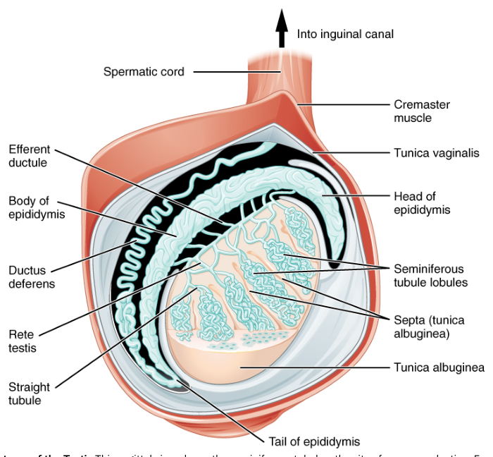
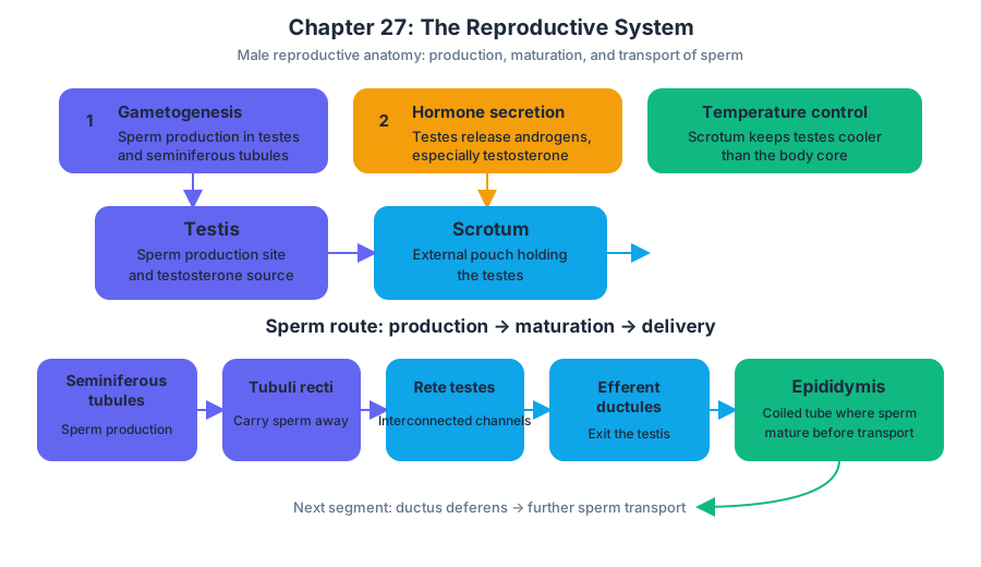
The testes, or male gonads, lie in the scrotum outside the pelvic cavity. Their position helps maintain a temperature slightly below core body temperature, which supports normal sperm production. Each testis is divided into lobules containing tightly coiled seminiferous tubules, the sites where sperm develop.
Between the seminiferous tubules are clusters of endocrine cells called Leydig cells, or interstitial cells. These cells secrete testosterone when stimulated by luteinizing hormone. Within the tubules, large supporting cells called Sertoli cells nourish developing sperm, help regulate the local environment, and release substances that coordinate spermatogenesis.
The testes therefore perform both major classes of glandular activity:
- Exocrine function: production of sperm, which are released into ducts.
- Endocrine function: secretion of testosterone and inhibin into the blood.
From testis to ejaculation
A newly formed sperm cell is not immediately capable of effective movement or fertilization. Its route through the male reproductive tract provides both transport and maturation.
Sperm develop within the seminiferous tubules and pass into progressively larger channels: the straight tubules, rete testis, and efferent ductules. These ductules carry sperm to the epididymis, a long, highly coiled tube attached to the posterior surface of each testis.
The epididymis
The epididymis is the site where sperm mature and are stored. During passage through its duct, sperm acquire improved motility and other functional properties. The epididymis is therefore more than a storage tube: it is a biochemical training environment.
A common misconception is that sperm are fully functional the instant they are produced in the testis. In reality, sperm formed in the seminiferous tubules must mature in the epididymis before they can move effectively through the female reproductive tract.
From the epididymis, sperm enter the ductus deferens, a muscular tube that transports them toward the pelvic cavity. During ejaculation, contractions of its smooth muscle propel sperm toward the ejaculatory duct. The ejaculatory duct passes through the prostate and empties into the urethra, which carries semen through the penis.
The route can be represented as:
Seminiferous tubules $\rightarrow$ rete testis $\rightarrow$ efferent ductules $\rightarrow$ epididymis $\rightarrow$ ductus deferens $\rightarrow$ ejaculatory duct $\rightarrow$ urethra
Accessory glands and semen
Sperm cells account for only a small fraction of semen. Accessory glands add fluids that nourish sperm, protect them, and support their movement.
The seminal vesicles contribute a fluid containing nutrients and other substances that help support sperm metabolism. The prostate gland adds a secretion that contributes to semen and helps create an environment in which sperm can function. The bulbourethral glands secrete mucus-like fluid that helps lubricate the urethra.
Together, sperm and glandular secretions form semen. The penis delivers semen during ejaculation, while the urethra serves as the final passageway through which semen exits the body.
Gonads, gametes, and reproductive hormones
A gonad is a reproductive organ that produces gametes and hormones. The testes produce sperm and testosterone; the ovaries produce oocytes and secrete ovarian hormones. Gametes are haploid cells, meaning that each contains one set of $23$ chromosomes rather than the paired chromosome sets found in most body cells.
Testosterone supports several processes, including:
- development and maintenance of male reproductive structures;
- promotion of sperm production, in cooperation with FSH;
- development of secondary sex characteristics during puberty;
- maintenance of libido and other reproductive functions.
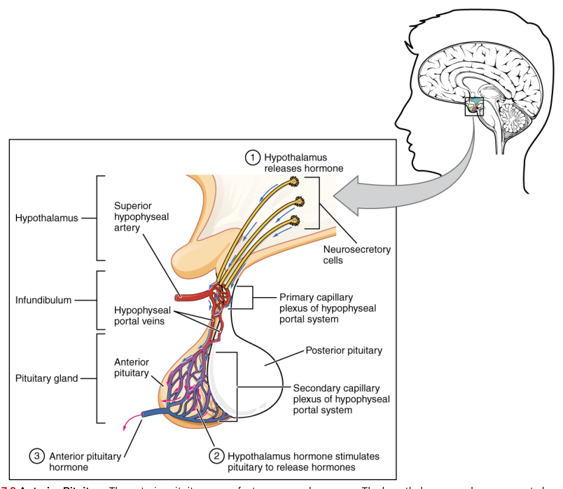
The reproductive endocrine axis begins in the hypothalamus. The hypothalamus releases gonadotropin-releasing hormone (GnRH), which stimulates the anterior pituitary gland to secrete the two gonadotropins: follicle-stimulating hormone and luteinizing hormone.
Gonadotropins are anterior-pituitary hormones that act on the gonads. The two gonadotropins are follicle-stimulating hormone (FSH) and luteinizing hormone (LH).
FSH, LH, and the testis
In the male reproductive system, LH and FSH act on different testicular target cells.
LH binds to receptors on Leydig cells. This stimulates testosterone production and secretion. Testosterone then diffuses through nearby tissue and enters the blood, where it can act on distant target cells.
FSH binds to receptors on Sertoli cells. Sertoli cells support developing sperm and produce androgen-binding protein (ABP). ABP binds testosterone within the seminiferous tubules, maintaining a high local testosterone concentration needed for spermatogenesis.
This division of labor is essential:
- LH primarily stimulates Leydig cells to produce testosterone.
- FSH primarily stimulates Sertoli-cell functions that support sperm development.
- Testosterone and FSH act together to sustain spermatogenesis.
FSH is not limited to the male system. In the ovaries, it promotes follicular growth and supports the development of oocytes within ovarian follicles. The growing follicles produce estrogens. Thus, the same pituitary hormone participates in gamete production in both sexes, although it acts on different gonadal structures.
Negative feedback stabilizes reproductive function
The hypothalamic-pituitary-testicular axis is regulated by negative feedback, in which a downstream product reduces the signals that produced it.
GnRH stimulates pituitary secretion of LH and FSH. LH stimulates testosterone secretion by Leydig cells, while FSH stimulates Sertoli-cell activity. When testosterone levels rise sufficiently, testosterone inhibits GnRH release from the hypothalamus and reduces LH and FSH secretion from the anterior pituitary.
Sertoli cells also secrete inhibin when sperm production is sufficient. Inhibin acts primarily on the anterior pituitary to reduce FSH secretion. This creates a more specific feedback loop: testosterone restrains the broader reproductive axis, while inhibin provides information about the state of sperm production.
The system is not an on-off switch. Its feedback loops continuously adjust hormone secretion so that testosterone concentration and sperm production remain within a functional range.
Hormone target cells and receptor regulation
A hormone affects only cells that possess the appropriate receptor, a protein that recognizes and binds that hormone. The presence of a hormone in the blood does not guarantee an effect in every tissue. A cell must be a target cell with the correct receptor and the intracellular machinery needed to respond.
Receptors also change in number and sensitivity. Up-regulation occurs when a target cell increases its receptor expression, making it more responsive to a given hormone. Down-regulation occurs when receptor number decreases, reducing responsiveness despite the continued presence of the hormone.
This explains why hormone effects depend on more than hormone concentration. A reproductive hormone may be abundant, yet its effect can weaken if target cells reduce receptor availability. Conversely, increased receptor expression can make a tissue respond strongly to a smaller hormonal signal.
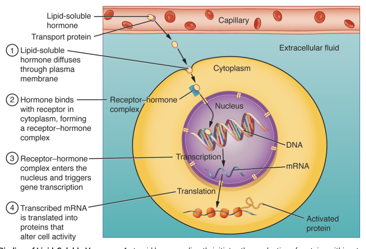
The receptor’s location reflects the hormone’s chemical properties. Peptide hormones such as FSH and LH cannot readily cross the lipid bilayer of the plasma membrane, so they bind to receptors on the cell surface and activate intracellular signaling pathways. Steroid hormones such as testosterone are lipid-soluble and can cross the plasma membrane; their receptors may be located inside the cell, where the hormone-receptor complex can influence gene expression.
Puberty and reproductive maturation
Before puberty, the reproductive axis is relatively quiescent. During puberty, the hypothalamus increases its release of GnRH. The anterior pituitary then releases more LH and FSH, and the gonads increase their production of sex hormones.
In males, rising testosterone supports growth and maturation of the testes and accessory reproductive organs and contributes to secondary sex characteristics. FSH and testosterone promote the onset and maintenance of spermatogenesis. In females, FSH and LH stimulate ovarian activity, follicular development, ovulation, and ovarian hormone secretion.
Puberty therefore reflects coordinated maturation of the hypothalamus, pituitary gland, gonads, reproductive ducts, and target tissues. It is not caused by a single hormone; it emerges from a regulated signaling network.
The reproductive system ultimately depends on precise coordination between anatomy and endocrine control: the testes produce sperm and testosterone, the epididymis matures and stores sperm, accessory glands build semen, and the hypothalamic-pituitary-gonadal axis adjusts the entire process through GnRH, FSH, LH, testosterone, and inhibin.
Source Materials
Study Anatomy and Physiology on Lykke
This course page is free to open: signed out, you can read its first two sections, and signing in, on any plan, opens the rest. The free Lykke Chrome extension syncs your own Canvas courses, so ChatGPT, Claude and Muse can answer from your classes and deadlines. It only reads Canvas.
Add the free extensionSign in to open every sectionAll sections of Anatomy and Physiology · Browse more course pages