From the free course Anatomy and Physiology on Lykke
Chapter 4: The Tissue Level of Organization
Key concepts: Organs are anatomically distinct structures composed of two or more tissue types and performing one or more specific physiological functions. · The first law of thermodynamics states that energy cannot be created or destroyed, only changed in form. · Stretch-sensitive nerve cells detect the degree of stretching and send signals to the brain, which can influence pituitary gland activity. · The cranial and spinal cavities are continuous and house the brain and spinal cord. · The brain and spinal cord are protected by the bones of the skull and vertebral column. · The parietal membrane lines the walls of a body cavity, whereas the visceral membrane covers the organs within it. · The pleura is the serous membrane associated with the pleural cavity, which surrounds the lungs. · X-rays and CT scans use ionizing radiation; CT exposes patients to substantially more radiation than conventional X-rays. · MRI uses magnetic fields and radio waves to produce images without ionizing radiation. · Ultrasound uses reflected sound waves, has no electromagnetic radiation, and is used to monitor pregnancies; the cell is the smallest independently functioning biological unit, and tritium has a half-life of about 12 years.
By the end of this chapter the student can: Identify the main tissue types and discuss their roles in the human body; Identify the four types of tissue membranes and the characteristics of each that make them functional; Explain the functions of various epithelial tissues and how their forms enable their functions; Explain the functions of various connective tissues and how their forms enable their functions; Describe the characteristics of muscle tissue and how these enable function; Discuss the characteristics of nervous tissue and how these enable information processing and control of muscular and glandular activities.
Chapter 4: The Tissue Level of Organization
An MRI can distinguish tissues without cutting into the body because organized tissues differ in their physical and chemical properties. That visible organization begins with a simple fact: the human body contains at least 200 distinct cell types, built from broadly similar cellular components but arranged into specialized groups called tissues.
A tissue is a group of similar cells, together with their extracellular products, that performs a specific function. Cells are therefore not merely individual units; they are coordinated members of a larger structure. A layer of epithelial cells can form a protective barrier, collagen-producing connective-tissue cells can reinforce a tendon, muscle cells can generate force, and neurons can transmit information.
Tissue is an organized group of cells and the materials surrounding them that performs one or more related functions.
The four primary tissue types are epithelial tissue, connective tissue, muscle tissue, and nervous tissue. They are distinguished less by the names of their constituent cells than by their arrangement, the amount of extracellular material between cells, and the work the tissue performs.
From cells to organs
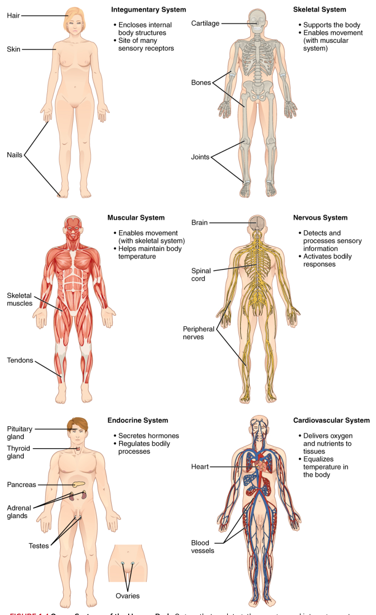
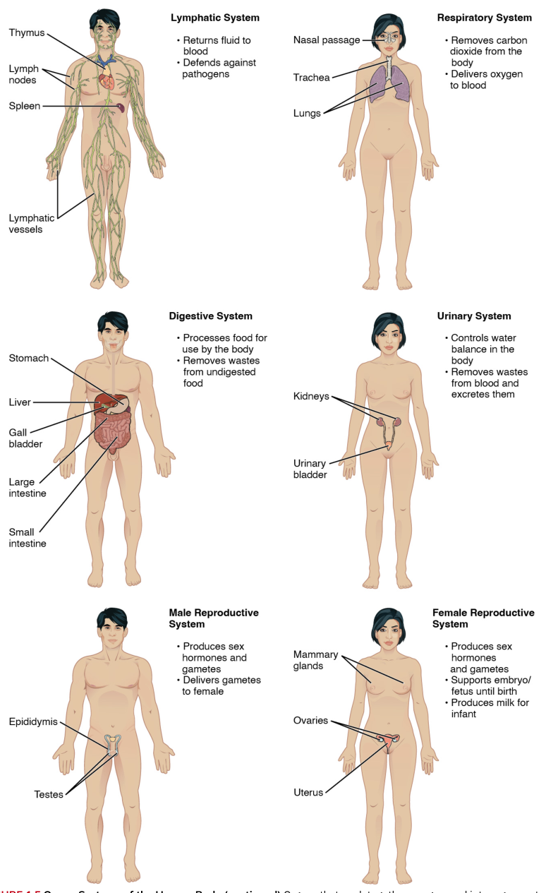
Biological organization builds upward: specialized cells form tissues, tissues combine to form organs, and organs cooperate in organ systems. An organ is an anatomically distinct structure composed of two or more tissue types that performs one or more specific physiological functions. The stomach, for example, contains epithelial tissue for secretion and protection, connective tissue for support, muscle tissue for mixing, and nervous tissue for regulation.
This definition corrects a common misconception: an organ is not simply a large mass of one tissue. The four tissue types are integrated within organs, and the organ’s function emerges from their cooperation. A failure in any one component can compromise the whole structure—for example, damage to the epithelial lining can expose deeper tissues, while loss of smooth-muscle activity can prevent normal movement of contents.
Tissue organization and energy
Every tissue requires energy to maintain its organization. Cells use chemical energy to transport substances across membranes, synthesize proteins, repair damage, contract, and communicate. The first law of thermodynamics states that energy cannot be created or destroyed; it can only be changed from one form into another.
First law of thermodynamics: Energy is conserved. It may be transferred or transformed, but the total amount of energy remains constant.
In human tissues, chemical energy stored in nutrients is transformed into cellular work and heat. Muscle converts chemical energy into mechanical force and heat. Glandular epithelial cells use chemical energy to synthesize and release secretions. Nervous tissue uses electrochemical gradients to transmit signals. The body does not create energy from nothing; it continually transforms and distributes it.
Epithelial tissue: covering, lining, and secretion
Epithelial tissue covers body surfaces, lines internal passageways and cavities, and forms glands. Its cells are tightly packed, leaving very little extracellular space. This arrangement makes epithelium well suited for barriers, controlled exchange, absorption, and secretion.
Epithelial tissue has several defining features:
- Cells are closely joined by specialized connections, helping the tissue resist passage between cells.
- The tissue has an exposed apical surface facing the outside, a body cavity, or a passageway, and a basal surface attached to a basement membrane.
- Epithelial tissue is usually avascular, meaning it contains no blood vessels of its own. Nutrients and oxygen diffuse from vessels in underlying connective tissue.
- It can regenerate rapidly when stem or dividing cells replace damaged cells.
- Its cells show polarity, meaning the apical and basal regions have different structures and functions.
Epithelial classification uses two ideas: the number of cell layers and the shape of the cells at the apical surface. Simple epithelium has one layer and is generally suited for diffusion, absorption, or secretion. Stratified epithelium has multiple layers and is better suited for protection. Cells may be squamous and flat, cuboidal and roughly cube-shaped, or columnar and taller than they are wide. Pseudostratified epithelium appears layered because nuclei lie at different heights, but every cell contacts the basement membrane.
A simple squamous epithelium is thin enough for rapid diffusion, as in alveoli and the lining of blood vessels. A simple columnar epithelium provides more cell volume for absorption and secretion, as in much of the digestive tract. Stratified squamous epithelium protects against abrasion; its keratinized form makes the skin resistant to water loss, whereas its nonkeratinized form lines moist regions such as the mouth and esophagus.
Some epithelial cells are specialized for secretion. Goblet cells, for example, are unicellular exocrine glands embedded among epithelial cells. They produce mucin, which combines with water to form mucus. Mucus protects and lubricates epithelial surfaces and can trap particles in respiratory passages.
Connective tissue: support, binding, storage, and transport
Connective tissue supports, connects, protects, stores, and transports. Unlike epithelium, connective tissue usually contains substantial extracellular matrix—the noncellular material surrounding its cells. The matrix may include protein fibers and a fluid, gel-like, or solid ground substance.
The three major fiber types are collagen fibers, which provide tensile strength; elastic fibers, which allow stretch and recoil; and reticular fibers, which form delicate supportive networks. The relative amounts and arrangement of cells, fibers, and ground substance determine whether a connective tissue behaves like a flexible packing material, a strong cable, a resilient cushion, or a rigid framework.
Loose connective tissue includes areolar tissue, adipose tissue, and reticular tissue. Areolar tissue wraps and cushions structures, binds tissues, and holds tissue fluid. Adipose tissue stores energy, insulates the body, and cushions organs. Reticular tissue forms a supporting framework in organs such as lymph nodes and bone marrow.
Dense connective tissue contains more fibers and fewer cells. Dense regular connective tissue, with parallel collagen fibers, forms tendons and ligaments that resist pulling in one main direction. Dense irregular connective tissue has fibers arranged in many directions, making it useful in structures such as the dermis, which experiences stress from multiple angles. Elastic connective tissue contains abundant elastic fibers and permits recoil.
Cartilage, bone, and blood are also connective tissues. Cartilage provides flexible support and reduces friction at joints. Bone provides a rigid framework and protects organs. Blood has a fluid matrix that permits transport of gases, nutrients, wastes, hormones, and immune cells. Their appearances differ dramatically, but each is defined by cells embedded in an extracellular matrix.
Muscle tissue: converting chemical energy into motion
Muscle tissue is specialized for contraction. Its elongated cells contain proteins that interact to produce tension. This tension can move the skeleton, propel contents through hollow organs, alter the diameter of blood vessels, and drive the pumping action of the heart.
The three types are skeletal muscle, cardiac muscle, and smooth muscle. Skeletal muscle is attached to bones and is usually under voluntary control. Cardiac muscle forms the heart wall and contracts rhythmically. Smooth muscle forms the walls of hollow organs and vessels and generally operates involuntarily. Skeletal and cardiac muscle are striated because their contractile proteins are arranged in repeating patterns; smooth muscle lacks this visible striation.
Nervous tissue: rapid communication and control
Nervous tissue detects changes, processes information, and coordinates responses. Its principal cells are neurons, which transmit electrical signals, and glial cells, which support, protect, nourish, and regulate neurons.
Nervous tissue controls muscular and glandular activity through communication. A stimulus may be detected by a receptor, converted into a signal, processed by the nervous system, and followed by a response in muscle or glandular tissue. This makes nervous tissue central to perception, movement, secretion, and homeostasis.
One example is stretch detection. Stretch-sensitive nerve cells detect how much a structure is being stretched and send signals to the brain. The brain can then influence pituitary gland activity, linking mechanical information from the body to endocrine regulation. The important distinction is that the receptor detects the physical change, while neural processing determines how the body responds.
Tissue membranes and body cavities
A tissue membrane is a thin sheet of tissue that covers a surface, lines a cavity, or separates structures. Membranes usually combine an epithelial layer with underlying connective tissue, although synovial membranes are formed primarily from connective tissue.
The four major types are cutaneous, mucous, serous, and synovial membranes:
- Cutaneous membrane forms the skin. It consists of keratinized stratified squamous epithelium attached to connective tissue and is exposed to the external environment.
- Mucous membranes line passageways that open to the exterior, including the digestive, respiratory, urinary, and reproductive tracts. They are coated with mucus from mucous glands.
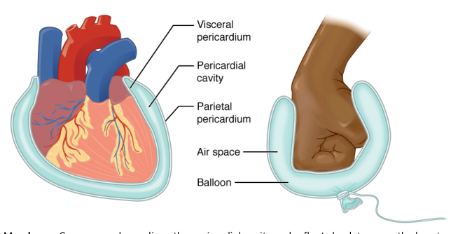
- Serous membranes line closed body cavities and cover the organs within them. They produce a watery serous fluid that reduces friction.
- Synovial membranes line freely movable joint cavities. They secrete synovial fluid, which lubricates the joint and helps nourish the articular cartilage.
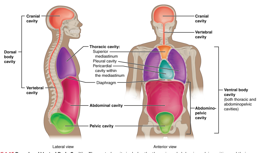
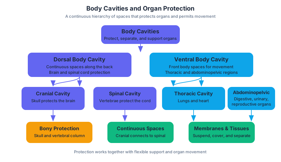
The cranial and spinal cavities are continuous spaces within the dorsal body cavity. Together they house the brain and spinal cord. The bones of the skull and vertebral column protect these organs, while connective tissues and membranes provide additional support and compartmentalization.
The terms parietal and visceral describe two surfaces of a serous membrane. The parietal membrane lines the wall of a body cavity; the visceral membrane covers the organs within it. For example, the parietal pleura lines the thoracic cavity, while the visceral pleura covers the lungs. The narrow space between them contains serous fluid, allowing the organs to move with minimal friction.
Parietal membrane lines the cavity wall; visceral membrane covers the organ.
Development, repair, and aging
During embryonic development, cells become specialized through differentiation, the process by which cells acquire distinct structures and functions. The three primary germ layers—ectoderm, mesoderm, and endoderm—give rise to the tissues and organs of the body. Ectoderm contributes to epidermis and nervous tissue; mesoderm contributes to connective tissue, muscle, and many internal structures; endoderm contributes to epithelial linings of the digestive and respiratory tracts and associated glands.
Stem cells differ in how many cell types they can produce. Totipotent cells can form all body cell types as well as extraembryonic tissues. Pluripotent cells can form derivatives of the three germ layers but not extraembryonic tissues. Multipotent adult stem cells can produce a more limited family of related cells. Specialized adult cells can also be reprogrammed into induced pluripotent stem cells, giving them embryonic-like developmental potential.
Tissue injury triggers inflammation, cell replacement, and remodeling. Some tissues regenerate effectively because they retain dividing stem or progenitor cells; others heal mainly by forming scar tissue. Aging can reduce regenerative capacity, alter extracellular matrix structure, and impair tissue function. Cancer illustrates the danger of disrupted tissue organization: cells lose normal control of division, invade surrounding structures, and replace orderly architecture with disorganized growth.
The central principle is therefore structural as well as chemical: healthy function depends on cells being in the right kinds of groups, in the right positions, with the right connections. Tissue architecture is not decoration—it is the physical organization that makes physiology possible.
Source Materials
Study Anatomy and Physiology on Lykke
This course page is free to open: signed out, you can read its first two sections, and signing in, on any plan, opens the rest. The free Lykke Chrome extension syncs your own Canvas courses, so ChatGPT, Claude and Muse can answer from your classes and deadlines. It only reads Canvas.
Add the free extensionSign in to open every sectionAll sections of Anatomy and Physiology · Browse more course pages