From the free course Anatomy and Physiology on Lykke
Chapter 22: The Respiratory System
Key concepts: Intercostal muscles · External intercostals · Internal intercostals · Respiratory muscles and general anesthesia · Airway management during anesthesia · Glycolysis · Aerobic respiration · Lactic acid production without oxygen
By the end of this chapter the student can: List the structures of the respiratory system; List the major functions of the respiratory system; Outline the forces that allow for air movement into and out of the lungs; Outline the process of gas exchange; Summarize the process of oxygen and carbon dioxide transport within the respiratory system; Create a flow chart illustrating how respiration is controlled; Discuss how the respiratory system responds to exercise; Describe the development of the respiratory system in the embryo.
Chapter 22: The Respiratory System
Breathing is a pressure problem powered by muscle: when the thoracic cavity expands, pressure inside the lungs falls below atmospheric pressure and air flows inward. That simple relationship keeps oxygen entering the body and carbon dioxide leaving it, while cellular metabolism uses oxygen to extract energy from nutrients.
The respiratory system’s job
The respiratory system performs two closely linked tasks. Pulmonary ventilation is the movement of air into and out of the lungs. Gas exchange is the diffusion of oxygen and carbon dioxide between air and blood. Together, these processes deliver oxygen to the blood for metabolic reactions and remove carbon dioxide generated as a metabolic byproduct.
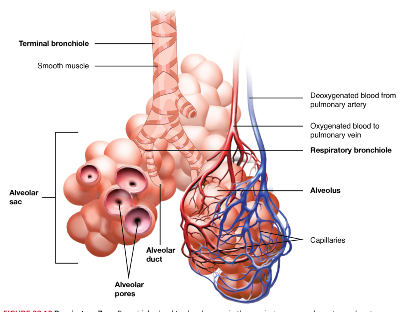
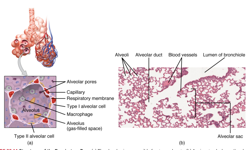
Its pathway begins with the nasal passages, where incoming air enters, and continues through the pharynx, larynx, trachea, bronchi, and progressively smaller bronchioles. The lungs contain alveoli, microscopic air sacs surrounded by pulmonary capillaries. The thin respiratory membrane separating alveolar air from blood is the crucial exchange surface: oxygen diffuses into blood, while carbon dioxide diffuses toward the alveolar air to be exhaled.
A useful distinction prevents a common misconception: breathing is not the same as cellular respiration. Breathing moves air; external respiration exchanges gases between alveoli and blood; internal respiration exchanges gases between blood and tissues; cellular respiration uses oxygen and nutrients inside cells to produce ATP. The respiratory system supplies the oxygen and removes the carbon dioxide, but mitochondria perform the oxygen-dependent reactions.
The mechanics of ventilation
Air moves because of a pressure gradient. Boyle’s law states that, at a constant temperature, pressure varies inversely with volume:
$$P \propto \frac{1}{V}$$
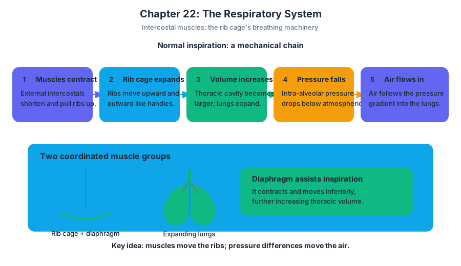
When the volume of the thoracic cavity increases, intra-alveolar pressure decreases. If it falls below atmospheric pressure, air flows into the lungs. When thoracic volume decreases, intra-alveolar pressure rises above atmospheric pressure and air flows out. The lungs do not actively pull air inward; the expanding thoracic cage lowers pressure, and the atmosphere pushes air into the lungs.
During normal inspiration, the diaphragm contracts and moves inferiorly toward the abdominal cavity. At the same time, the external intercostal muscles contract, moving the ribs upward and outward. These actions enlarge the thoracic cavity. Adhesive forces in the pleural fluid couple the lungs to the expanding thoracic wall, so the lungs stretch with it. Lung volume increases, intra-alveolar pressure falls, and air enters.
Normal expiration is primarily passive. The diaphragm and external intercostals relax, the elastic tissues of the lungs recoil, thoracic volume decreases, and intra-alveolar pressure rises. Air then leaves the lungs. Additional muscles become important when a person needs a larger breath or must force air out rapidly.
Intercostal muscles: three layers, two major actions
The intercostal muscles occupy the spaces between adjacent ribs. They are arranged in three overlapping layers whose fiber directions differ: the external intercostals are superficial, the internal intercostals lie beneath them, and the innermost intercostals are deepest.
The eleven pairs of external intercostal muscles assist inspiration. When they contract, they raise the ribs and expand the rib cage. This increases thoracic volume and contributes to the pressure drop that draws air into the lungs.
The eleven pairs of internal intercostal muscles lie just deep to the external intercostals and are associated primarily with expiration, especially forced expiration. Their contraction draws the ribs together, reducing the dimensions of the rib cage. The innermost intercostals are deepest and act as synergists, meaning they assist the internal intercostals.
The important qualification is that “external equals inspiration” and “internal equals expiration” describes their principal actions, not an isolated on/off rule for every breathing condition. Quiet breathing depends mainly on the diaphragm and external intercostals; vigorous breathing recruits additional respiratory muscles and makes active expiration more important.
Gas exchange and transport
Gas exchange occurs by diffusion down partial-pressure gradients. In pulmonary exchange, oxygen moves from alveolar air into pulmonary capillary blood, while carbon dioxide moves from blood into alveoli. The efficiency of this exchange depends on the enormous surface area of the alveoli, the thin respiratory membrane, and adequate matching of ventilation with pulmonary blood flow.
Most oxygen is transported bound to hemoglobin inside erythrocytes; a small amount is dissolved directly in plasma. Carbon dioxide travels in three forms. About $7$ to $10%$ is dissolved in plasma. A much larger fraction, about $70%$, is transported as bicarbonate. The remainder is carried mainly bound to proteins, including hemoglobin.
Bicarbonate transport begins when carbon dioxide enters an erythrocyte and reacts with water. The enzyme carbonic anhydrase accelerates formation of carbonic acid:
$$CO_2 + H_2O \rightleftharpoons H_2CO_3 \rightleftharpoons H^+ + HCO_3^-$$
The bicarbonate ion travels in the plasma to the lungs. There, the reaction reverses: bicarbonate and hydrogen ions form carbonic acid, which becomes carbon dioxide and water. Carbon dioxide diffuses across the respiratory membrane into the alveoli and leaves the body during exhalation. This reaction also links respiratory function to acid-base balance, because changing carbon dioxide levels changes hydrogen-ion concentration.
Respiratory control and exercise
Breathing is controlled involuntarily by respiratory centers in the brainstem. These centers adjust the rate and depth of ventilation in response primarily to changes in carbon dioxide, hydrogen-ion concentration, and oxygen. Rising carbon dioxide increases hydrogen ions through the bicarbonate reaction, making the blood more acidic and stimulating ventilation.
The urge to breathe during breath-holding is therefore driven chiefly by accumulating carbon dioxide and increasing acidity, not simply by an immediate disappearance of oxygen. Eventually, autonomic control can override voluntary breath-holding. This protects the brain and other tissues that depend continuously on oxygen delivery.
Exercise increases ATP demand in skeletal muscle. Ventilation rises to bring in more oxygen and remove the additional carbon dioxide produced by metabolism. Respiratory muscles themselves also require ATP, so heavy breathing increases the work performed by the diaphragm, external intercostals, and accessory muscles.
Glycolysis and the energy cost of breathing
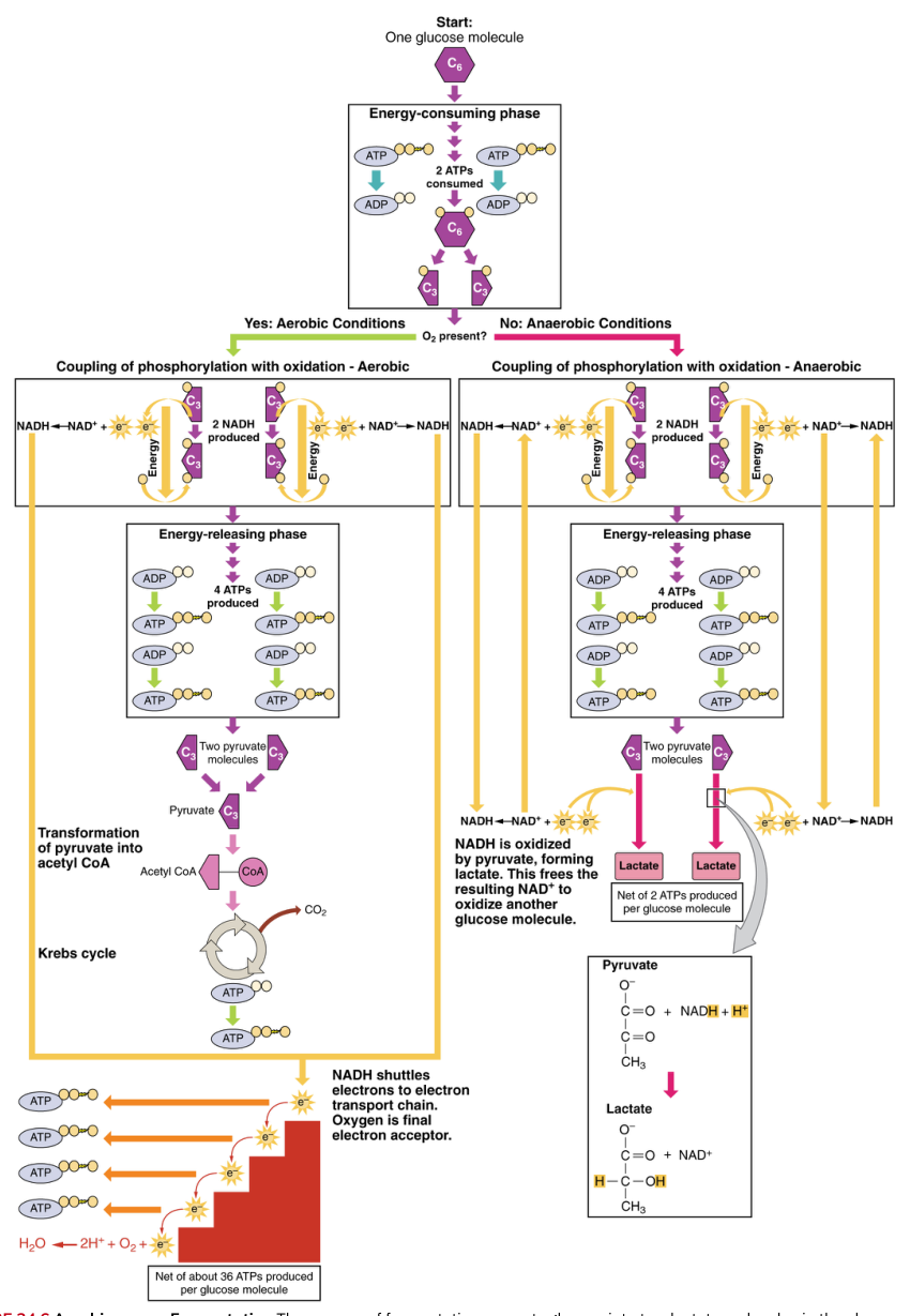
Glycolysis is the cytoplasmic pathway that breaks glucose into two molecules of pyruvate. Blood glucose or muscle glycogen supplies the starting glucose. Glycolysis produces a net yield of $2$ ATP and $2$ pyruvate:
$$\text{Glucose} \rightarrow 2\text{ pyruvate} + 2\text{ ATP}$$
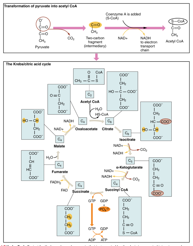
Glycolysis does not require oxygen. If oxygen is available, pyruvate enters aerobic pathways in mitochondria, where nutrients are more completely broken down and produce a much larger ATP yield, along with carbon dioxide, water, and heat. Aerobic respiration supplies about $95%$ of the ATP needed by resting or moderately active muscles.
If oxygen delivery cannot meet demand, pyruvate is converted to lactic acid, which can enter the blood and is associated with the metabolic conditions of intense exertion and muscle fatigue. This is not an alternative form of breathing; it is an alternative fate for pyruvate when aerobic processing cannot keep pace.
General anesthesia and airway management
General anesthesia changes respiratory physiology because it depresses central nervous system activity and reduces the patient’s ability to maintain ventilation and protect the airway. An anesthetized person may breathe less deeply, lose coordinated muscle tone, and fail to respond normally to obstruction or aspiration. Respiratory muscles may still contract, but their activity can become insufficient or poorly coordinated for reliable gas exchange.
Airway management therefore establishes and preserves a passage for air. Positioning the head and neck, removing obstruction, and using airway devices can keep the upper airway open. In situations requiring greater control, an endotracheal tube is placed through the larynx into the trachea and connected to mechanical ventilation. The tube allows clinicians to deliver oxygen-containing gas, control ventilation, and reduce the risk that secretions or stomach contents enter the lungs.
The key clinical principle is mechanical: if the airway is blocked, respiratory muscles cannot move useful air into the alveoli; if ventilation is inadequate, oxygen delivery falls and carbon dioxide accumulates. Airway patency, chest movement, gas exchange, and circulation must therefore be assessed together rather than treated as separate problems.
Development of the respiratory system
The respiratory system develops from embryonic tissues that form the conducting passages and the branching lung structures. The respiratory tree repeatedly branches, producing bronchi, bronchioles, and eventually alveolar regions. Functional maturity depends not merely on the presence of lungs but on the development of thin exchange surfaces and adequate surfactant.
Surfactant is a substance that reduces surface tension at the air-liquid interface of the alveoli. Without sufficient surfactant, alveoli are more difficult to expand and more likely to collapse during expiration. Thus, respiratory development must produce both a branching airway system and alveoli capable of remaining open for repeated cycles of ventilation.
Worked synthesis: what happens during a deep breath?
- The respiratory centers increase motor output to the diaphragm and external intercostals.
- The diaphragm contracts inferiorly, while the external intercostals elevate the ribs.
- Thoracic and lung volume increase.
- Intra-alveolar pressure falls below atmospheric pressure.
- Air flows through the respiratory passages into the alveoli.
- Oxygen diffuses into pulmonary capillary blood, while carbon dioxide diffuses into the alveoli.
- Hemoglobin carries most oxygen onward, and carbon dioxide returns partly as bicarbonate.
- During expiration, inspiratory muscles relax, elastic recoil reduces lung volume, and carbon dioxide-rich air leaves the body.
The entire sequence depends on cooperation among anatomy, pressure mechanics, muscle contraction, diffusion, blood transport, neural control, and cellular metabolism. A failure at any link—from airway obstruction to weak intercostal activity to inadequate alveolar exchange—can compromise the oxygen supply required for ATP production.
Source Materials
Study Anatomy and Physiology on Lykke
This course page is free to open: signed out, you can read its first two sections, and signing in, on any plan, opens the rest. The free Lykke Chrome extension syncs your own Canvas courses, so ChatGPT, Claude and Muse can answer from your classes and deadlines. It only reads Canvas.
Add the free extensionSign in to open every sectionAll sections of Anatomy and Physiology · Browse more course pages