From the free course Anatomy and Physiology on Lykke
Chapter 12: The Nervous System and Nervous Tissue
Key concepts: Three components of the cytoskeleton · Embryonic origins of tissues and major organs · Long-bone anatomy and organization · Bone coverings and vascular supply · Bone surface markings and their functions · Thumb movement in multiple planes · Elbow and knee ligament or movement mechanics · Apocrine sweat glands · Electrical excitability of neurons and muscle cells · Cell membrane potential and action potentials
By the end of this chapter the student can: Name the major divisions of the nervous system, both anatomical and functional; Describe the functional and structural differences between gray matter and white matter structures; Name the parts of the multipolar neuron in order of polarity; List the types of glial cells and assign each to the proper division of the nervous system, along with their function(s); Distinguish the major functions of the nervous system: sensation, integration, and response; Describe the components of the membrane that establish the resting membrane potential; Describe the changes that occur to the membrane that result in the action potential; Explain the differences between types of graded potentials
Chapter 12: The Nervous System and Nervous Tissue
A neuron can detect a stimulus, integrate thousands of incoming signals, and produce a precisely timed response because its membrane, cytoskeleton, and supporting glial cells work as one system. The nervous system is therefore not merely a collection of “wires”: it is living tissue specialized for rapid communication, information processing, and coordinated action.
Nervous-system organization
The nervous system has two anatomical divisions. The central nervous system (CNS) consists of the brain and spinal cord; the peripheral nervous system (PNS) consists of nerves, ganglia, and sensory receptors outside the CNS. Functionally, sensory, or afferent, pathways carry information toward the CNS, while motor, or efferent, pathways carry commands away from it.
The efferent division separates into the somatic nervous system, which controls skeletal muscle and includes conscious sensation and voluntary motor control, and the autonomic nervous system, which regulates smooth muscle, cardiac muscle, glands, and many visceral functions. The autonomic system is subdivided into sympathetic, parasympathetic, and enteric components. These divisions describe function, not separate physical organs: one peripheral nerve may contain both sensory and motor axons.
Key distinction: The CNS and PNS are anatomical divisions; sensory and motor pathways are functional divisions. Mixing these two classification systems is a common source of confusion.
Neurons: the information-processing cells
A neuron is an excitable cell specialized to receive, process, and transmit information. In a typical multipolar neuron, signals move through a recognizable sequence: dendrites receive input, the cell body maintains the cell and integrates signals, the axon hillock initiates an action potential when threshold is reached, and the axon conducts the signal toward its terminals. Axon terminals release chemical messengers at synapses.
Neurons vary in shape according to their function. Multipolar neurons have many dendrites and one axon and are common in the CNS. Unipolar neurons, characteristic of many sensory neurons in the PNS, have a single process that divides into peripheral and central branches. Bipolar neurons have one dendrite and one axon and occur in specialized sensory systems.
Glial cells and the working environment of neurons
Glia are nervous-system cells that support, protect, nourish, and electrically organize neurons. They do not simply fill empty space. In the CNS, astrocytes help regulate the extracellular environment and contribute to the blood-brain barrier; oligodendrocytes form myelin around CNS axons; microglia provide immune defense; and ependymal cells line cavities associated with cerebrospinal fluid.
The PNS contains satellite cells and Schwann cells. Satellite cells surround neuronal cell bodies in peripheral ganglia and provide structural and chemical support. Schwann cells wrap around peripheral axons, forming myelin in successive segments. A single oligodendrocyte can myelinate portions of several CNS axons, whereas a Schwann cell forms a myelin segment around one axon.
Myelin increases conduction speed by electrically insulating the axon. Gaps between myelin segments are nodes of Ranvier, where voltage-gated channels are concentrated. The action potential appears to “jump” from node to node, a process called saltatory conduction. Damage to myelin can therefore disrupt communication even when the neuron’s cell body remains alive.
Gray matter, white matter, and the cytoskeleton
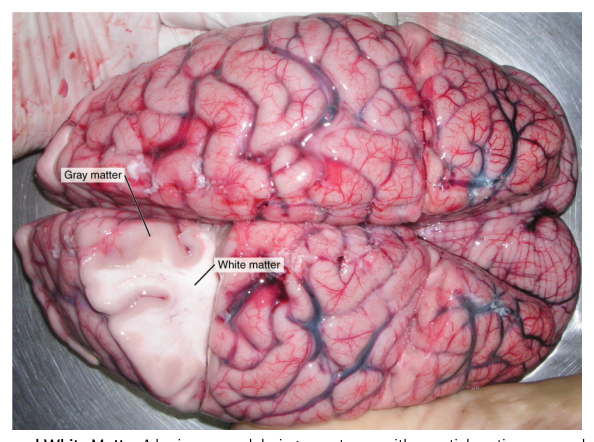
Gray matter contains many neuronal cell bodies, dendrites, synapses, and unmyelinated portions of axons; it is where much integration occurs. White matter contains large numbers of myelinated axons, whose lipid-rich myelin gives the tissue its pale appearance. In the spinal cord, gray matter is centrally located and white matter surrounds it; in the brain, the arrangement is more complex, with gray matter forming an outer cortex and also occurring in deep nuclei.
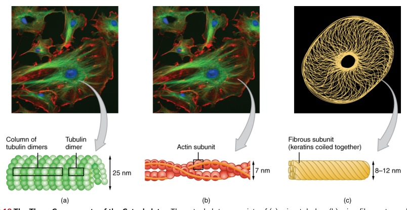
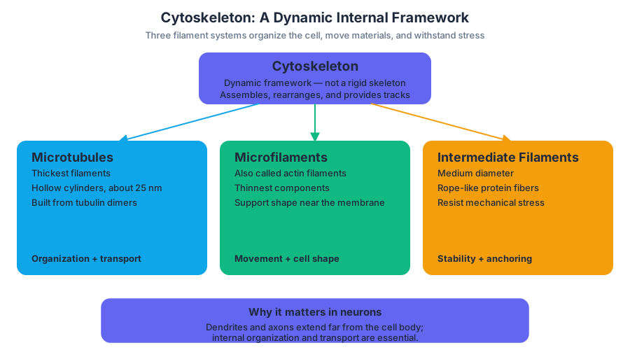
The neuron’s shape and transport system depend on its cytoskeleton, an internal network of protein fibers. Its three components differ in diameter, composition, and mechanical role:
| Cytoskeletal component | Main structural feature | Major functions |
|---|---|---|
| Microtubules | Hollow tubes made from tubulin dimers arranged into protofilaments; approximately $25\ \text{nm}$ in diameter | Maintain cell shape and provide tracks for intracellular transport |
| Intermediate filaments | Rope-like fibers assembled from fibrous proteins | Resist tension and strengthen cells |
| Microfilaments | Thin actin filaments | Support the cell cortex, shape changes, and movement |
Microtubules are the thickest of the three filament systems. Their hollow cylindrical structure makes them suitable for long-distance transport within the axon. Motor proteins move vesicles and organelles along microtubules, allowing materials produced in the cell body to reach axon terminals and allowing worn components to return for recycling. Intermediate filaments provide tensile strength, while actin microfilaments participate in membrane shape, growth-cone behavior, and synaptic structure.
Embryonic origins of nervous tissue and organs
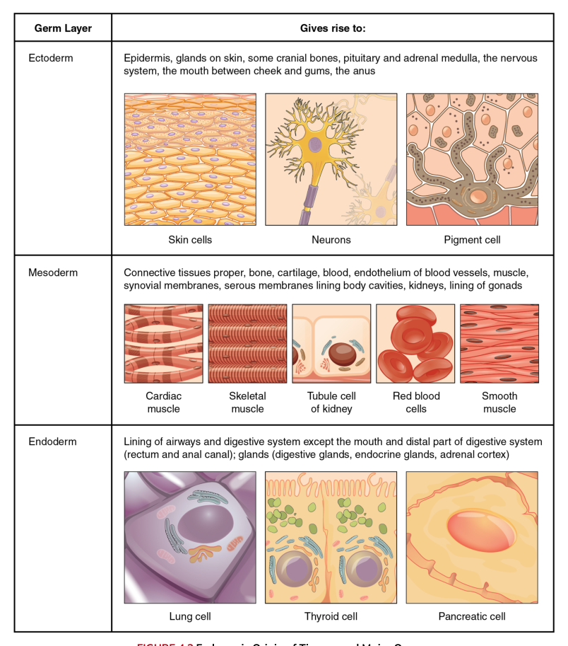
During embryonic development, the three primary germ layers establish the tissue plan of the body. Ectoderm gives rise to the epidermis, skin glands, some cranial bones, the pituitary, adrenal medulla, nervous system, and epithelial linings of the mouth between the cheek and gums and of the anus. Neurons and many associated nervous structures therefore have an ectodermal origin.
Mesoderm produces connective tissues proper, bone, cartilage, blood, the endothelium lining blood vessels, muscle, and synovial membranes. Endoderm forms much of the epithelial lining of the digestive and respiratory tracts and contributes to organs associated with those systems. The germ layers are developmental origins, not mature anatomical compartments: an adult organ commonly contains tissues derived from more than one layer.
Membrane potentials: how neurons encode change
A neuron’s plasma membrane separates electrically different fluids. The resting membrane potential is typically about $-70\ \text{mV}$, meaning the inside is more negative than the outside. This voltage depends on unequal ion concentrations, selective membrane permeability, and the sodium-potassium pump, which moves $3$ sodium ions out of the cell for every $2$ potassium ions moved in.
A graded potential is a local change in membrane voltage whose size varies with stimulus strength. A depolarization makes the membrane less negative, moving it toward $0\ \text{mV}$; a hyperpolarization makes it more negative. Graded potentials can weaken as they spread and can combine through spatial or temporal summation.
If depolarization at the axon hillock reaches threshold, approximately $-55\ \text{mV}$, voltage-gated sodium channels open and an action potential begins. Sodium enters rapidly, producing further depolarization. Sodium channels then inactivate while voltage-gated potassium channels open; potassium leaves the cell, repolarizing and briefly hyperpolarizing the membrane. The refractory period helps ensure one-way propagation and limits the maximum firing frequency.
All-or-none principle: A sufficiently strong stimulus produces an action potential of standard amplitude; a stronger stimulus does not create a larger action potential. It can instead increase firing frequency or recruit more neurons.
At a synapse, an arriving action potential causes neurotransmitter release from the presynaptic terminal. The neurotransmitter binds receptors on the postsynaptic membrane and produces either an excitatory postsynaptic potential, which moves the membrane toward threshold, or an inhibitory postsynaptic potential, which moves it away from threshold. The axon hillock integrates these simultaneous inputs before deciding whether to fire.
Bone as living connective tissue
Bone belongs to the mesoderm-derived connective tissues, but it is not an inert scaffold. Its matrix stores minerals, supports the body, protects organs, houses marrow, and provides levers for skeletal muscle. A typical long bone illustrates how living tissue is organized around both mechanical strength and internal supply.
A long bone has a shaft called the diaphysis, expanded ends called the proximal and distal epiphyses, and transitional regions called metaphyses. The shaft is dominated by compact bone surrounding a medullary cavity. The epiphyses contain more spongy bone, whose trabeculae distribute forces while keeping the bone relatively light. Red marrow in spongy bone produces blood cells; yellow marrow in the medullary cavity stores energy-rich fat.
The external surface is covered by the periosteum, a tough membrane with an outer fibrous layer and an inner cellular layer containing osteogenic cells and bone-forming cells. The internal surfaces of the medullary cavity and spaces within spongy bone are lined by the endosteum, which contains cells involved in bone maintenance and remodeling. At a synovial joint, articular cartilage covers the bone surface and reduces friction while distributing pressure.
Bone receives blood through vessels that enter nutrient foramina. A nutrient artery passes into the shaft and supplies internal bone and marrow; additional vessels supply the periosteum and epiphyses. Because bone is vascular and continuously remodeled, fractures can heal, although healing depends on adequate blood supply and stable alignment.
Bone markings: anatomy with a mechanical purpose
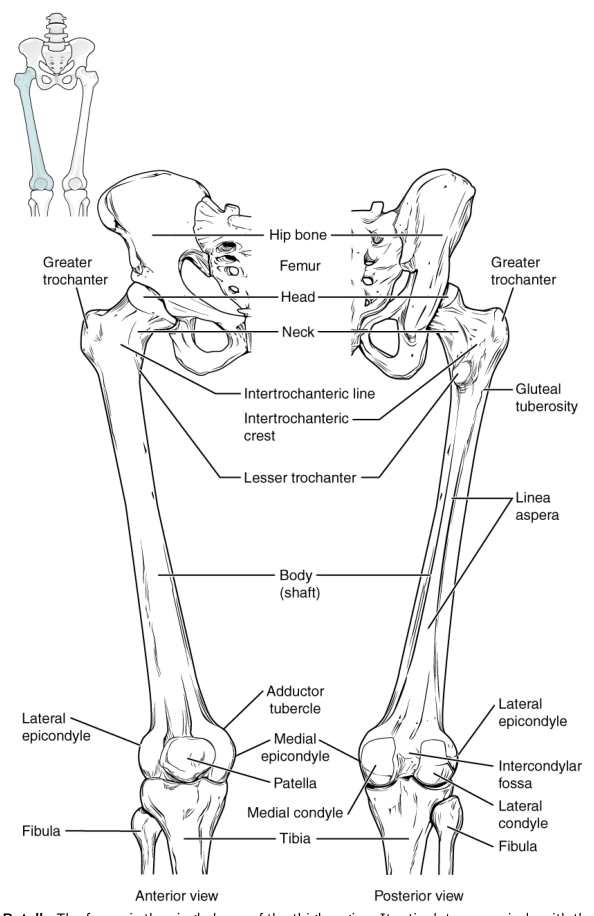
A bone marking is a recognizable surface feature. Projections such as tubercles, tuberosities, crests, spines, and processes provide attachment sites for muscles, tendons, and ligaments. Depressions and openings, including fossae, grooves, foramina, canals, and fissures, accommodate other structures or allow nerves and blood vessels to pass through. The marking is therefore functional anatomy: its shape records the forces and structures acting on the bone.
Thumb movement in multiple planes
The thumb is unusual because its carpometacarpal joint allows movement in more than one anatomical plane. Flexion moves the thumb across the palm, whereas extension returns it away from the palm. Abduction moves it anteriorly, away from the plane of the palm, and adduction brings it back. Opposition combines abduction, flexion, and medial rotation so that the thumb pad can face the fingers. Reposition reverses opposition.
Opposition is not a single-axis movement. Touching the thumb to the little finger requires a coordinated sequence across several planes, followed by slight rotation at the thumb’s carpometacarpal joint. This mobility explains both the hand’s precision grip and the joint’s vulnerability to repetitive stress.
Unifying idea: Nervous tissue supplies rapid control, cytoskeletal fibers preserve cellular architecture and transport, and bone supplies the living mechanical framework. Effective movement requires all three: a neural command, a structurally supported neuron, and a skeletal system shaped to transmit force.

Source Materials
Study Anatomy and Physiology on Lykke
This course page is free to open: signed out, you can read its first two sections, and signing in, on any plan, opens the rest. The free Lykke Chrome extension syncs your own Canvas courses, so ChatGPT, Claude and Muse can answer from your classes and deadlines. It only reads Canvas.
Add the free extensionSign in to open every sectionAll sections of Anatomy and Physiology · Browse more course pages