From the free course Anatomy and Physiology on Lykke
Chapter 14: The Somatic Nervous System
Key concepts: Somatic nervous system (SNS) · Basic functions of the nervous system · Sensory receptors and sensory input · Sensory pathways and brain processing · Upper and lower motor neurons · Somatic motor responses and skeletal muscle contraction · Action potentials · Neuronal integration · Chemical synaptic transmission · Axonal structures and insulation, including nodes of Ranvier and Schwann cells
By the end of this chapter the student can: Describe the components of the somatic nervous system; Name the modalities and submodalities of the sensory systems; Distinguish between general and special senses; Describe regions of the central nervous system that contribute to somatic functions; Explain the stimulus-response motor pathway.
Chapter 14: The Somatic Nervous System
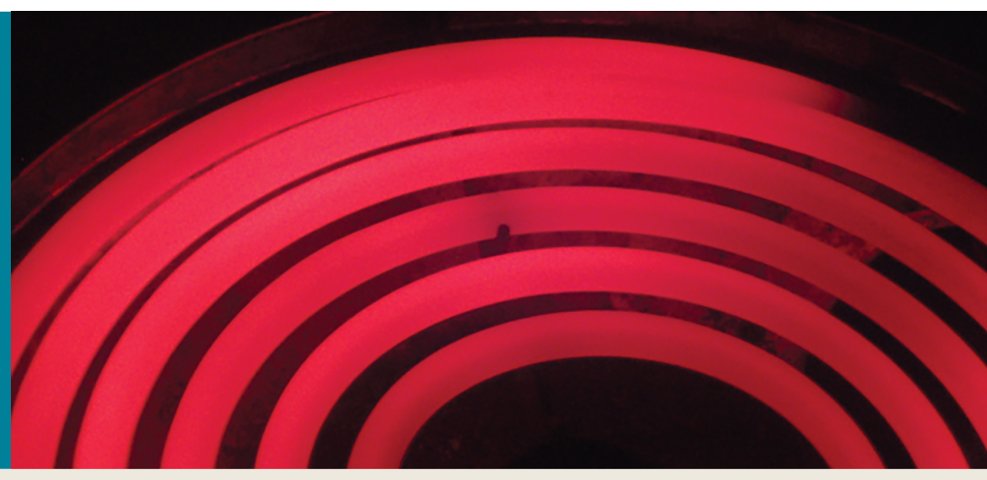
A hand touching water that is too hot can withdraw before conscious thought catches up: sensory receptors detect the stimulus, the nervous system processes the information, and motor neurons activate skeletal muscle. The somatic nervous system (SNS) is the functional division responsible for conscious sensory perception and responses involving skeletal muscle. It can produce deliberate movement, such as reaching for a shower handle, and rapid reflexes, such as pulling the hand away from damaging heat.
The SNS is distinguished from the autonomic nervous system (ANS) primarily by the type of effector it controls and the nature of the response. Somatic motor pathways control skeletal muscles, which produce voluntary movement and certain rapid reflexes. Autonomic pathways regulate cardiac muscle, smooth muscle, and glands without requiring conscious direction. The distinction is functional rather than a simple division between “conscious” and “unconscious”: a somatic reflex may occur before conscious awareness, while the brain can influence many autonomic functions.
The Basic Functions of the Nervous System
Nervous-system activity can be organized into three linked functions: sensory input, integration, and motor output. Sensory input gathers information from inside or outside the body. Integration, meaning the combination and interpretation of incoming information, determines an appropriate response. Motor output sends commands to an effector, such as a skeletal muscle.
For the somatic system, peripheral sensory neurons receive information from receptors in the skin, muscles, joints, and special sensory organs. The sensory information enters the central nervous system, where processing occurs in the spinal cord and brain. Motor commands originate in the central nervous system and travel outward through motor neurons to skeletal muscle.
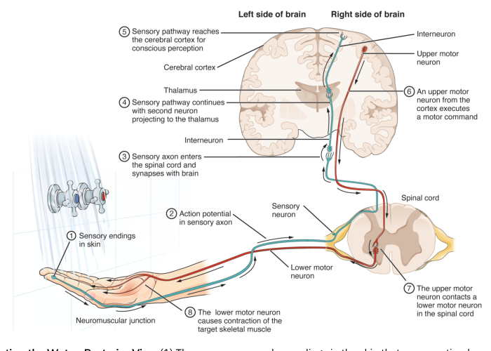
The somatic stimulus–response pathway is: sensory receptor detects a stimulus → sensory neuron carries an electrical signal toward the central nervous system → central processing integrates the information → motor command descends through motor neurons → skeletal muscle contracts.
Sensory Receptors and Sensory Input
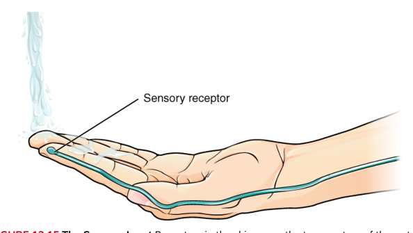
A sensory receptor is a structure that detects a physical or chemical change called a stimulus. For example, receptors in the skin of the palm detect the temperature of water contacting the hand. Receptors do not merely “notice” a stimulus in a vague sense; they convert stimulus energy into an electrical change in the nervous system, allowing neurons to communicate the information.
Receptors can be classified by their structure. Some are free nerve endings, in which sensory nerve fibers have relatively simple exposed endings. Others are encapsulated nerve endings, in which connective tissue surrounds the sensory ending and helps determine how mechanical forces are detected. A third group consists of specialized receptor cells that communicate with sensory neurons.
Receptors may also be classified by the type of stimulus they transduce. Mechanoreceptors respond to mechanical deformation, such as pressure, vibration, stretch, or movement. Thermoreceptors respond to temperature. Chemoreceptors respond to chemicals, and receptors involved in vision respond to light. These functional categories describe what kind of energy the receptor converts into a neural signal.
A useful distinction is the difference between sensation and perception. Sensation is the activation of a receptor and the transmission of information into the nervous system. Perception is the central nervous system’s meaningful interpretation of that information. A temperature receptor can signal that the skin is being heated, but the brain must process the signal to interpret the water as comfortably warm, unpleasantly hot, or potentially damaging.
Sensory Pathways and Brain Processing
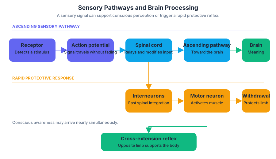
When a receptor is activated strongly enough, the sensory neuron generates an action potential, a rapidly propagating electrical signal in the membrane of a neuron. Action potentials are central to nervous-system communication because they carry information over long distances without fading in the way a local electrical change would.
The action potential travels along the sensory neuron into an ascending sensory pathway. Ascending means traveling toward the brain from the peripheral nervous system or spinal cord. Along the way, information may be relayed through synapses and modified by spinal-cord circuits. Ultimately, the brain receives and processes the sensory input, determining what the stimulus means and what response is appropriate.
Processing does not always wait for conscious perception. In a withdrawal reflex, sensory information enters the spinal cord and is integrated rapidly through interneurons. Motor neurons then activate muscles that withdraw the limb. The brain may receive information about the painful stimulus at nearly the same time, producing conscious awareness, but the immediate protective movement can begin through spinal processing.
A related response is the cross-extension reflex. When one limb withdraws from a damaging stimulus, the opposite limb can be activated to help support the body. This illustrates that integration is not merely a relay of sensory information; it is the nervous system’s process of combining incoming signals and coordinating a useful pattern of muscle activity.
Synapses and Neuronal Integration
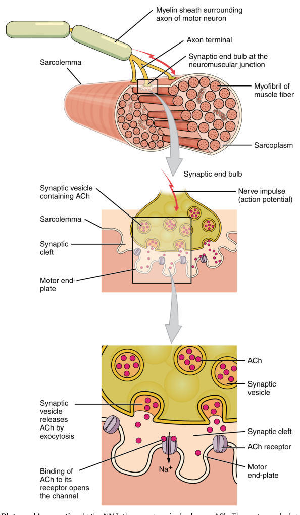
Neurons communicate at specialized junctions called synapses. At many synapses, an electrical signal arriving at the end of one neuron causes the release of chemical messenger molecules called neurotransmitters. The neurotransmitters diffuse across a small gap and bind to receptors on the next neuron or target cell. Thus, the signal is electrical within the neuron but chemical between neurons.
The swollen endings at the tips of axons are called synaptic end bulbs. These swellings contain synaptic vesicles filled with neurotransmitter molecules. When an action potential reaches a synaptic end bulb, it triggers neurotransmitter release into the synaptic cleft. The neurotransmitter then changes the electrical state of the receiving cell, either making a new action potential more likely or less likely.
Neuronal integration occurs when a neuron combines signals arriving from multiple inputs. Some incoming signals may be excitatory, promoting action-potential generation, while others may be inhibitory, reducing that likelihood. The neuron’s response depends on the combined effect of these inputs rather than on one isolated signal. This mechanism allows the nervous system to select, coordinate, and refine responses.
Myelin helps action potentials travel efficiently along many axons. In the peripheral nervous system, Schwann cells wrap around and insulate axons, forming myelin. The insulated regions are interrupted by small gaps called nodes of Ranvier. Electrical changes are regenerated at these nodes, allowing the action potential to appear to jump from node to node, a process that increases conduction speed.
Descending Motor Commands
After sensory information has been processed, the brain formulates a descending motor command. Descending means traveling from the brain toward the spinal cord and peripheral effectors. The command may produce a voluntary action, such as moving the hand to adjust water temperature, or contribute to a coordinated postural response.
Motor commands pass through two functional levels of motor neurons. An upper motor neuron begins in the brain and descends through pathways of the central nervous system. It influences a lower motor neuron, whose cell body is located in the brainstem or spinal cord and whose axon extends through the peripheral nervous system to skeletal muscle.
The lower motor neuron is the final common pathway to the skeletal muscle. Its axon reaches the muscle fiber and forms a neuromuscular junction, the synapse between a motor neuron and a skeletal-muscle fiber. At this junction, the motor neuron releases the neurotransmitter acetylcholine. The resulting electrical change in the muscle fiber initiates the events that produce contraction.
Somatic Motor Responses and Skeletal-Muscle Contraction
A somatic motor response therefore follows a continuous functional sequence: the brain or spinal cord determines the response, an upper motor neuron carries the command within the central nervous system, a lower motor neuron carries it to the muscle, and the skeletal muscle generates force. The physical result may be movement of a joint, maintenance of posture, withdrawal from injury, or stabilization of the body.
At the neuromuscular junction, acetylcholine binds to receptors on the muscle-fiber membrane and produces an electrical response. If the response reaches threshold, an action potential spreads along the muscle fiber. This activates calcium release inside the muscle cell. Calcium permits the contractile proteins actin and myosin to interact, and myosin pulls on actin to generate tension and shortening. ATP is required for the repeated cycling of the contractile machinery.
The motor response ends when neural stimulation stops and acetylcholine is removed from the neuromuscular junction. Calcium is returned to storage within the muscle cell, and the contractile interaction declines. A single lower motor neuron can branch to control multiple muscle fibers; the neuron and all of the fibers it activates form a motor unit. Coordinated recruitment of motor units allows the nervous system to adjust the strength of skeletal-muscle contraction.
The central idea is that somatic movement is not produced by muscle acting alone. Receptors provide information, ascending pathways carry it to the central nervous system, integration determines the response, descending pathways transmit the command, and the neuromuscular junction converts neural activity into skeletal-muscle contraction. Damage at any point can disrupt the final movement even when the muscle itself remains structurally intact.
Content adapted from OpenStax Anatomy and Physiology 2e by J. Gordon Betts et al., licensed CC BY 4.0.

Source Materials
Study Anatomy and Physiology on Lykke
This course page is free to open: signed out, you can read its first two sections, and signing in, on any plan, opens the rest. The free Lykke Chrome extension syncs your own Canvas courses, so ChatGPT, Claude and Muse can answer from your classes and deadlines. It only reads Canvas.
Add the free extensionSign in to open every sectionAll sections of Anatomy and Physiology · Browse more course pages