From the free course Anatomy and Physiology on Lykke
Chapter 20: The Cardiovascular System: Blood Vessels and Circulation
Key concepts: Cerebral circulation and protection of CNS blood supply · Circle of Willis · Internal carotid artery and carotid canal · Vertebral and basilar arteries · Major cerebral arterial branches · Cerebrospinal fluid circulation through the ventricular system · Osmoregulation and antidiuretic hormone (ADH/vasopressin) · Vasoconstriction, peripheral resistance, and blood pressure · Orthostatic hypotension · Blood flow through erectile tissue
By the end of this chapter the student can: Compare and contrast the anatomical structure of arteries, arterioles, capillaries, venules, and veins; Accurately describe the forces that account for capillary exchange; List the major factors affecting blood flow, blood pressure, and resistance; Describe how blood flow, blood pressure, and resistance interrelate; Discuss how the neural and endocrine mechanisms maintain homeostasis within the blood vessels; Describe the interaction of the cardiovascular system with other body systems; Label the major blood vessels of the pulmonary and systemic circulations; Identify and describe the hepatic portal system
Chapter 20: The Cardiovascular System: Blood Vessels and Circulation
Blood vessels are not passive pipes: they are living organs that distribute blood, regulate pressure, exchange materials with tissues, and redirect flow when local conditions change. Their branching architecture allows the cardiovascular system to move blood rapidly through large vessels while slowing it enough at microscopic exchange surfaces for oxygen, nutrients, wastes, and hormones to cross between blood and tissues.
20.1 Structure and Function of Blood Vessels
A useful way to picture the vascular system is as a branching transport network. The arteries carry blood away from the heart, progressively dividing into smaller arterioles. Arterioles deliver blood to microscopic capillaries, where exchange occurs. Blood then enters venules, which merge into larger veins that return it to the heart. The direction of flow—not oxygen content—defines an artery or vein. Thus, pulmonary arteries carry oxygen-poor blood away from the heart, while pulmonary veins carry oxygen-rich blood toward it.
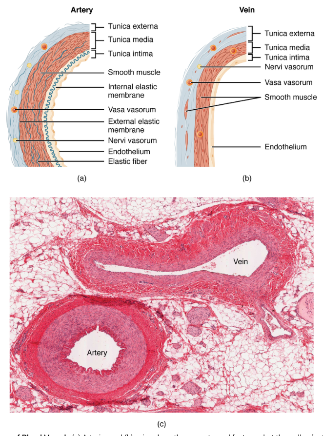
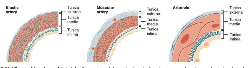
Most blood vessels contain three concentric layers, or tunics. The innermost tunica interna, also called the tunica intima, includes a thin endothelium made of simple squamous epithelial cells. This smooth lining reduces friction, forms a selectively permeable barrier, and participates in clotting, inflammation, and regulation of vascular diameter. Beneath it lies a thin layer of connective tissue and, in many vessels, an internal elastic layer.
The tunica media is the middle layer. It contains circular layers of smooth muscle and elastic fibers. Smooth muscle contraction causes vasoconstriction, a decrease in vessel diameter; relaxation causes vasodilation, an increase in diameter. Elastic fibers allow arteries to stretch when the heart ejects blood and recoil between beats. The outer tunica externa, or tunica adventitia, consists primarily of connective tissue that anchors and protects the vessel. Large vessels may contain tiny vessels called vasa vasorum, which supply the outer layers because diffusion from the lumen is insufficient.
Arteries have thick walls, especially a substantial tunica media, because they receive blood directly from the heart under relatively high pressure. Large elastic arteries, including the aorta, contain abundant elastic tissue and smooth the pulsatile output of the ventricles. Muscular arteries distribute blood to specific organs. Arterioles have smaller diameters and a proportionally important smooth-muscle layer, making them the principal resistance vessels. By changing their diameter, they determine how much blood enters a tissue and contribute greatly to systemic blood pressure.
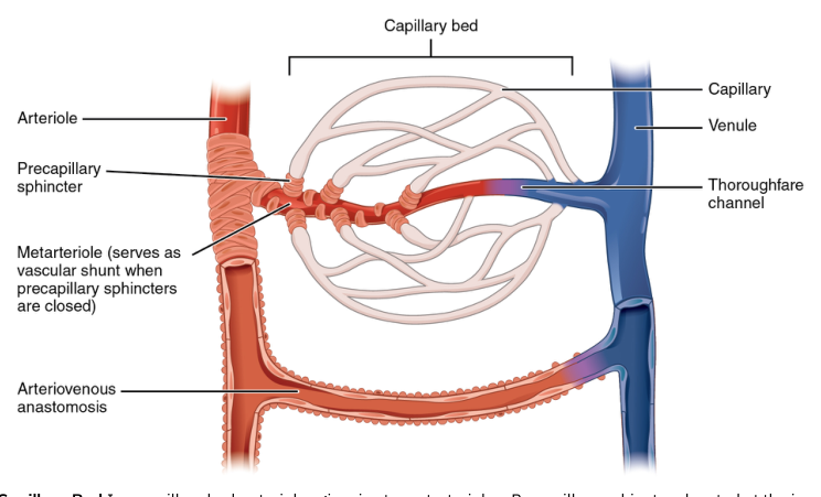
Capillaries are the smallest blood vessels and are specialized for exchange. Their walls consist almost entirely of a single endothelial layer and a basement membrane. This short diffusion distance allows gases, nutrients, hormones, and wastes to move between plasma and interstitial fluid. Capillaries are organized into capillary beds, supplied by a metarteriole and regulated by precapillary sphincters. When sphincters relax, blood enters more capillaries; when they contract, blood is routed through a vascular shunt or preferential channel.
Capillaries vary in permeability. Continuous capillaries have uninterrupted endothelial walls and are common in skeletal muscle, skin, lungs, and the central nervous system. Fenestrated capillaries contain pores that increase exchange and occur in organs such as the kidneys, endocrine glands, and intestines. Sinusoidal capillaries have large gaps and a discontinuous basement membrane, permitting passage of large proteins and even cells; they occur in the liver, spleen, and bone marrow.
Veins operate at lower pressure than arteries and therefore have thinner walls, less smooth muscle, and less elastic tissue. Their larger lumens allow them to hold substantial blood volume, so veins are often called capacitance vessels or blood reservoirs. Many medium-sized veins contain venous valves, folds of the tunica interna that prevent backflow. Venous return is assisted by contraction of surrounding skeletal muscles, pressure changes during breathing, and sympathetic venoconstriction.
A concrete example is the change in skin blood flow during exercise. Arterioles supplying active skeletal muscle dilate, increasing delivery of oxygen and glucose. At the same time, sympathetic control can constrict selected vessels supplying less immediately necessary regions. In warm conditions, cutaneous vessels dilate so more heat reaches the skin; in cold conditions, they constrict to conserve core heat. The vessels are therefore both transport routes and active regulators of homeostasis.
Key distinction: arteries and veins are classified by the direction of blood flow relative to the heart, whereas vessel wall structure reflects pressure, resistance, storage, and exchange requirements.
20.2 Blood Flow, Blood Pressure, and Resistance
Blood flow is the movement of blood through a vessel, organ, or the entire circulation. Flow depends on the pressure difference between two points and the resistance opposing movement. The basic relationship is:
$$ Q = \frac{\Delta P}{R} $$
where $Q$ is blood flow, $\Delta P$ is the pressure gradient, and $R$ is resistance. Blood moves from higher pressure toward lower pressure. The heart creates the pressure gradient, while vessel diameter, blood viscosity, and vessel length determine much of the resistance.
Blood pressure is the force exerted by circulating blood against vessel walls. In the systemic circuit, pressure is highest in the aorta and large arteries, falls across arterioles, and becomes very low in veins. Systolic pressure is the arterial pressure during ventricular contraction; diastolic pressure is the pressure during ventricular relaxation. Pulse pressure is:
$$ P_{\text{pulse}} = P_{\text{systolic}} - P_{\text{diastolic}} $$
Mean arterial pressure is approximated by:
$$ P_{\text{mean}} \approx P_{\text{diastolic}} + \frac{1}{3}P_{\text{pulse}} $$
This approximation applies because the cardiac cycle spends more time in diastole than systole.
Resistance is the opposition to blood flow. The most powerful short-term determinant is vessel radius. For a cylindrical vessel, resistance is described by a relationship based on Poiseuille’s law:
$$ R \propto \frac{\eta L}{r^4} $$
Here, $\eta$ is blood viscosity, $L$ is vessel length, and $r$ is vessel radius. The fourth-power relationship means that a small change in radius produces a much larger change in resistance. If radius decreases by one-half, resistance theoretically increases sixteenfold, assuming other variables remain constant.
This is why arterioles are so important. Their smooth muscle can narrow or widen the lumen, changing both local blood flow and total peripheral resistance. Vasoconstriction raises resistance and tends to increase arterial pressure, while vasodilation lowers resistance and tends to decrease pressure. The common misconception is that pressure is greatest in capillaries because they are narrow. In fact, resistance is greatest across arterioles, and pressure progressively declines as blood travels through the systemic circuit.
Blood viscosity depends mainly on the amount of formed elements, especially red blood cells, and on plasma proteins. Increased viscosity raises resistance. Polycythemia, an abnormally high red blood cell concentration, can therefore make blood more difficult to move. Reduced viscosity, as may occur when red blood cell concentration is low, lowers resistance but also reduces the blood’s oxygen-carrying capacity. Vessel length also matters: longer vessels create more friction, although length usually changes less rapidly than radius.
Flow through an organ is also controlled locally. Accumulation of metabolic wastes such as carbon dioxide, hydrogen ions, potassium ions, and adenosine generally promotes arteriolar dilation. Reduced oxygen availability has a similar effect in many systemic tissues. These changes increase perfusion where cellular activity is high. In contrast, local vasoconstrictors and sympathetic stimulation can reduce flow when blood must be redirected.
A concrete example occurs during vigorous exercise. Contracting skeletal muscle consumes oxygen and produces carbon dioxide, hydrogen ions, heat, and other metabolites. Local arterioles dilate, reducing resistance and increasing muscle blood flow. Although cardiac output rises, the fall in resistance in active muscle helps deliver the increased flow without requiring every systemic vessel to dilate. At the same time, sympathetic vasoconstriction can preserve pressure by reducing flow to selected organs.
Key relationship: increasing cardiac output tends to increase blood pressure, whereas increasing total peripheral resistance also tends to increase blood pressure. The body regulates both variables continuously rather than treating pressure as a fixed quantity.
20.3 Capillary Exchange
Capillaries exchange materials between blood and interstitial fluid by diffusion, transcytosis, and bulk flow. Diffusion is the most important mechanism for oxygen, carbon dioxide, glucose, amino acids, and many other small solutes. Lipid-soluble substances can pass through endothelial cell membranes, whereas water-soluble substances move through intercellular clefts or fenestrations. Large proteins and some particles may cross by vesicular transport called transcytosis.
Bulk flow moves water and dissolved substances together because of pressure differences. Movement out of a capillary is called filtration; movement into a capillary is called reabsorption. Two opposing pressures dominate this exchange:
- Capillary hydrostatic pressure pushes fluid out of the capillary.
- Blood colloid osmotic pressure pulls fluid into the capillary because plasma proteins, especially albumin, remain in the blood and attract water.
The interstitial fluid also contributes pressures. Interstitial hydrostatic pressure can oppose filtration when positive, although it is often near zero or slightly negative. Interstitial fluid colloid osmotic pressure draws water out of the capillary when proteins are present in the interstitial space. The net filtration pressure can be represented as:
$$ \text{NFP} = (P_c - P_i) - (\pi_c - \pi_i) $$
where $P_c$ is capillary hydrostatic pressure, $P_i$ is interstitial hydrostatic pressure, $\pi_c$ is capillary colloid osmotic pressure, and $\pi_i$ is interstitial colloid osmotic pressure.
At the arteriole end of a typical capillary, capillary hydrostatic pressure is relatively high. Filtration therefore tends to exceed reabsorption, moving water and small solutes into the interstitial space. As blood travels toward the venule end, capillary pressure falls, while plasma colloid osmotic pressure remains relatively stable. Reabsorption becomes more likely. The traditional description emphasizes filtration at the arterial end and reabsorption at the venous end, although a portion of the filtered fluid is normally returned to the circulation through lymphatic vessels.
The balance can fail when hydrostatic pressure rises, plasma protein concentration falls, lymphatic drainage is obstructed, or capillary permeability increases. Excess fluid accumulation in the interstitial space is edema. For example, heart failure can raise venous pressure. That increased back-pressure is transmitted to capillaries, raising capillary hydrostatic pressure and promoting filtration. If lymphatic return cannot compensate, swelling develops in dependent tissues such as the ankles.
Exchange is selective rather than indiscriminate. A capillary in the brain has tight endothelial junctions that help form the blood-brain barrier, limiting the movement of many substances. A sinusoid in the liver is much more permeable, allowing plasma proteins and larger molecules to move between blood and hepatic tissue. Thus, capillary structure matches the function of the organ it supplies.
A concrete example is oxygen delivery to a skeletal muscle fiber. Oxygen concentration is higher in capillary blood than in metabolically active tissue, so oxygen diffuses across the thin endothelial wall and interstitial fluid into the cell. Carbon dioxide follows the opposite gradient, moving from the cell into the blood. Meanwhile, filtration delivers water and dissolved nutrients to the interstitial space, and lymphatic vessels recover excess fluid and return it to the venous circulation.
Key insight: capillary exchange depends on both permeability and pressure. A capillary can be highly permeable yet exchange little if the pressure gradient is small; conversely, high pressure can drive excessive filtration even when the wall is structurally normal.
20.4 Homeostatic Regulation of the Vascular System
The cardiovascular system must adjust blood flow and blood pressure from moment to moment. Regulation occurs through local autoregulation, neural mechanisms, and endocrine mechanisms. These systems work together: local tissues match flow to metabolism, while the nervous and endocrine systems coordinate changes across the body.
In local autoregulation, a tissue changes the diameter of its arterioles and precapillary sphincters according to its needs. Increased metabolic activity produces vasodilating conditions, including reduced oxygen and increased carbon dioxide, hydrogen ions, potassium ions, and adenosine. Arterioles dilate, capillary sphincters relax, and blood flow increases. This response is especially important in skeletal muscle, cardiac muscle, and the brain. Local vasoconstrictors can produce the opposite effect when flow must be restricted.
The vasomotor center in the medulla oblongata helps regulate vascular tone. Sensory information from baroreceptors in the carotid sinuses and aortic arch reports changes in arterial stretch. Increased pressure stretches these receptors more strongly. Signals travel to cardiovascular centers in the brainstem, which alter sympathetic and parasympathetic output. Reduced pressure decreases baroreceptor firing and increases sympathetic activity.
Sympathetic stimulation increases heart rate and contractility while constricting many systemic arterioles and veins. Arteriolar constriction raises total peripheral resistance, and venoconstriction increases venous return, helping maintain cardiac output and arterial pressure. Parasympathetic influence acts primarily on the heart, reducing heart rate. Selected vessels, including some in skeletal muscle, can dilate in response to local signals involving acetylcholine and nitric oxide.
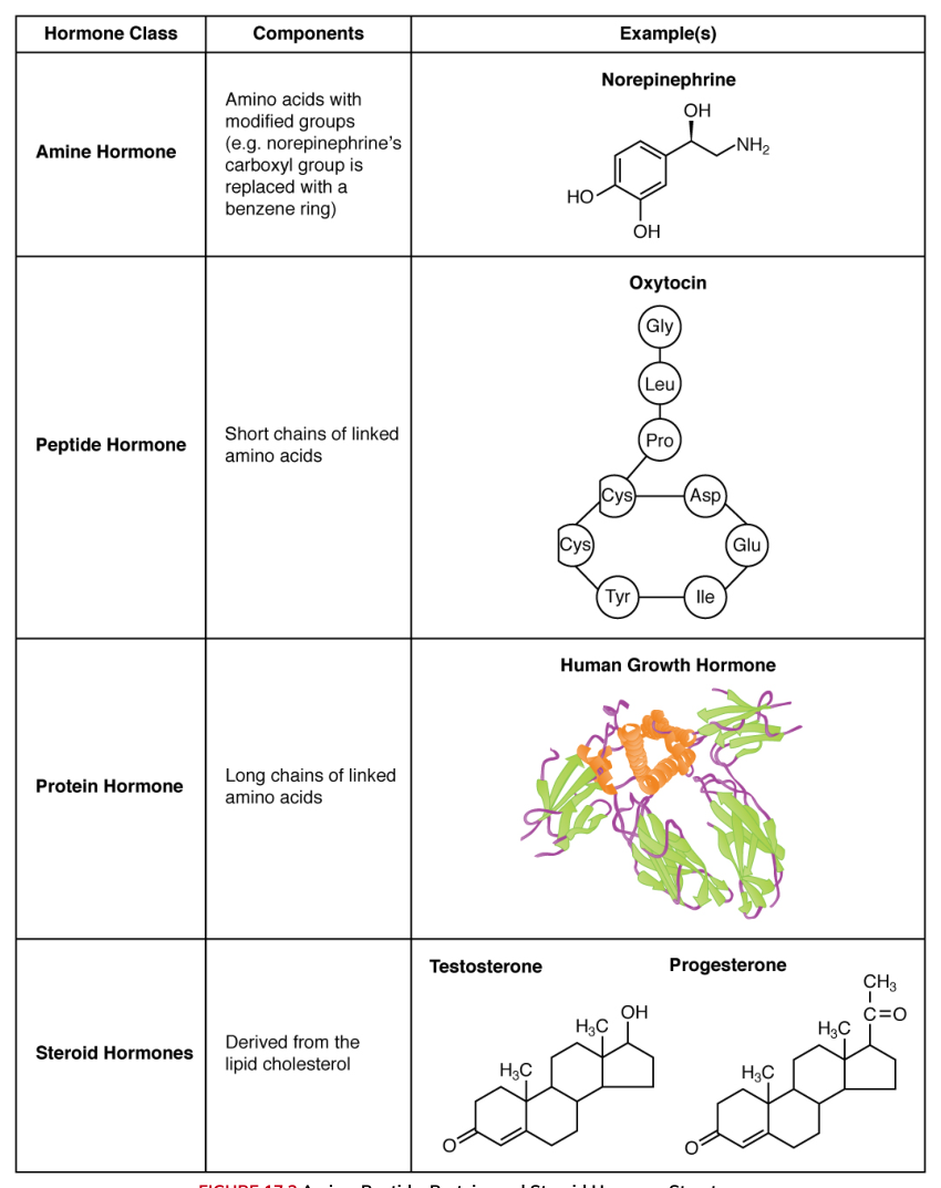
Endocrine regulation provides slower or longer-lasting control. The kidneys release renin, initiating the renin-angiotensin-aldosterone mechanism. Renin ultimately promotes formation of angiotensin II, a powerful vasoconstrictor. Angiotensin II also stimulates aldosterone release, increasing sodium reabsorption by the kidneys; water follows sodium, increasing blood volume and pressure. The posterior pituitary releases antidiuretic hormone, or ADH, which promotes water reabsorption by the kidneys and can contribute to vasoconstriction at higher concentrations.
The kidneys also produce erythropoietin, which stimulates red blood cell production when tissue oxygen delivery is inadequate. The adrenal medulla releases catecholamines, including epinephrine and norepinephrine, which alter heart activity and vascular diameter. The heart releases atrial natriuretic hormone, also called atrial natriuretic peptide, when atrial stretch indicates increased blood volume. It promotes sodium and water loss, opposing mechanisms that elevate pressure.
A concrete example follows sudden blood loss. Reduced blood volume lowers venous return and cardiac output, causing arterial pressure to fall. Baroreceptor firing decreases; sympathetic activity rises, producing tachycardia, stronger ventricular contraction, arteriolar vasoconstriction, and venoconstriction. The kidneys release renin, while ADH promotes water retention. These responses do not instantly replace the lost blood, but they help preserve perfusion of vital organs until volume can be restored.
Common misconception: ADH is not only a “water hormone,” and the renin-angiotensin-aldosterone mechanism is not merely a blood-pressure pathway. Both regulate fluid balance, vascular tone, and circulatory volume together.
20.5 Circulatory Pathways
The cardiovascular system consists of two connected circuits. The pulmonary circuit carries blood from the right ventricle to the lungs and back to the left atrium. The systemic circuit carries blood from the left ventricle through the body and back to the right atrium. Because the circuits are arranged in series, the volume pumped by the right and left ventricles must remain closely matched.
The pulmonary pathway is:
$$ \text{right ventricle} \rightarrow \text{pulmonary trunk} \rightarrow \text{right and left pulmonary arteries} \rightarrow \text{lung arterioles} \rightarrow \text{pulmonary capillaries} \rightarrow \text{pulmonary veins} \rightarrow \text{left atrium} $$
In pulmonary capillaries, carbon dioxide diffuses out of the blood and oxygen diffuses into it. Pulmonary arteries are unusual because they carry oxygen-poor blood, while pulmonary veins carry oxygen-rich blood.
The systemic pathway begins at the left ventricle:
$$ \text{left ventricle} \rightarrow \text{aorta} \rightarrow \text{systemic arteries} \rightarrow \text{arterioles} \rightarrow \text{systemic capillaries} \rightarrow \text{venules} \rightarrow \text{veins} \rightarrow \text{superior and inferior venae cavae} \rightarrow \text{right atrium} $$
The aorta gives rise to major branches that supply the head, upper limbs, thorax, abdominal organs, and lower limbs. Venous blood from the head and upper limbs returns through the superior vena cava; blood from the lower body returns through the inferior vena cava.
The hepatic portal system is a specialized systemic pathway. Blood from capillaries in the stomach, intestines, pancreas, and spleen enters the hepatic portal vein rather than returning directly to the heart. It then flows through liver sinusoids before reaching the hepatic veins and inferior vena cava. This arrangement allows the liver to process absorbed nutrients, store or modify them, detoxify substances, and regulate their release into the general circulation.
The kidneys receive a large fraction of resting cardiac output because renal circulation supports filtration and regulation of blood volume, electrolytes, and pressure. Blood enters through the renal arteries, passes through glomerular capillaries, and exits through renal veins. The digestive organs, skin, skeletal muscles, and brain each receive blood through branching arterial networks that regulate flow according to function.
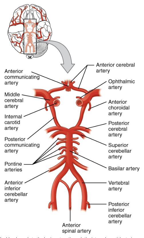
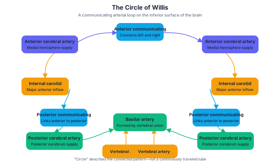
Cerebral circulation is especially protected because nervous tissue depends continuously on oxygen and glucose. The brain receives blood through two paired inputs: the internal carotid arteries and the vertebral arteries. Each internal carotid artery enters the skull through the carotid canal and gives rise to branches including the ophthalmic artery, anterior cerebral artery, middle cerebral artery, and posterior communicating artery. The vertebral arteries ascend through openings in the cervical vertebrae and unite to form the basilar artery.
The basilar artery gives rise to branches serving the brainstem and cerebellum, including the anterior inferior cerebellar, superior cerebellar, and posterior cerebral arteries. The vertebral arteries contribute posterior inferior cerebellar arteries. The circle of Willis, or cerebral arterial circle, links the anterior and posterior arterial systems through anterior and posterior communicating arteries. This collateral arrangement can preserve cerebral perfusion if one contributing vessel becomes narrowed or blocked.
For example, if blood flow is obstructed in a vessel just posterior to the left middle cerebral artery, collateral blood may reach the affected territory through connections involving the circle of Willis and the communicating arteries, depending on the exact location and severity of the blockage. The circle does not guarantee that a stroke will be prevented: small distal branches and regions with limited collateral supply may still become ischemic. Its importance is that it provides alternate routes rather than a single unavoidable pathway.
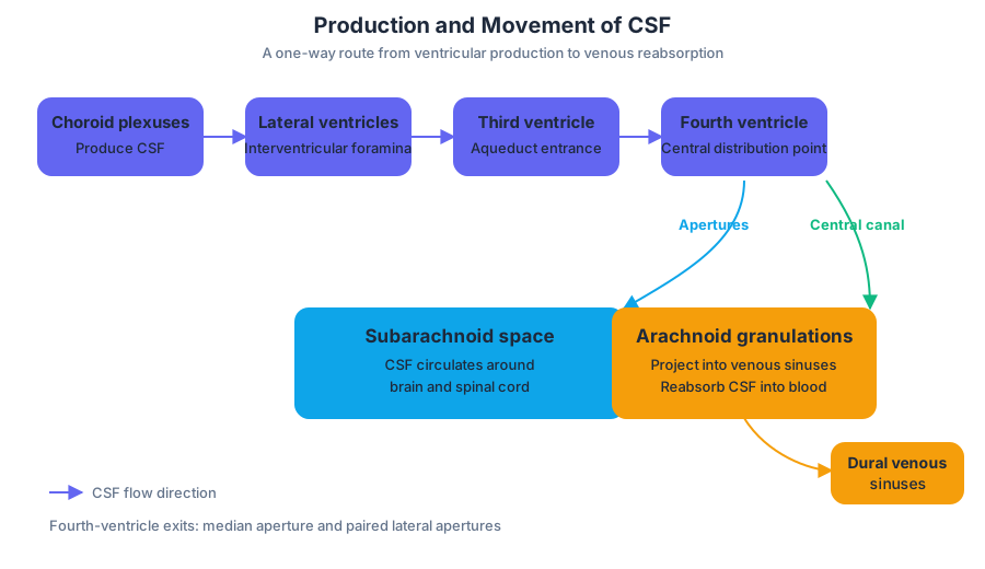
Cerebrospinal fluid, or CSF, supports and protects the central nervous system and circulates through a separate but related pathway. The choroid plexuses in the ventricles produce CSF. It flows from the lateral ventricles through the interventricular foramina into the third ventricle, passes through the cerebral aqueduct into the fourth ventricle, and then exits through the median and lateral apertures. From there it enters the subarachnoid space around the brain and spinal cord. Some CSF also enters the central canal of the spinal cord.
CSF is eventually reabsorbed into venous blood through arachnoid granulations, which project into the dural venous sinuses, especially the superior sagittal sinus. The sequence is therefore:
$$ \text{choroid plexus} \rightarrow \text{lateral ventricles} \rightarrow \text{interventricular foramina} \rightarrow \text{third ventricle} \rightarrow \text{cerebral aqueduct} \rightarrow \text{fourth ventricle} \rightarrow \text{median and lateral apertures} \rightarrow \text{subarachnoid space} \rightarrow \text{arachnoid granulations} \rightarrow \text{dural sinuses} $$
20.6 Development of Blood Vessels and Fetal Circulation
Blood vessels develop early because the growing embryo requires a transport system before the organs are mature. Vasculogenesis forms new vessels from mesoderm-derived precursor cells called angioblasts. Angiogenesis expands the vascular network by sprouting new vessels from existing ones. The developing heart and blood vessels therefore arise in close coordination, creating an embryonic circulation that can transport oxygen, nutrients, hormones, and wastes.
Fetal circulation differs from postnatal circulation because the fetal lungs are not yet performing gas exchange. Oxygen and nutrients arrive through the placenta from maternal blood, while carbon dioxide and wastes move from the fetus to the placenta. The umbilical cord contains one umbilical vein, which carries relatively oxygen-rich blood toward the fetus, and two umbilical arteries, which carry oxygen-poor blood toward the placenta.
Several fetal shunts direct blood away from the lungs and much of the liver:
- The ductus venosus allows much of the umbilical venous blood to bypass hepatic circulation and enter the inferior vena cava.
- The foramen ovale permits blood to flow directly from the right atrium to the left atrium.
- The ductus arteriosus connects the pulmonary trunk to the aorta, allowing much of the right ventricular output to bypass the high-resistance fetal lungs.
A simplified fetal pathway is:
$$ \text{placenta} \rightarrow \text{umbilical vein} \rightarrow \text{ductus venosus} \rightarrow \text{inferior vena cava} \rightarrow \text{right atrium} \rightarrow \text{foramen ovale} \rightarrow \text{left atrium} \rightarrow \text{left ventricle} \rightarrow \text{aorta} $$
Other right atrial blood enters the right ventricle and pulmonary trunk, but most is diverted through the ductus arteriosus into the aorta. Blood returns to the placenta through the internal iliac arteries and umbilical arteries.
At birth, the first breaths expand the lungs and sharply lower pulmonary vascular resistance. Pulmonary blood flow increases, and pressure in the left atrium rises relative to the right atrium. The foramen ovale functionally closes as its flap is pressed shut. Increased oxygen concentration and reduced circulating prostaglandins promote closure of the ductus arteriosus, which later becomes the ligamentum arteriosum. The ductus venosus closes and becomes the ligamentum venosum, while the umbilical vessels become fibrous remnants.
A concrete example is the transition immediately after delivery. Before birth, the placenta—not the lungs—oxygenates blood, so the fetal shunts are essential. After the umbilical cord is clamped and the newborn begins breathing, the pressure pattern reverses: blood must now travel from the right ventricle through the pulmonary arteries to the lungs, return through pulmonary veins to the left atrium, and then enter systemic circulation. Failure of a shunt to close can produce abnormal mixing or excessive pulmonary blood flow.
Key distinction: fetal circulation is not an incomplete version of adult circulation; it is a specialized arrangement built around placental gas exchange and then remodeled rapidly when pulmonary respiration begins.
Content adapted from OpenStax Anatomy and Physiology 2e by J. Gordon Betts et al., licensed CC BY 4.0.
Source Materials
Study Anatomy and Physiology on Lykke
This course page is free to open: signed out, you can read its first two sections, and signing in, on any plan, opens the rest. The free Lykke Chrome extension syncs your own Canvas courses, so ChatGPT, Claude and Muse can answer from your classes and deadlines. It only reads Canvas.
Add the free extensionSign in to open every sectionAll sections of Anatomy and Physiology · Browse more course pages