From the free course Anatomy and Physiology on Lykke
Chapter 17: The Endocrine System
Key concepts: Endocrine signaling and hormones · Comparison of endocrine and nervous system signaling · Hormone transport through the bloodstream · Target-cell hormone receptors and responses · Endocrine regulation of homeostasis and reproduction · Rapid adrenal stress responses · Endocrine glands and their major hormones · Hormonal regulation of blood calcium · cAMP and IP3 intracellular signaling pathways · Factors influencing target-cell responses
By the end of this chapter the student can: Identify the contributions of the endocrine system to homeostasis; Discuss the chemical composition of hormones and the mechanisms of hormone action; Summarize the site of production, regulation, and effects of the hormones of the pituitary, thyroid, parathyroid, adrenal, and pineal glands; Discuss the hormonal regulation of the reproductive system; Explain the role of the pancreatic endocrine cells in the regulation of blood glucose; Identify the hormones released by the heart, kidneys, and other organs with secondary endocrine functions; Discuss several common diseases associated with endocrine system dysfunction; Discuss the embryonic development of, and the effects of aging on, the endocrine system.
Chapter 17: The Endocrine System
Hormones coordinate growth, metabolism, temperature regulation, stress responses, reproduction, and many other processes by carrying chemical messages between cells. A child reaching for a falling leaf, a person maintaining blood glucose between meals, and an athlete responding instantly to danger all depend on endocrine signaling working with the nervous system.
The endocrine system is the body’s system of glands and cells that release hormones into interstitial fluid and usually into the bloodstream, allowing chemical signals to regulate distant target cells.
Endocrine signaling is one form of intercellular communication, meaning communication between cells. Unlike a signal that acts only on a neighboring cell, an endocrine hormone may travel through the circulation to a distant organ. The message is effective only if the receiving cell has the appropriate receptor, a protein that recognizes and binds that hormone.
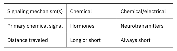
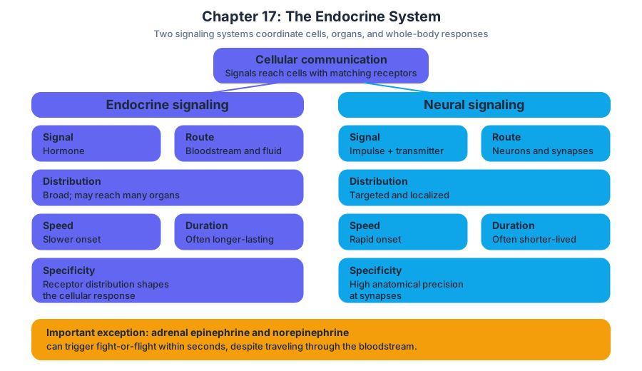
The endocrine system and nervous system are the body’s major coordinating systems. The nervous system commonly uses electrical impulses and neurotransmitters to produce rapid, brief, and highly localized responses. The endocrine system uses hormones carried through body fluids, producing responses that are often slower to begin but longer lasting and more widespread. The two systems are not separate competitors: the hypothalamus links neural control with endocrine control, and many physiological responses require both systems.
For example, an unexpected threat activates sympathetic neurons almost immediately, increasing heart rate and redirecting blood flow. At the same time, the hypothalamus and pituitary gland stimulate the adrenal glands to release glucocorticoids, which support metabolism and help the body sustain the stress response. The nervous system starts the response quickly; endocrine signaling helps maintain and coordinate it.
17.1 An Overview of the Endocrine System
A hormone is like a message released into a city-wide transportation network: it may circulate throughout the body, but only buildings with the correct address can respond. The bloodstream distributes the signal broadly; receptor proteins provide the biological address.
Endocrine glands are ductless glands that release their secretions into the interstitial fluid and bloodstream. Their secretions, called hormones, regulate target cells elsewhere in the body.
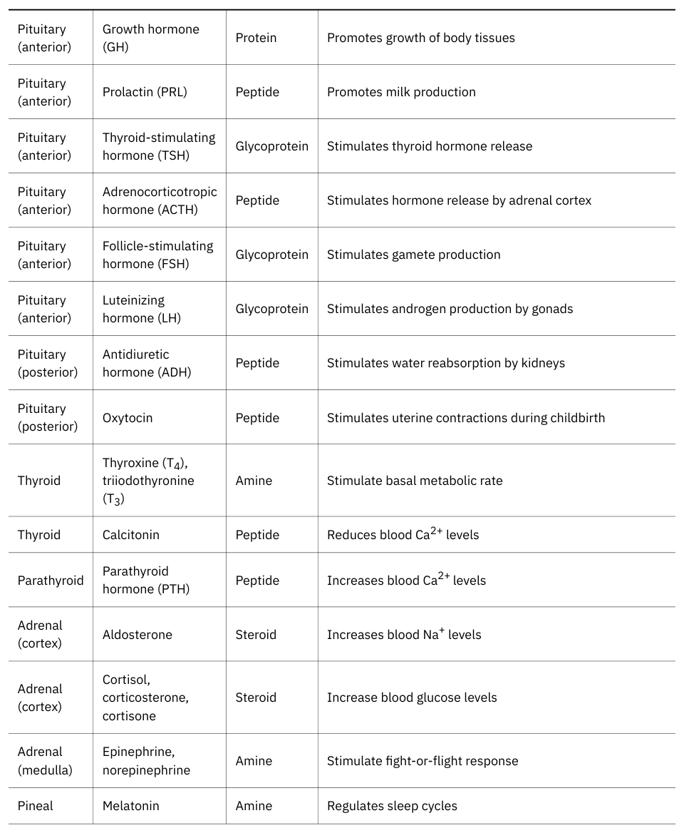
Major endocrine glands include the hypothalamus, pituitary gland, thyroid gland, parathyroid glands, adrenal glands, pineal gland, and the endocrine portions of the pancreas, gonads, and placenta. Several organs—including the heart, kidneys, stomach, small intestine, liver, thymus, and adipose tissue—also release hormones even though endocrine secretion is not their primary function.
Endocrine regulation is essential for homeostasis, the maintenance of relatively stable internal conditions. Hormones help regulate blood glucose, blood calcium, fluid and electrolyte balance, metabolism, growth, development, the stress response, and reproductive functions. A hormone may alter the activity of an existing protein, change gene expression, stimulate secretion, or influence cell division and differentiation.
Endocrine signaling follows a general sequence:
- A stimulus changes an internal or external condition.
- An endocrine cell detects the stimulus or receives a signal from another regulatory center.
- The cell secretes a hormone.
- The hormone travels through interstitial fluid and often through the bloodstream.
- The hormone binds to receptors on or in target cells.
- Target cells change their activity.
- Feedback from the response modifies further hormone release.
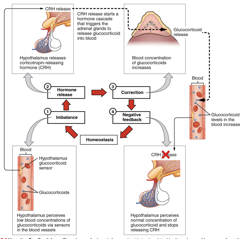
Most endocrine pathways use negative feedback, in which the response reduces the original stimulus. For example, when blood glucocorticoid concentrations rise, the hypothalamus and pituitary gland reduce the signals that stimulate the adrenal glands. This prevents excessive glucocorticoid secretion and keeps the hormone concentration within a functional range.
A concrete example is blood calcium regulation. When blood calcium rises above approximately $10\ \text{mg/dL}$, the thyroid gland releases calcitonin. Calcitonin inhibits osteoclast activity and decreases calcium reabsorption by the kidneys, promoting movement of calcium out of the blood. When blood calcium falls below approximately $10\ \text{mg/dL}$, the parathyroid glands release parathyroid hormone, or PTH. PTH stimulates calcium release from bone, increases renal calcium reabsorption, and promotes vitamin $D$ synthesis, which increases calcium absorption in the small intestine.
A common misconception is that only organs labeled “glands” have endocrine functions. In reality, endocrine cells are distributed throughout many organs. The heart releases atrial natriuretic peptide, the kidneys release erythropoietin and renin, and adipose tissue releases hormones such as leptin. Endocrine function is defined by what a cell secretes and how the secretion acts, not only by the organ’s name.
17.2 Hormones
A hormone can travel through the entire circulation without affecting most cells because a target cell must possess the correct receptor. This selectivity allows one chemical signal to circulate widely while producing a response in only particular tissues.
A hormone is a chemical messenger released by an endocrine cell that travels through body fluids to regulate the activity of target cells.
Chemical classes of hormones
The three major chemical classes of hormones are peptide hormones, steroid hormones, and amino acid derivatives. Their chemical structures determine how they are transported in blood, where their receptors are located, and how quickly their effects develop.
Peptide hormones consist of chains of amino acids. They include short peptides and larger proteins such as insulin and growth hormone. Because they are water-soluble, they generally cannot cross the hydrophobic interior of the plasma membrane. They therefore bind to receptors on the cell membrane and activate intracellular signaling pathways.
Steroid hormones are lipid-soluble molecules derived from cholesterol. Examples include cortisol, aldosterone, estrogen, progesterone, and testosterone. Because they are lipid-soluble, they can cross the plasma membrane. In the blood, many steroid hormones bind to transport proteins, which make them more soluble and extend their time in circulation.
Amino acid derivatives are modified molecules derived from amino acids. Epinephrine and norepinephrine are derived from tyrosine and act mainly through cell-membrane receptors. Thyroid hormones are also derived from tyrosine, but their lipid-soluble behavior allows them to enter target cells and bind intracellular receptors.
Hormone transport and receptors
Water-soluble hormones dissolve readily in blood plasma and circulate mostly in an unbound form. Lipid-soluble hormones do not dissolve well in plasma, so they commonly travel bound to carrier proteins. Only the unbound fraction can readily leave the bloodstream and bind to receptors, although bound hormone serves as a circulating reserve.
Intracellular receptors are located in the cytoplasm or nucleus. A lipid-soluble hormone crosses the plasma membrane and binds its receptor. The hormone-receptor complex then binds specific regions of DNA and changes gene transcription. The resulting messenger RNA is translated into proteins that alter cell activity.
The intracellular pathway proceeds as follows:
- A lipid-soluble hormone diffuses through the plasma membrane.
- The hormone binds an intracellular receptor.
- The hormone-receptor complex enters the nucleus or acts there.
- The complex binds DNA and stimulates or inhibits transcription.
- Messenger RNA is produced.
- Ribosomes translate the messenger RNA into proteins.
- The new proteins change the target cell’s function.
Because this pathway requires gene transcription and protein synthesis, its effects often develop more slowly but may last longer. A steroid hormone such as cortisol can alter the production of proteins involved in metabolism, immune regulation, and the stress response.
Cell-membrane receptors bind water-soluble hormones and some amino acid derivatives. Binding activates a signal-transduction pathway inside the cell. The receptor may activate an enzyme directly or activate a membrane-associated G protein, which then stimulates an effector enzyme.
One important pathway uses cyclic adenosine monophosphate, or cAMP, as a second messenger:
- The hormone binds to a membrane receptor.
- The receptor activates a G protein.
- The G protein activates adenylyl cyclase.
- Adenylyl cyclase converts adenosine triphosphate into cAMP.
- cAMP activates protein kinases.
- Protein kinases phosphorylate target proteins.
- The target cell changes its activity.
A second important pathway uses inositol trisphosphate, or IP$_3$, and diacylglycerol, or DAG:
- A hormone binds to a membrane receptor.
- The receptor activates a G protein.
- The G protein activates phospholipase C.
- Phospholipase C splits a membrane phospholipid into IP$_3$ and DAG.
- IP$_3$ stimulates the release of calcium ions from intracellular stores.
- Calcium ions and DAG activate protein kinase C.
- Protein phosphorylation changes cell activity.
The same hormone can produce different effects in different tissues because target cells may have different receptors, different intracellular signaling proteins, or different genes available for regulation. For example, epinephrine increases heart rate through one receptor-mediated response but promotes airway smooth-muscle relaxation through another.
17.3 The Pituitary Gland and Hypothalamus
The hypothalamus and pituitary gland form a command center in which neural information is translated into hormonal instructions. The hypothalamus monitors internal conditions and controls the pituitary; the pituitary then regulates other endocrine glands and tissues.
The hypothalamus is a region of the brain that maintains homeostasis and controls the pituitary gland through releasing and inhibiting hormones as well as neural connections.
The pituitary gland, or hypophysis, lies in the sella turcica of the sphenoid bone and connects to the hypothalamus by the infundibulum. It has two major regions: the posterior pituitary, or neurohypophysis, and the anterior pituitary, or adenohypophysis. Their embryological origins and mechanisms of secretion differ.
Posterior pituitary
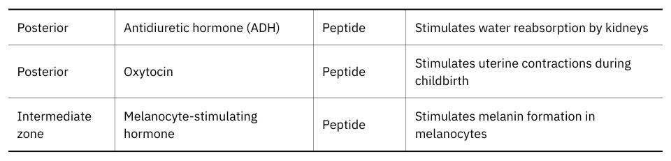
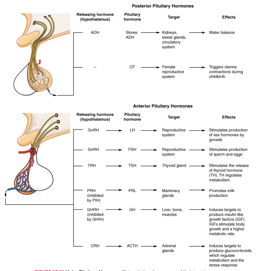
The posterior pituitary does not synthesize its two major hormones. Instead, hypothalamic neurons synthesize antidiuretic hormone, or ADH, and oxytocin. Their axons extend through the infundibulum, and the hormones are stored in and released from posterior pituitary terminals.
ADH, also called vasopressin, acts primarily on the kidneys. It increases water reabsorption by stimulating the insertion of aquaporin water channels into the membranes of kidney collecting-duct cells. At higher concentrations it also causes vasoconstriction. ADH release increases when blood osmolarity rises or blood volume falls.
Oxytocin stimulates uterine contractions during labor and milk ejection during breastfeeding. In both situations, the response can reinforce further hormone release: uterine stretching promotes more oxytocin release, and suckling promotes oxytocin release that causes milk ejection. These are examples of positive feedback, in which the response intensifies the original stimulus until a defined event ends it.
Anterior pituitary
The anterior pituitary synthesizes and secretes hormones in response to hypothalamic releasing or inhibiting hormones delivered through the hypophyseal portal system. This specialized blood pathway carries hypothalamic hormones directly to anterior pituitary cells, allowing small quantities of regulatory hormones to control pituitary secretion.
The major anterior pituitary hormones include:
- Thyroid-stimulating hormone, or TSH, stimulates the thyroid gland to release thyroid hormones.
- Adrenocorticotropic hormone, or ACTH, stimulates the adrenal cortex to release glucocorticoids.
- Follicle-stimulating hormone, or FSH, supports ovarian follicle development and estrogen secretion in females and stimulates sperm production in males.
- Luteinizing hormone, or LH, triggers ovulation and supports ovarian hormone secretion in females; in males it stimulates testosterone secretion by interstitial cells.
- Growth hormone, or GH, promotes growth directly and indirectly through insulin-like growth factors.
- Prolactin, or PRL, promotes milk production by mammary glands.
The anterior pituitary also produces melanocyte-stimulating hormone, or MSH, although its role in adult humans is less prominent than in many other vertebrates. The hypothalamus regulates anterior pituitary secretion using hormones such as thyrotropin-releasing hormone, corticotropin-releasing hormone, gonadotropin-releasing hormone, growth hormone-releasing hormone, and prolactin-releasing hormone. Inhibiting hormones, including somatostatin and dopamine, suppress particular pituitary secretions.
A typical hypothalamic-pituitary-target-gland pathway works in three steps. First, the hypothalamus releases a releasing hormone. Second, the anterior pituitary releases a tropic hormone. Third, the tropic hormone stimulates a peripheral endocrine gland. Rising concentrations of the peripheral gland’s hormone then inhibit both hypothalamic and pituitary signaling through negative feedback.
For example, low thyroid hormone levels stimulate hypothalamic thyrotropin-releasing hormone. This causes anterior pituitary release of TSH, which stimulates the thyroid gland to release thyroid hormones. As thyroid hormone concentration rises, it inhibits further release of thyrotropin-releasing hormone and TSH.
Excessive or deficient pituitary signaling can disrupt several body systems at once. Abnormal GH secretion before epiphyseal plates close can cause gigantism, whereas GH deficiency can cause pituitary dwarfism. Excess GH after epiphyseal plates close causes acromegaly, characterized by enlargement of bones and soft tissues.
17.4 The Thyroid Gland
The thyroid gland is a metabolic thermostat and a calcium-regulating organ. Its hormones influence how quickly cells use energy, while its calcitonin-producing cells participate in calcium homeostasis.
The thyroid gland lies in the anterior neck, inferior to the larynx and adjacent to the trachea. It has two lobes connected by an isthmus. The gland is composed largely of spherical thyroid follicles, which contain colloid and are surrounded by follicular cells. Parafollicular cells, or C cells, lie between follicles.
Thyroid hormones
Follicular cells produce thyroxine, or T$_4$, and triiodothyronine, or T$_3$. Both are derived from the amino acid tyrosine and require iodine. T$_4$ contains four iodine atoms, whereas T$_3$ contains three. Although the thyroid releases more T$_4$, many tissues convert T$_4$ into the more biologically active T$_3$.
Thyroid hormone synthesis occurs in a sequence:
- Follicular cells transport iodide from the blood into the cell.
- Iodide moves into the colloid.
- Thyroglobulin, a protein made by follicular cells, is secreted into the colloid.
- Iodide is oxidized and attached to tyrosine residues on thyroglobulin.
- Iodinated tyrosines combine to form T$_3$ and T$_4$.
- Thyroglobulin containing T$_3$ and T$_4$ is taken back into follicular cells.
- Lysosomes release T$_3$ and T$_4$ from thyroglobulin.
- Thyroid hormones enter the bloodstream, where most bind carrier proteins.
TSH from the anterior pituitary stimulates nearly every step of thyroid hormone synthesis and secretion. Thyroid hormones enter target cells and bind intracellular receptors, altering gene expression. They increase basal metabolic rate, support normal cardiovascular function, influence protein and carbohydrate metabolism, and are essential for normal growth and nervous-system development.
Thyroid hormone production is regulated by negative feedback. Increasing concentrations of T$_3$ and T$_4$ inhibit hypothalamic thyrotropin-releasing hormone and pituitary TSH. Low thyroid hormone production may result in hypothyroidism, which can cause fatigue, cold intolerance, weight gain, and slowed metabolism. Excess production causes hyperthyroidism, which may produce weight loss, heat intolerance, rapid heartbeat, and nervousness.
Calcitonin
Parafollicular cells release calcitonin when blood calcium concentration rises. Calcitonin lowers blood calcium partly by inhibiting osteoclast activity and reducing calcium reabsorption by the kidneys. Its role in adult calcium regulation is less dominant than that of PTH, but it contributes to the overall balance between calcium deposition and release.
For example, after calcium-rich food is absorbed and blood calcium begins to rise, calcitonin supports the movement of calcium into bone and increases urinary calcium loss. This response helps oppose a rise away from the normal concentration.
17.5 The Parathyroid Glands
Small parathyroid glands perform a large task: they defend the calcium concentration required for nerve conduction, muscle contraction, blood clotting, and many intracellular processes.
Parathyroid hormone, or PTH, is the principal hormone that raises blood calcium concentration.
Usually four parathyroid glands are embedded on the posterior surface of the thyroid gland. Their chief cells monitor blood calcium through calcium-sensing receptors. When blood calcium falls, the glands release PTH.
PTH raises blood calcium through three coordinated actions:
- Bone: PTH indirectly increases osteoclast activity. Osteoclasts resorb bone matrix and release calcium and phosphate into the blood.
- Kidneys: PTH increases calcium reabsorption, reducing calcium loss in urine. It decreases phosphate reabsorption, promoting phosphate excretion.
- Small intestine: PTH stimulates the kidneys to produce the active form of vitamin $D$. Active vitamin $D$ increases intestinal absorption of calcium.
The response to low blood calcium is a negative-feedback loop. Low calcium stimulates PTH release; PTH increases calcium movement into the blood; rising calcium then reduces stimulation of the parathyroid glands.
A concrete example occurs when inadequate dietary calcium or vitamin $D$ limits calcium absorption. PTH secretion rises, conserving calcium through the kidneys and mobilizing calcium from bone. If this stimulation persists, excessive bone resorption can weaken the skeleton.
Hyperparathyroidism produces abnormally high PTH secretion and can cause elevated blood calcium, bone loss, kidney stones, abdominal discomfort, and changes in nervous-system function. Hypoparathyroidism lowers PTH and may produce abnormally low blood calcium, causing muscle spasms or tetany because excitable tissues become more responsive.
17.6 The Adrenal Glands
The adrenal glands can respond to danger on two time scales: the adrenal medulla releases catecholamines within seconds, while the adrenal cortex releases steroid hormones that sustain and organize the longer stress response.
The adrenal glands sit on the superior poles of the kidneys. Each gland has an outer adrenal cortex and an inner adrenal medulla. These regions differ in embryological origin, structure, hormones, and regulation.
Adrenal cortex
The adrenal cortex has three zones. The outer zona glomerulosa produces mineralocorticoids, especially aldosterone. The middle zona fasciculata produces glucocorticoids, especially cortisol. The inner zona reticularis produces adrenal androgens.
Aldosterone regulates sodium, potassium, and water balance. It acts mainly on kidney tubules to increase sodium reabsorption and potassium secretion. Water follows reabsorbed sodium, helping maintain blood volume and pressure. Aldosterone release is stimulated chiefly by the renin-angiotensin-aldosterone system and elevated blood potassium; ACTH has a smaller supporting role.
Cortisol supports the long-term response to stress. It increases blood glucose by promoting gluconeogenesis, helps mobilize energy stores, and suppresses inflammatory and immune activity. Cortisol secretion is controlled by the hypothalamic-pituitary-adrenal axis: corticotropin-releasing hormone stimulates ACTH, and ACTH stimulates the adrenal cortex. Rising cortisol inhibits both hypothalamic and pituitary signals.
Adrenal androgens contribute to sex-hormone activity, especially before puberty and in females. Their effects are weaker than those of gonadal androgens but can contribute to pubic and axillary hair development.
Adrenal medulla
The adrenal medulla contains modified sympathetic neurons called chromaffin cells. Sympathetic preganglionic neurons stimulate these cells to release epinephrine and norepinephrine into the bloodstream.
The rapid adrenal stress response proceeds as follows:
- A threatening stimulus is processed by the brain.
- The hypothalamus activates sympathetic preganglionic neurons.
- Sympathetic fibers stimulate chromaffin cells.
- The adrenal medulla releases epinephrine and norepinephrine.
- Heart rate, cardiac output, and blood glucose rise; blood flow is redirected toward skeletal muscle; and airways widen.
- The body is prepared for rapid action.
For example, a person who nearly steps into traffic may experience a pounding heart, rapid breathing, sweating, and tremor before consciously analyzing the event. These rapid effects are primarily sympathetic and adrenal-medullary responses, not the slower genomic effects of cortisol.
Excess cortisol can produce Cushing syndrome, associated with abnormal fat distribution, muscle weakness, hyperglycemia, and impaired immune function. Insufficient adrenal cortex activity produces Addison disease, which may cause fatigue, low blood pressure, weight loss, and impaired ability to respond to stress.
17.7 The Pineal Gland
The daily rhythm of sleep and wakefulness is influenced by light entering the eyes, neural signals reaching the brain, and hormonal output from the pineal gland.
The pineal gland is a small endocrine gland located in the roof of the third ventricle of the brain. It secretes melatonin, a hormone derived from serotonin. Melatonin secretion generally increases during darkness and decreases in bright light.
The light-dark pathway operates in steps:
- Photoreceptors in the retina detect environmental light.
- Neural signals travel to the suprachiasmatic nucleus of the hypothalamus, a major biological-clock center.
- The hypothalamus influences sympathetic pathways to the pineal gland.
- Darkness permits increased melatonin secretion.
- Melatonin acts on the brain and other tissues to help coordinate circadian timing.
Circadian rhythms are approximately $24$-hour cycles in physiological activity. Melatonin does not simply “turn sleep on,” but it helps signal biological night and coordinate sleep timing, body temperature rhythms, and other daily cycles.
A concrete example is jet lag. Rapid travel across time zones exposes the body’s internal clock to a new light-dark schedule. Melatonin secretion and sleepiness may initially occur at the old local time, producing difficulty sleeping at night and fatigue during the day until the rhythm adjusts.
The pineal gland has also been associated with the timing of reproductive development, particularly in animals with strong seasonal breeding patterns. In humans, its most established endocrine role concerns biological timing rather than direct control of reproductive function.
17.8 Gonadal and Placental Hormones
Reproduction requires coordinated endocrine communication among the hypothalamus, pituitary gland, gonads, and—during pregnancy—the placenta. The same hypothalamic-pituitary pathway supports both gamete production and reproductive hormone secretion.
Gonadal regulation
The hypothalamus releases gonadotropin-releasing hormone, or GnRH, in pulses. GnRH stimulates the anterior pituitary to release FSH and LH, collectively called gonadotropins. FSH and LH act on the ovaries or testes, and gonadal hormones feed back to the hypothalamus and pituitary.
In the testes, LH stimulates interstitial, or Leydig, cells to produce testosterone. FSH acts on Sertoli cells and supports spermatogenesis, the production of sperm. Sertoli cells also release inhibin, which suppresses FSH secretion. Testosterone supports development and maintenance of male reproductive structures, muscle and bone growth, sperm production, and secondary sex characteristics.
In the ovaries, FSH promotes follicle development. Developing follicles secrete estrogens, especially estradiol. LH contributes to follicle maturation and triggers ovulation. After ovulation, LH supports the corpus luteum, which secretes progesterone and estrogen.
Estrogen supports development of female reproductive structures and secondary sex characteristics and stimulates growth of the uterine lining during the menstrual cycle. Progesterone prepares and maintains the uterine lining for implantation and helps support pregnancy. Inhibin from ovarian cells reduces FSH secretion.
Most of the menstrual cycle is regulated by negative feedback from estrogen, progesterone, and inhibin. Near ovulation, however, sustained high estrogen produces positive feedback on the pituitary, generating the LH surge that triggers ovulation.
Placental hormones
The placenta is a temporary endocrine organ formed during pregnancy. It produces hormones that maintain the uterine environment, support fetal development, and prepare the mother’s body for childbirth and lactation.
Human chorionic gonadotropin, or hCG, is secreted early in pregnancy by placental tissue. It maintains the corpus luteum, allowing continued progesterone and estrogen secretion before the placenta can produce sufficient amounts itself. Pregnancy tests detect hCG or its products in urine or blood.
The placenta also produces estrogens and progesterone. Estrogens promote growth of the uterus and breasts and increase blood flow to reproductive tissues. Progesterone maintains the uterine lining, reduces uterine contractions, and supports the conditions required for pregnancy.
Human placental lactogen, or hPL, contributes to mammary-gland development and changes maternal metabolism so that more glucose remains available to the fetus. The placenta also releases relaxin, which helps prepare the pelvis and cervix for childbirth.
For example, early pregnancy can be maintained by the sequence: implantation leads placental tissue to release hCG; hCG preserves the corpus luteum; the corpus luteum continues releasing progesterone; progesterone maintains the uterine lining; and later the placenta becomes the major source of progesterone and estrogen.
17.9 The Endocrine Pancreas
The pancreas continuously adjusts blood glucose so that cells have fuel without allowing glucose to reach damaging concentrations. Its endocrine cells form small clusters called pancreatic islets.
The pancreas lies posterior to the stomach and has both exocrine and endocrine functions. Its exocrine portion secretes digestive enzymes into ducts. Its endocrine portion consists primarily of the pancreatic islets, which release hormones into the bloodstream.
The major endocrine cell types are:
- Alpha cells, which secrete glucagon.
- Beta cells, which secrete insulin.
- Delta cells, which secrete somatostatin.
- PP cells, which secrete pancreatic polypeptide.
When blood glucose rises, beta cells release insulin. Insulin promotes glucose uptake by many cells, stimulates glycogen synthesis in liver and skeletal muscle, and promotes storage of excess energy as fat. It therefore lowers blood glucose.
When blood glucose falls, alpha cells release glucagon. Glucagon stimulates the liver to break down glycogen and produce glucose through gluconeogenesis. The liver releases glucose into the blood, raising blood glucose.
The regulation of blood glucose follows a negative-feedback sequence:
- A meal increases blood glucose.
- Beta cells detect the increase and release insulin.
- Target cells take up glucose, and the liver stores glucose as glycogen.
- Blood glucose falls toward its set range.
- Insulin secretion decreases.
Between meals, the reverse occurs:
- Blood glucose begins to fall.
- Alpha cells release glucagon.
- The liver breaks down glycogen and produces glucose.
- Blood glucose rises toward its normal range.
- Glucagon secretion decreases.
Somatostatin inhibits the release of both insulin and glucagon and helps regulate digestive and absorptive processes. Pancreatic polypeptide influences pancreatic secretory activity and gastrointestinal function.
A concrete example is the response to a carbohydrate-rich meal. Rising glucose stimulates insulin, which promotes glucose uptake by skeletal muscle and storage as liver glycogen. In diabetes mellitus, insulin secretion, insulin action, or both are inadequate. Persistent hyperglycemia can damage blood vessels, nerves, kidneys, eyes, and other tissues.
17.10 Organs with Secondary Endocrine Functions
Endocrine communication is distributed throughout the body. Several organs release hormones that regulate fluid balance, blood-cell production, digestion, appetite, and tissue growth even though endocrine secretion is not their primary role.
The heart releases atrial natriuretic peptide, or ANP, when atrial walls stretch because blood volume or pressure is elevated. ANP promotes sodium and water excretion by the kidneys and suppresses mechanisms that raise blood volume and pressure.
The kidneys release erythropoietin, or EPO, when oxygen delivery is inadequate. EPO stimulates red bone marrow to increase red blood cell production. The kidneys also release renin, an enzyme that begins the renin-angiotensin-aldosterone system, which increases blood pressure and supports blood volume. They also activate vitamin $D$ into a hormone that promotes intestinal calcium absorption.
The stomach and small intestine release hormones that coordinate digestion. Gastrin stimulates gastric secretion and motility. Secretin stimulates pancreatic bicarbonate secretion and helps regulate gastric activity. Cholecystokinin, or CCK, stimulates pancreatic enzyme secretion and gallbladder contraction. Gastric inhibitory peptide, or GIP, helps regulate insulin secretion and gastrointestinal function.
The liver produces insulin-like growth factors, or IGFs, in response to GH. IGFs mediate many of GH’s growth-promoting effects on bone, cartilage, and muscle. The liver also produces angiotensinogen, a precursor in blood-pressure regulation.
The thymus secretes hormones such as thymosins that support the development and maturation of T lymphocytes. Although thymic activity is especially important during childhood, the gland gradually becomes smaller and more fatty after puberty.
Adipose tissue is an endocrine organ. It secretes leptin, which signals the hypothalamus about energy stores and tends to reduce food intake, as well as other signaling molecules involved in metabolism and inflammation.
A concrete example is reduced oxygen availability at high altitude. The kidneys detect lower oxygen delivery and increase EPO secretion. EPO stimulates erythrocyte production in bone marrow, increasing the blood’s oxygen-carrying capacity over time. This response is slower than a nervous reflex but useful for long-term adaptation.
17.11 Development and Aging of the Endocrine System
Endocrine organs develop from several embryonic tissue sources, and their functions change throughout life. Developmental timing is itself hormonally regulated, so endocrine disorders can affect both body structure and the schedule of maturation.
The pituitary gland develops from two embryonic sources. The anterior pituitary develops from an upward growth of oral ectoderm called Rathke’s pouch. The posterior pituitary develops from a downward extension of neuroectoderm from the diencephalon. This dual origin explains why the two regions differ in structure and function.
The thyroid gland begins as an outgrowth from the embryonic pharyngeal region and migrates to its adult position in the neck. The parathyroid glands develop from pharyngeal pouch tissue. The adrenal cortex develops from mesoderm, whereas the adrenal medulla develops from neural-crest cells associated with the sympathetic nervous system.
The pancreas develops from endodermal buds of the embryonic duodenum. The gonads develop from the genital ridge, and their endocrine activity becomes especially important during puberty. The pineal gland develops as an outgrowth of the diencephalon.
Puberty and reproductive maturation
During childhood, hypothalamic GnRH secretion is relatively low. At puberty, pulsatile GnRH secretion increases, stimulating the pituitary to release FSH and LH. Gonadal hormone production rises, producing maturation of reproductive organs, gamete production, growth spurts, and secondary sex characteristics.
In males, increased LH stimulates testosterone production and FSH supports sperm production. In females, FSH and LH coordinate follicle development, estrogen secretion, ovulation, and menstrual cycling. Adrenal androgens also contribute to the development of pubic and axillary hair.
Endocrine aging
Aging is associated with changes in endocrine secretion and tissue responsiveness. Growth hormone and IGF activity generally decline, contributing to reduced muscle mass, changes in bone maintenance, and altered body composition. Melatonin secretion may decline, contributing to changes in sleep patterns. The thyroid may become less responsive or produce less thyroid hormone, although substantial variation exists among individuals.
Reproductive endocrine function also changes. In females, ovarian follicles become depleted and ovarian estrogen and progesterone production decline during menopause. The resulting loss of negative feedback increases FSH and LH concentrations and may produce hot flashes, sleep disturbance, and changes in bone maintenance. In males, testosterone generally declines more gradually, with considerable individual variation.
Aging may also reduce pancreatic beta-cell reserve and alter insulin sensitivity, increasing the risk of type 2 diabetes. Vascular and kidney changes can modify hormone clearance and fluid regulation. These are trends, not inevitable diseases: nutrition, physical activity, medical treatment, and inherited factors influence endocrine health.
A concrete developmental example is congenital hypothyroidism. If thyroid hormone production is severely inadequate during early development, brain development and body growth can be impaired. Early detection and thyroid-hormone replacement can prevent much of this damage, illustrating why endocrine hormones are essential not only for adult homeostasis but also for normal development.
Across the lifespan, endocrine function depends on coordinated feedback. Hormone concentrations, receptor sensitivity, gland structure, and target-tissue responsiveness all matter. A disorder can therefore arise from excessive secretion, deficient secretion, abnormal transport, defective receptors, or failure of feedback regulation.
Content adapted from OpenStax Anatomy and Physiology 2e by J. Gordon Betts et al., licensed CC BY 4.0.

Source Materials
Study Anatomy and Physiology on Lykke
This course page is free to open: signed out, you can read its first two sections, and signing in, on any plan, opens the rest. The free Lykke Chrome extension syncs your own Canvas courses, so ChatGPT, Claude and Muse can answer from your classes and deadlines. It only reads Canvas.
Add the free extensionSign in to open every sectionAll sections of Anatomy and Physiology · Browse more course pages