From the free course Anatomy and Physiology on Lykke
Chapter 3: The Cellular Level of Organization
Key concepts: Cell structure and function · Cell membrane structure and function · Phospholipid bilayer and membrane fluidity · Transport of materials across the cell membrane · Cytoplasmic organelles · Nucleus and DNA replication · Cell differentiation and specialization · Cell division and mitosis · History of cell observation and the term “cell”
By the end of this chapter the student can: Describe the structure and function of the cell membrane, including its regulation of materials into and out of the cell; Describe the functions of the various cytoplasmic organelles; Explain the structure and contents of the nucleus, as well as the process of DNA replication; Explain the process by which a cell builds proteins using the DNA code; List the stages of the cell cycle in order, including the steps of cell division in somatic cells; Discuss how a cell differentiates and becomes more specialized; List the morphological and physiological characteristics of some representative cell types in the human body.
Chapter 3: The Cellular Level of Organization
A cell is the smallest unit of life capable of carrying out the functions necessary for survival. Robert Hooke coined the term cell in 1665 after observing the boxlike subdivisions of cork, which reminded him of the small rooms occupied by monks. The cork compartments he saw were not living cells, but the name endured because every living cell is enclosed by a boundary and contains organized internal structures.
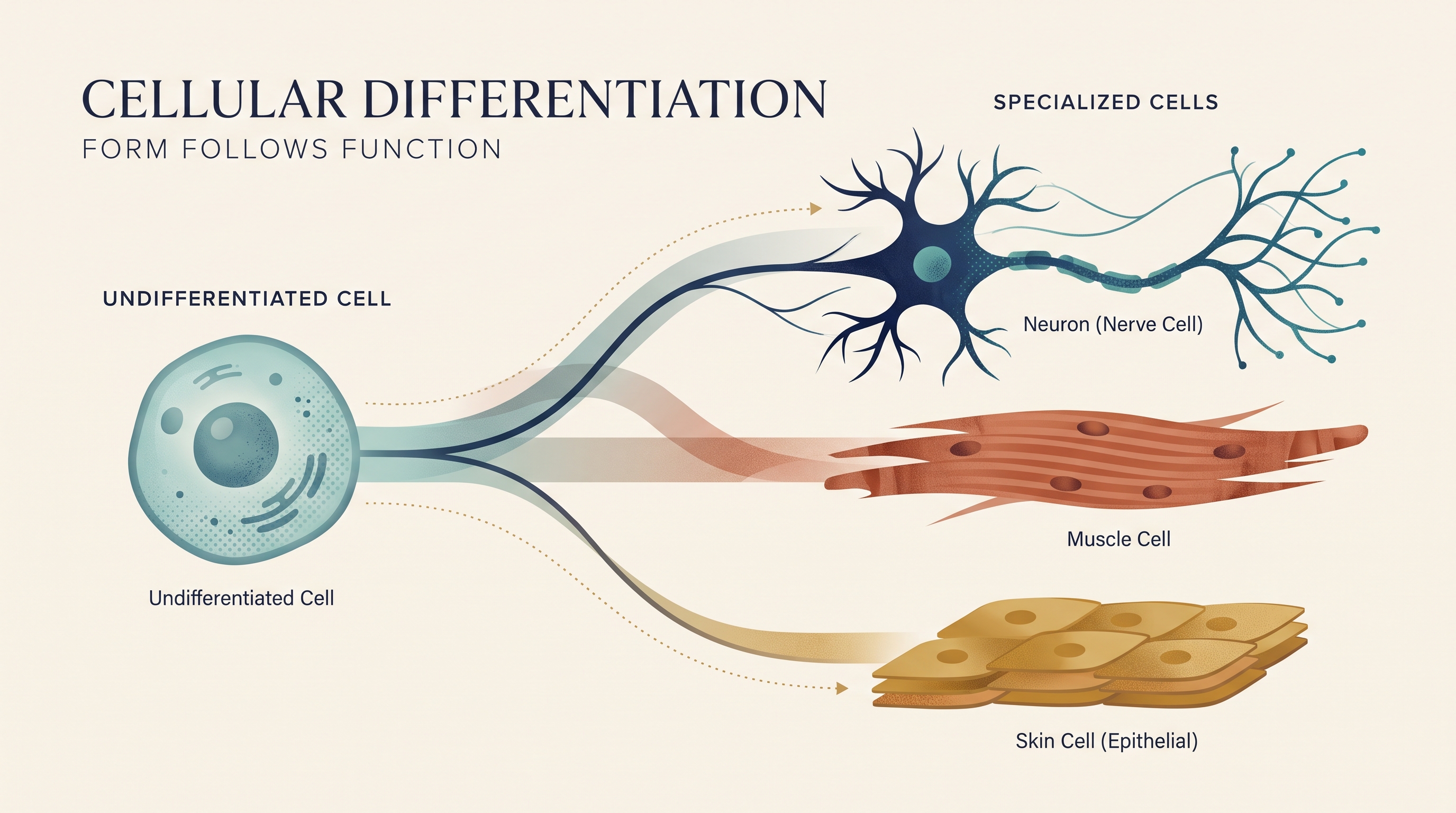
All living cells in a multicellular organism have a surrounding cell membrane, even though cells differ dramatically in shape, size, internal organization, and function. A flat skin cell forms a protective surface, a long skeletal-muscle cell generates force, and a neuron may extend processes over considerable distances to communicate with other cells. Their shared cellular plan provides a common foundation; their specialized structures make different jobs possible.
During development, early cells are initially relatively undifferentiated, meaning they have not yet acquired a specialized structure or function. Through cellular differentiation, cells activate particular genetic instructions and become specialized. Differentiation allows cells to cooperate in tissues and organs: the DNA of a muscle cell and a nerve cell is fundamentally the same, but the cells use different portions of that information.
A useful principle is form follows function. A cell’s shape, surface features, organelles, and internal arrangement are related to what the cell must do. The fluorescence-stained newt lung cell shown undergoing mitosis illustrates that a cell is not static: its membrane, cytoplasm, chromosomes, and mitotic spindle participate in an organized process that produces two new cells.
3.1 The Cell Membrane
The cell membrane is like a flexible border checkpoint rather than a rigid wall. It separates the cell’s internal environment from the extracellular fluid while allowing selected substances to enter or leave. This selective control is essential for maintaining the different chemical conditions required inside the cell.
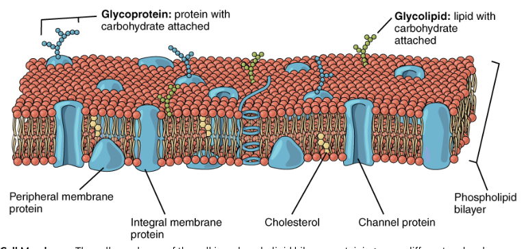
The cell membrane, also called the plasma membrane, is an extremely pliable boundary composed primarily of a phospholipid bilayer with embedded proteins and other molecules. It regulates the movement of materials into and out of the cell.
The phospholipid bilayer
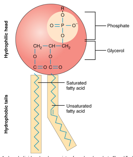
A phospholipid has a polar, hydrophilic head that interacts with water and two nonpolar, hydrophobic fatty-acid tails that avoid water. Because the cell contains watery fluid both inside and outside, phospholipids spontaneously arrange themselves into two back-to-back layers: the hydrophilic heads face the watery environments, while the hydrophobic tails face one another away from water.
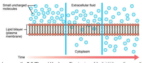
This arrangement creates the phospholipid bilayer. Its interior is hydrophobic, so ions and most large or polar molecules cannot cross it easily. Small nonpolar molecules, such as oxygen and carbon dioxide, can pass through the lipid portion more readily. The bilayer is therefore both a physical boundary and a selective barrier.
The membrane is described by the fluid mosaic model. Fluid means that many phospholipids and proteins can move laterally within the membrane. Mosaic means that the membrane contains a varied collection of proteins, lipids, carbohydrates, and cholesterol arranged together. The membrane is not a frozen shell; it is a dynamic surface whose components move and interact.
Cholesterol is interspersed among the phospholipids. It contributes to membrane fluidity by preventing phospholipids from packing too closely together when temperatures are low, while also restraining excessive movement when temperatures are high. Thus, cholesterol helps stabilize the membrane across changing conditions rather than simply making it uniformly more or less fluid.
Membrane proteins perform many specialized tasks. Channel proteins form selective passageways; carrier proteins bind substances and change shape to move them; receptor proteins bind signaling molecules; enzymes catalyze reactions at the membrane; and cell-identity proteins help cells recognize one another. Carbohydrate chains attached to proteins or lipids contribute to cell recognition and communication.
Movement across the membrane
Transport across the membrane depends on the properties of the material, the concentration gradient, the membrane’s permeability, and whether cellular energy is required. A concentration gradient is a difference in the concentration of a substance between two regions. Diffusion tends to move particles from higher concentration toward lower concentration until the gradient is reduced.
Passive transport does not require the cell to expend metabolic energy. The movement is driven by an existing concentration or electrochemical gradient. Active transport requires energy, usually from ATP, to move substances against a gradient or to move large quantities of material using membrane vesicles.
| Passive transport | Active transport |
|---|---|
| Does not directly require cellular energy | Requires cellular energy |
| Moves substances down a concentration or electrochemical gradient | Can move substances against a gradient |
| Includes simple diffusion, osmosis, and facilitated diffusion | Includes membrane pumps, endocytosis, and exocytosis |
| Example: oxygen diffusing through the lipid bilayer | Example: the sodium-potassium pump moving ions against their gradients |
Passive transport
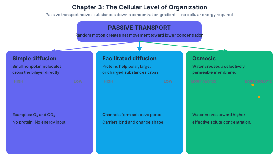
Simple diffusion is the net movement of small, nonpolar molecules directly through the phospholipid bilayer. Oxygen and carbon dioxide can cross in this way. The cell does not push these molecules across; random molecular motion produces a net movement down the concentration gradient.
Facilitated diffusion is passive transport through a membrane protein. It is needed for polar, large, or charged substances that cannot cross the hydrophobic membrane interior on their own. Channel proteins create selective pores through which particular ions or molecules can move. Carrier proteins bind specific substances and change conformation to transport them across the membrane.
Osmosis is the diffusion of water across a selectively permeable membrane. Water moves toward the side with the higher effective solute concentration, where the water concentration is lower. The key misconception is that osmosis is the movement of solute; it is the movement of water. If a membrane permits water to cross but not a dissolved solute, water shifts until the opposing forces reach equilibrium.
Active transport and bulk transport
The sodium-potassium pump, or $\text{Na}^+/\text{K}^+$ ATPase, is a major example of active transport. Using energy released when ATP is hydrolyzed to ADP and inorganic phosphate, each pump moves three $\text{Na}^+$ ions out of the cell and two $\text{K}^+$ ions into the cell. Both movements occur against their concentration gradients. This helps maintain high extracellular sodium and high intracellular potassium, conditions important for cell volume regulation and nerve-cell signaling.
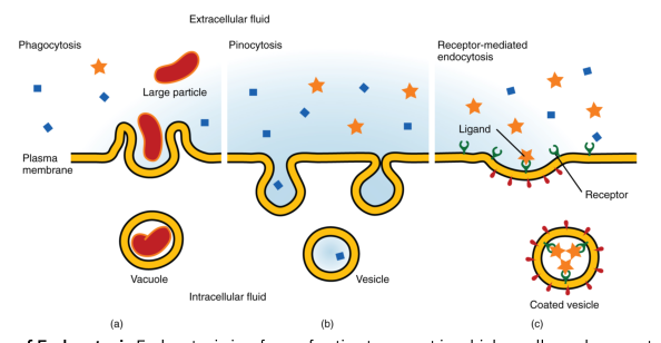
Endocytosis brings material into the cell by engulfing it with the membrane and enclosing it in a vesicle. Phagocytosis engulfs large particles, such as cellular debris or microorganisms. Pinocytosis takes in extracellular fluid and dissolved substances. Receptor-mediated endocytosis uses membrane receptors to concentrate particular molecules before the membrane forms a vesicle around them.
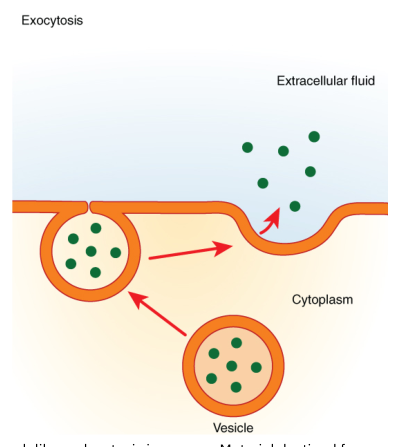
Exocytosis moves material out of the cell. An intracellular vesicle fuses with the cell membrane and releases its contents into the extracellular space. Cells use exocytosis to secrete substances such as hormones, neurotransmitters, digestive enzymes, and mucus. Both endocytosis and exocytosis require energy because the cell actively reshapes its membrane.
3.2 The Cytoplasm and Cellular Organelles
The cytoplasm includes everything inside the cell membrane but outside the nucleus. It consists of the cytosol, organelles, and various suspended particles. The cytosol is the watery internal fluid in which many chemical reactions occur. Organelles divide cellular labor into specialized compartments, much as different work areas in a large facility support different operations.
Organelles and their functions
The cytoskeleton is an internal framework made of protein filaments. Microfilaments help support the cell and produce movement; intermediate filaments provide mechanical strength; and microtubules help maintain cell shape, position organelles, and form the mitotic spindle during cell division. The cytoskeleton also supports movement of materials within the cell.
Centrosomes organize microtubules. Before mitosis, the centrosome duplicates, and the resulting structures move toward opposite sides of the cell. Microtubules extending between them form the mitotic spindle, which attaches to chromosomes and helps separate duplicated genetic material.
Cilia are short, numerous projections that move substances across a cell surface, such as mucus along the respiratory tract. A flagellum is a longer projection that propels an entire cell; the human sperm cell is a familiar example. Microvilli are small projections that increase surface area for absorption rather than beating to move fluid.
Ribosomes are the sites of protein synthesis. Some float freely in the cytosol and produce proteins used inside the cell. Others attach to the rough endoplasmic reticulum and produce proteins destined for secretion, insertion into membranes, or delivery to certain organelles.
The endoplasmic reticulum, or ER, is a membranous network. Rough ER is covered with ribosomes and synthesizes or begins processing proteins. Smooth ER lacks ribosomes and participates in lipid synthesis, carbohydrate metabolism, detoxification, and storage of calcium ions in certain cells.
The Golgi apparatus modifies, sorts, and packages proteins and lipids received from the ER. It may attach carbohydrate groups, concentrate products into vesicles, and direct them to their destinations. In a goblet cell, rough ER and the Golgi apparatus help produce and process mucin, which is packaged into secretory vesicles and released as mucus.
Mitochondria produce most of the cell’s ATP through aerobic cellular respiration. They have an outer membrane and a highly folded inner membrane. Cells with high energy demands, such as muscle cells, generally contain many mitochondria.
Lysosomes contain digestive enzymes that break down worn-out organelles, particles, and cellular debris. Peroxisomes participate in oxidation reactions and help detoxify harmful substances. Vesicles transport materials within the cell or between the cell and its environment.
3.3 The Nucleus and DNA Replication
The nucleus is the membrane-bound organelle that stores most of the cell’s genetic material. Its double membrane, the nuclear envelope, contains nuclear pores that regulate movement between the nucleus and cytoplasm. The nucleus directs cellular activity by controlling which genetic instructions are used.
Inside the nucleus, DNA is associated with proteins to form chromatin. When a cell prepares to divide, chromatin condenses into visible chromosomes. The nucleolus is a dense region within the nucleus where components of ribosomes are produced and assembled before leaving through nuclear pores.
DNA replication
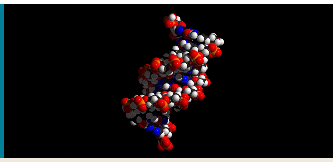
Before a cell divides, it must copy its DNA so that each daughter cell receives a complete set of genetic information. DNA replication is semiconservative: each original DNA strand serves as a template for a new complementary strand. The result is two DNA molecules, each containing one original strand and one newly synthesized strand.
Replication begins when enzymes separate the two strands of the DNA double helix. Free nucleotides pair with exposed bases according to complementary base pairing: adenine pairs with thymine, and cytosine pairs with guanine. DNA polymerase joins the incoming nucleotides into new strands. Because the original strands determine the sequence of the new strands, the genetic information is copied with high accuracy.
DNA replication does not itself make a protein. It duplicates the information so it can be passed to daughter cells. Protein synthesis uses that information in two stages: transcription produces messenger RNA from a DNA template, and translation uses the messenger RNA sequence to assemble amino acids into a protein at a ribosome. Differences in which genes are expressed help produce differentiated cell types.
3.4 Cell Division and Specialization
Mitosis is nuclear division that produces two genetically identical daughter nuclei. After DNA replication, duplicated chromosomes become organized and attached to spindle fibers. During anaphase, sister chromatids separate and move toward opposite poles, as shown by the light-blue chromosomes and green mitotic spindles in the fluorescence-stained newt lung cell. Cytoplasmic division, called cytokinesis, then separates the cell into two daughter cells.
The major biological purpose of mitosis is to support growth, tissue repair, replacement of worn-out cells, and asexual cell reproduction in some organisms. Accurate chromosome separation is essential: if genetic material is distributed incorrectly, daughter cells may receive abnormal chromosome numbers or damaged instructions.
Cell differentiation gives multicellular organisms many specialized cell types while preserving a shared genetic foundation. A cell becomes specialized when regulatory mechanisms control which genes are active and which are inactive. The resulting proteins shape the cell’s structure and behavior. In this way, the membrane, organelles, cytoskeleton, and gene-expression pattern work together: cellular structure supports cellular function.
The central cellular pattern is coordinated organization: the membrane controls exchange, organelles divide metabolic labor, the nucleus stores and regulates genetic information, DNA replication preserves that information, and differentiation turns shared instructions into specialized living cells.
The cell membrane is therefore not merely an outer covering, and the nucleus is not simply a storage compartment. Together with the cytoplasm and organelles, they form an integrated system capable of regulating its internal environment, exchanging materials, producing energy and proteins, responding to signals, dividing, and specializing.
Content adapted from OpenStax Anatomy and Physiology 2e by J. Gordon Betts et al., licensed CC BY 4.0.
Source Materials
Study Anatomy and Physiology on Lykke
This course page is free to open: signed out, you can read its first two sections, and signing in, on any plan, opens the rest. The free Lykke Chrome extension syncs your own Canvas courses, so ChatGPT, Claude and Muse can answer from your classes and deadlines. It only reads Canvas.
Add the free extensionSign in to open every sectionAll sections of Anatomy and Physiology · Browse more course pages