From the free course Anatomy and Physiology on Lykke
Chapter 1: An Introduction to the Human Body
Key concepts: Anatomy and physiology · Gross anatomy · Microscopic anatomy · Regional anatomy · Systemic anatomy · Physiology and homeostasis · Relationship between structure and function · Anatomical terminology and body directions · Medical imaging techniques · Structural organization of the human body
By the end of this chapter the student can: Distinguish between anatomy and physiology, and identify several branches of each; Describe the structure of the body, from simplest to most complex, in terms of the six levels of organization; Identify the functional characteristics of human life; Identify the four requirements for human survival; Define homeostasis and explain its importance to normal human functioning; Use appropriate anatomical terminology to identify key body structures, body regions, and directions in the body; Compare and contrast at least four medical imaging techniques in terms of their function and use in medicine.
Chapter 1: An Introduction to the Human Body
A broken bone, a racing heart, and a CT scan all become easier to understand once one distinction is clear: anatomy describes the body’s structures, whereas physiology explains how those structures work. The two subjects are inseparable. A structure’s shape, location, and organization usually reveal something about its function, while its function helps explain why it has that structure.
A useful example is the opposable thumb. Its position and specialized joints allow the thumb to move across the palm, helping the hand pinch, grasp objects, and type. This is the central relationship between form and function: anatomy asks, “What is it, where is it, and what is it shaped like?” Physiology asks, “What does it do, and how does it work with other structures?”
Anatomy is the scientific study of body structures and the relationships among them. Physiology is the study of the functions of those structures and how they work together.
Anatomy: studying body structure
The word anatomy comes from a Greek root meaning “to cut apart,” reflecting the importance of dissection, the careful examination of a body by separating or exposing its parts. Dissection can reveal the size, shape, position, and physical relationships of organs, muscles, nerves, blood vessels, and other structures. Modern anatomy also examines living people through medical imaging, allowing clinicians to observe structures without physically opening the body.
Gross anatomy
Gross anatomy, also called macroscopic anatomy, examines body structures large enough to be seen without magnification. The heart, lungs, bones, skeletal muscles, and major blood vessels are examples. Gross anatomy can be organized by different methods depending on the question being asked.
Regional anatomy studies all structures within a particular body region. For example, an examination of the neck may include its muscles, nerves, blood vessels, glands, bones, and connective tissues together. This approach is especially useful in clinical practice because injuries and surgical procedures usually affect a specific region containing many interacting structures.
Systemic anatomy studies the structures that make up a discrete body system. A study of the nervous system considers the brain, spinal cord, nerves, and related structures; a study of the cardiovascular system considers the heart and blood vessels. Systemic anatomy emphasizes how anatomically related structures form an integrated system.
Microscopic anatomy
Microscopic anatomy examines structures too small to see without magnification. It includes cytology, the study of cells, and histology, the study of tissues. Cells, tissues, and molecular structures cannot be understood adequately by unaided observation because their important features occur at a scale requiring microscopes or other specialized methods.
The distinction between gross and microscopic anatomy is a matter of scale, not importance. A visible organ such as the pancreas has a gross structure, but its function depends on microscopic arrangements of cells and tissues. Similarly, the wall of a blood vessel has a visible overall form, while its behavior depends on the microscopic organization of its cells.
Physiology and the relationship between structure and function
Physiology examines how living structures operate. It may focus on processes within a cell, the contraction of a muscle, the pumping of the heart, the movement of air through the lungs, or the coordinated activity of several organ systems. Because body functions depend on cooperation among structures, physiology often crosses the boundaries between organs and systems.
Form and function are closely related, but they are not identical. The opposable thumb illustrates how a structural feature supports several functions. The thumb’s position and range of movement make precise grasping possible; the hand’s function cannot be explained by muscle tissue alone, because bones, joints, nerves, and connective tissues also contribute. Conversely, the function of pinching helps explain why the thumb is structurally different from the other digits.
Homeostasis: keeping the internal environment stable
A major concern of physiology is homeostasis, the body’s tendency to maintain relatively stable internal conditions. Stability does not mean that the body is motionless or that every value remains perfectly constant. Instead, conditions fluctuate within limits that support normal cell and organ function.
Homeostasis is the maintenance of stable internal conditions despite changes inside or outside the body.
Body temperature, blood pressure, blood glucose concentration, and the composition of body fluids must be regulated. For example, after physical activity, heat production rises. Physiological responses such as sweating and increased blood flow near the skin help move the body back toward its normal temperature range. The body therefore maintains stability through continuous adjustment rather than through permanent stillness.
Most homeostatic regulation uses negative feedback, in which a change triggers responses that oppose or reduce the original change. A disturbance is detected, a control center evaluates it, and effectors produce a response. If body temperature rises, responses that promote heat loss reduce the rise. Negative feedback prevents a temporary disturbance from becoming progressively larger.
A common misconception is that homeostasis means “keeping everything exactly the same.” In reality, living systems are dynamic. Homeostasis permits controlled variation while preventing conditions from moving outside the range compatible with life. When regulation fails, cells may become damaged and signs of illness may appear.
Standard anatomical terminology
Anatomical terminology provides standardized terms for identifying body structures, regions, directions, positions, and planes. Standardization matters because ordinary words such as “front,” “back,” “above,” or “near the side” can be ambiguous. Anatomists describe the body as if it were in the anatomical position: standing upright, facing forward, with the arms at the sides and the palms facing forward.
Directional terms describe the location of one structure relative to another. Superior means toward the head, while inferior means toward the feet. Anterior means toward the front of the body, and posterior means toward the back. Medial means nearer the body’s midline, whereas lateral means farther from it. Proximal means nearer the point of attachment, and distal means farther from that point. These terms remain consistent even when the body is lying down or viewed from an unusual angle.
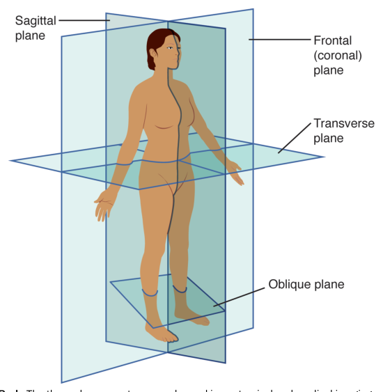
Anatomical planes are imaginary flat surfaces used to divide the body or interpret images. A sagittal plane divides the body into left and right portions; a midsagittal plane divides it into equal left and right halves. A frontal, or coronal, plane divides the body into anterior and posterior portions. A transverse plane divides the body into superior and inferior portions. An oblique plane passes through the body at an angle rather than parallel to the standard sagittal, frontal, or transverse planes.
These planes are not merely visual conveniences. Medical imaging often produces two-dimensional slices through a three-dimensional body. Knowing the plane allows a clinician to determine whether a structure lies toward the front or back, nearer the head or feet, or to the left or right. The same anatomical language therefore links physical examination, anatomy diagrams, injury descriptions, and scans.
Medical imaging: seeing structure in living patients
Medical imaging applies anatomical knowledge to diagnosis. Different techniques reveal different physical properties, so no single imaging method is best for every clinical question. The appropriate choice depends on whether the clinician needs to see dense tissues, soft tissues, motion, blood flow, or metabolic activity.
| Technique | What it uses or detects | Typical strengths and uses |
|---|---|---|
| X-ray radiography | X-rays passing through the body, absorbed differently by tissues | Produces images in which dense structures such as bones are prominent; commonly used to detect fractures |
| Computed tomography (CT) | Multiple X-ray measurements combined into cross-sectional images | Shows slices and detailed three-dimensional relationships; useful for examining bones, organs, bleeding, and complex injuries |
| Ultrasound | High-frequency sound waves reflected by internal structures | Produces real-time images and is useful for observing soft tissues, fluid-filled structures, blood flow, and fetal development |
| Magnetic resonance imaging (MRI) | Magnetic fields and radio waves | Provides detailed images of soft tissues, including the brain, spinal cord, muscles, joints, and many internal organs |
| Positron emission tomography (PET) | Signals from a radioactive tracer associated with tissue activity | Shows aspects of metabolic activity and can help identify areas functioning differently from surrounding tissue |
An X-ray is often effective when the question concerns bone, while MRI is generally more informative for soft-tissue detail. CT is valuable when rapid cross-sectional information is needed, especially in trauma. Ultrasound is useful when real-time movement or fluid flow matters and avoids the ionizing radiation used by X-ray-based methods. PET differs from these primarily because it emphasizes activity rather than simply anatomy.
Understanding anatomy and physiology has practical consequences beyond professional health care. Knowledge of normal structure and function helps a person recognize when something may be abnormal, understand why a diagnostic test is being ordered, communicate accurately with medical professionals, and make informed health choices. It also provides the foundation for recognizing how a change in one structure can affect the function of an entire body system.
Content adapted from OpenStax Anatomy and Physiology 2e by J. Gordon Betts et al., licensed CC BY 4.0.
Source Materials
Study Anatomy and Physiology on Lykke
This course page is free to open: signed out, you can read its first two sections, and signing in, on any plan, opens the rest. The free Lykke Chrome extension syncs your own Canvas courses, so ChatGPT, Claude and Muse can answer from your classes and deadlines. It only reads Canvas.
Add the free extensionSign in to open every sectionAll sections of Anatomy and Physiology · Browse more course pages