From the free course Anatomy and Physiology on Lykke
Chapter 15: The Autonomic Nervous System
Key concepts: Autonomic nervous system (ANS) and involuntary control of homeostasis · Sensory input, CNS integration, and motor responses · Graded potentials and action-potential initiation at the axon · Neurotransmitter systems and synaptic signaling · Serotonin reuptake and receptor specificity · Biogenic amines: dopamine, norepinephrine, and epinephrine · Acetylcholine, muscarinic receptors, and acetylcholinesterase · Neuron structure and glial-cell support of the CNS · Brain organization into forebrain, midbrain, and hindbrain · Basal nuclei, cerebellar coordination, and CNS circulation
By the end of this chapter the student can: Describe the components of the autonomic nervous system; Differentiate between the structures of the sympathetic and parasympathetic divisions in the autonomic nervous system; Name the components of a visceral reflex specific to the autonomic division to which it belongs; Predict the response of a target effector to autonomic input on the basis of the released signaling molecule; Describe how the central nervous system coordinates and contributes to autonomic functions.
Chapter 15: The Autonomic Nervous System
A sudden threat can make the heart accelerate, the pupils widen, the airways open, and digestion slow before conscious thought catches up. These coordinated changes are produced by the autonomic nervous system (ANS), the part of the nervous system that regulates visceral functions largely outside voluntary control. Its central problem is simple but profound: internal organs must adjust continuously to keep the body’s conditions within workable limits while the external world changes.
The ANS does not act as a completely independent system. Sensory input from internal organs travels to the central nervous system, where information is integrated with signals from the brain and spinal cord. The resulting motor response travels through autonomic pathways to smooth muscle, cardiac muscle, glands, and other visceral effectors. The ANS therefore participates in homeostasis, but it can also produce rapid, dramatic changes during stress, exercise, emotion, illness, and medication.
A common misconception is that “involuntary” means “unregulated” or “unmodifiable.” In reality, autonomic activity is precisely regulated. It is influenced by reflex circuits, the hypothalamus, the brain stem, the spinal cord, emotional centers of the limbic system, and conscious experiences. A person cannot usually command the heart to beat faster by willing it directly, but fear, focused breathing, temperature, posture, and physical activity can all alter autonomic output.
15.1 Divisions of the Autonomic Nervous System
The ANS controls smooth muscle, cardiac muscle, glands, and some adipose tissue. Its motor pathways differ from somatic motor pathways. A somatic motor neuron extends from the central nervous system directly to skeletal muscle. An autonomic motor pathway usually uses a chain of two neurons: a preganglionic neuron begins in the brain stem or spinal cord and synapses with a postganglionic neuron in an autonomic ganglion; the postganglionic axon then reaches the target effector.
That two-neuron arrangement creates a useful relay system. The first neuron carries instructions out of the central nervous system, while the second distributes the signal to the organ. The major divisions are the sympathetic division, the parasympathetic division, and the enteric nervous system. The sympathetic and parasympathetic divisions often act in opposition, but “opposition” does not mean that one division is always active while the other is always silent. Most organs receive a changing balance of both inputs.
Sympathetic division: thoracolumbar outflow
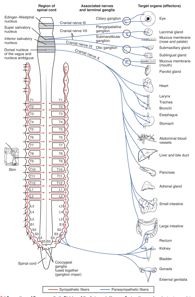
The sympathetic division is also called the thoracolumbar division because its preganglionic neurons arise from the thoracic and upper lumbar spinal cord, especially the lateral horns from approximately spinal cord levels $T_1$ through $L_2$. Sympathetic preganglionic axons are generally short because sympathetic ganglia lie close to the vertebral column. Postganglionic axons are generally longer and travel from those ganglia to the target organs.
Sympathetic preganglionic axons may follow several routes. They can synapse in a sympathetic chain ganglion at the same spinal level, ascend or descend in the chain before synapsing, or pass through the chain without synapsing. Axons that pass through become splanchnic nerves and synapse in prevertebral ganglia, which lie anterior to the vertebral column near major abdominal arteries. Some sympathetic preganglionic fibers travel directly to the adrenal medulla, where they stimulate chromaffin cells to release epinephrine and norepinephrine into the blood.
The sympathetic division is strongly associated with fight-or-flight physiology. Its activity redirects resources toward immediate action: heart rate and cardiac force increase, airways dilate, pupils enlarge, blood flow is adjusted, and digestive activity is generally reduced. These responses are not simply “good” or “bad.” They are appropriate when rapid movement or vigilance is required, but prolonged sympathetic activation can contribute to hypertension, sleep disruption, and digestive problems.
Parasympathetic division: craniosacral outflow
The parasympathetic division is also called the craniosacral division. Its preganglionic neurons arise from brain-stem nuclei associated with cranial nerves and from the sacral spinal cord. The major cranial nerves carrying parasympathetic preganglionic fibers are cranial nerves III, VII, IX, and X. The sacral outflow contributes to pelvic organs and the distal portion of the large intestine.
Parasympathetic ganglia are located close to, or within, the target organs. Consequently, parasympathetic preganglionic axons are usually long, whereas postganglionic axons are short. Cranial nerve III carries fibers to the ciliary ganglion, which controls smooth muscle involved in pupil constriction and lens accommodation. Cranial nerve VII reaches the pterygopalatine and submandibular ganglia, influencing glands of the head. Cranial nerve IX reaches the otic ganglion and affects the parotid gland. Cranial nerve X, the vagus nerve, supplies thoracic and abdominal viscera.
Parasympathetic activity supports rest-and-digest functions. It promotes salivation, digestion, intestinal motility, urination, and energy conservation. It also slows the heart through vagal input. For example, in bright light, parasympathetic fibers traveling with the oculomotor nerve synapse in the ciliary ganglion; postganglionic fibers then release acetylcholine onto the iris sphincter muscle, producing pupil constriction.
Enteric nervous system and autonomic signaling
The enteric nervous system consists of neuronal networks in the wall of the gastrointestinal tract. It can coordinate local digestive activity, including smooth-muscle contraction and glandular secretion, without requiring direct commands from the brain. Sympathetic and parasympathetic pathways nevertheless modify enteric activity. This local independence explains why a short reflex can remain within the gut wall, while a long reflex sends information to the central nervous system before returning an autonomic command.
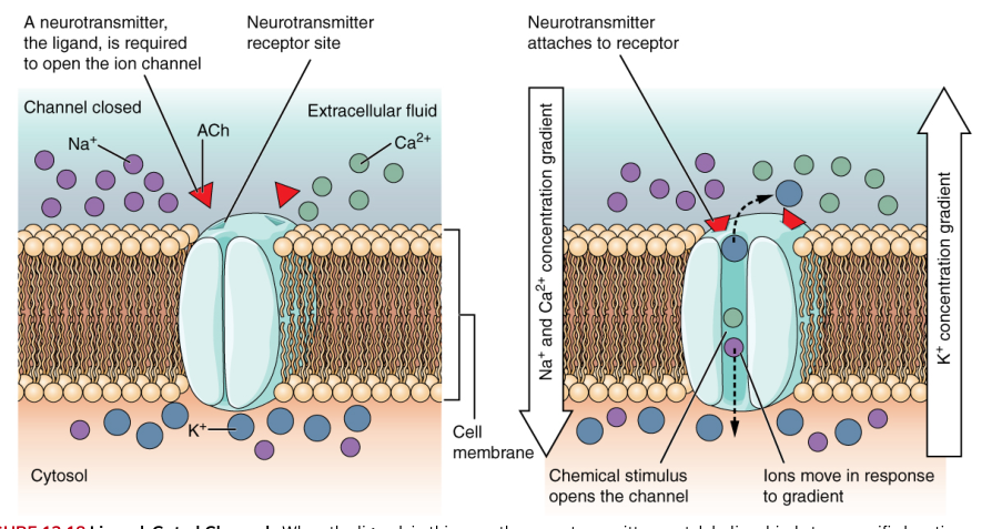
Both sympathetic and parasympathetic preganglionic neurons release acetylcholine (ACh). In autonomic ganglia, ACh binds nicotinic receptors, which are ligand-gated ion channels that produce rapid depolarization. At the effector, parasympathetic postganglionic neurons usually release ACh onto muscarinic receptors, which are G-protein-coupled receptors. Most sympathetic postganglionic neurons release norepinephrine (NE) onto adrenergic receptors. The important exception is sympathetic innervation of most sweat glands, whose postganglionic neurons release ACh onto muscarinic receptors.
The adrenal medulla functions as a modified sympathetic ganglion. Sympathetic preganglionic neurons release ACh onto chromaffin cells, and those cells release mostly epinephrine, with some norepinephrine, into the circulation. Blood-borne hormones can reach many organs at once, extending and broadening the sympathetic response beyond the direct pattern of neural synapses.
15.2 Autonomic Reflexes and Homeostasis
An autonomic reflex is an automatic response to a visceral stimulus. Its basic sequence is: a change is detected by a receptor; sensory information travels toward an integrating center; the central nervous system or a peripheral ganglion determines a response; autonomic motor output travels to an effector; and the effector changes its activity. The response is not conscious, although conscious sensations may accompany it.
A visceral reflex begins with sensory input. Receptors may detect stretch, pressure, chemical composition, temperature, or irritation in internal tissues. Sensory neurons carry that information either into the central nervous system or to a nearby autonomic ganglion. Integration then determines whether smooth muscle contracts or relaxes, whether a gland secretes, or whether cardiac activity changes. The final motor response helps restore an internal variable toward its normal range.
A long reflex includes integration in the spinal cord or brain. Its pathway can be summarized as follows:
- A visceral receptor detects a stimulus.
- A sensory neuron carries information to the spinal cord or brain stem.
- Interneurons and autonomic control centers interpret the input.
- Preganglionic autonomic neurons receive motor instructions.
- Preganglionic axons synapse in autonomic ganglia.
- Postganglionic axons carry the response to a visceral effector.
A short reflex bypasses the central nervous system. A sensory neuron synapses directly with an autonomic motor neuron in a peripheral ganglion, especially within an enteric plexus. Short reflexes are particularly useful for local control, because the digestive tract can respond rapidly to the presence, volume, or chemical composition of its contents without waiting for a signal from the brain.
Consider the baroreceptor reflex, a concrete example of autonomic homeostasis. Stretch-sensitive receptors in the walls of major arteries detect changes in blood pressure. When pressure rises, sensory information reaches cardiovascular centers in the brain stem. The response increases parasympathetic influence on the heart and reduces sympathetic influence on the heart and blood vessels. Heart rate and vascular resistance decrease, helping bring pressure downward. When pressure falls, the pattern reverses: sympathetic activity increases, the heart beats faster and more forcefully, and vascular smooth muscle constricts.
The pupillary reflex illustrates reciprocal autonomic control. Light activates retinal ganglion cells, and signals travel through the optic nerve to the brain. In bright light, parasympathetic output from the midbrain travels through the oculomotor nerve to the ciliary ganglion. Postganglionic fibers release ACh, constricting the iris and reducing the amount of light entering the eye. In dim light, sympathetic output through the superior cervical ganglion releases norepinephrine onto the iris dilator muscle, enlarging the pupil.
Autonomic control is often tonic, meaning that a baseline level of activity is continuously present. Blood vessels, for example, usually receive ongoing sympathetic stimulation that maintains partial vasoconstriction. Increasing or decreasing that tone changes vessel diameter. A second important principle is dual innervation: many organs receive both sympathetic and parasympathetic input. The final activity of the organ reflects the balance between those influences, not a simple on-or-off command.
Homeostasis does not require every organ to receive dual innervation. Some targets, such as sweat glands and many blood vessels, are controlled primarily by sympathetic pathways. In such cases, the nervous system adjusts the level of sympathetic activity. The absence of parasympathetic fibers does not mean the organ lacks regulation; it means regulation occurs by changing sympathetic tone.
15.3 Central Control
Autonomic activity is coordinated at several levels of the central nervous system. The hypothalamus is the principal integration center for autonomic function. It receives information about internal conditions and emotional states and sends output to brain-stem nuclei and spinal cord regions that control sympathetic and parasympathetic pathways. The hypothalamus also coordinates autonomic responses with endocrine activity, temperature regulation, hunger, thirst, and motivated behavior.
The hypothalamus communicates with lower autonomic centers through descending pathways, including the medial forebrain bundle and the dorsal longitudinal fasciculus. These pathways influence parasympathetic nuclei in the brain stem, such as the Edinger–Westphal nucleus associated with the oculomotor nerve and vagal nuclei involved in thoracic and abdominal regulation. Hypothalamic output also descends toward spinal cord regions containing sympathetic and sacral parasympathetic preganglionic neurons.
The brain stem contains important centers for basic visceral functions. Cardiovascular centers regulate heart rate, cardiac force, and vascular diameter. Respiratory centers coordinate breathing, while nuclei associated with cranial nerves control salivation, swallowing, pupil size, and other visceral actions. These centers can produce rapid reflex responses, but they remain subject to modulation by the hypothalamus and higher brain regions.
The limbic system links emotion and autonomic function. Structures including the amygdala, hippocampus, and cingulate gyrus connect with the hypothalamus. An emotionally threatening event can therefore trigger sympathetic activation even before a person consciously evaluates every detail. The familiar experience of a racing heart during fear is not imaginary: emotional processing changes hypothalamic output, which changes autonomic activity.
Higher cortical areas can influence autonomic responses indirectly. Seeing food can increase salivation; anticipating pain can increase heart rate; controlled breathing can alter cardiovascular and parasympathetic activity. Conscious control remains limited because the cortex does not normally send voluntary commands directly to visceral effectors. Instead, thoughts, perceptions, and emotions modify the hypothalamic and brain-stem centers that govern autonomic output.
Synaptic signaling and graded potentials
Autonomic signaling depends on the same basic electrical principles used elsewhere in the nervous system. A neurotransmitter released into a synaptic cleft binds to receptors on the postsynaptic membrane. The resulting ion movement produces a graded potential, a local change in membrane voltage whose size depends on the amount of neurotransmitter and the properties of the receptor.
A graded potential may be excitatory or inhibitory. If depolarization reaches threshold at the appropriate trigger zone, an action potential begins and travels along the axon. The sequence is therefore: neurotransmitter release, receptor binding, ion-channel or second-messenger activation, graded change in membrane potential, threshold achievement, action-potential initiation, and neurotransmitter release at the next synapse. The axon does not produce an action potential merely because a neurotransmitter is present; the postsynaptic voltage must reach threshold.
Receptor specificity explains why the same neurotransmitter can produce different effects in different tissues. ACh at a nicotinic receptor rapidly excites a postganglionic neuron in an autonomic ganglion. ACh at a muscarinic receptor may slow the heart, increase glandular secretion, or contract smooth muscle. Norepinephrine acting at different adrenergic receptor subtypes can constrict some blood vessels, relax other smooth muscle, or alter cardiac activity.
Serotonin and biogenic amines
Serotonin, also called $5$-hydroxytryptamine, is a neurotransmitter involved in central nervous system functions such as mood, sleep, appetite, and sensory processing. Its effects depend on the receptor subtype expressed by the target cell. After release, serotonin can be removed by enzymatic breakdown, diffusion, or reuptake, in which a transporter returns it to the presynaptic neuron.
A selective serotonin reuptake inhibitor (SSRI) reduces serotonin reuptake. More serotonin remains in the synaptic cleft for a longer time, increasing the opportunity for it to bind receptors. This does not mean serotonin produces one universal effect. Receptor specificity still determines the response, and changes in neural circuits develop over time rather than appearing as a simple instantaneous switch.
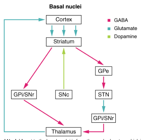
Important biogenic amines include dopamine, norepinephrine, and epinephrine. Dopamine participates in movement, motivation, reward, and cognition. Norepinephrine functions as both a neurotransmitter and a hormone, especially in sympathetic signaling and alertness. Epinephrine is released mainly by the adrenal medulla as a hormone during sympathetic activation. These molecules belong to related chemical families, but their physiological effects depend on where they are released, which receptors are present, and how the signal is terminated.
15.4 Drugs that Affect the Autonomic System
Autonomic drugs work by altering neurotransmitter release, receptor activation, receptor blockade, or neurotransmitter removal. A drug that produces an effect similar to a neurotransmitter is an agonist. A drug that prevents a neurotransmitter from producing its effect is an antagonist. Because autonomic receptors are distributed across many organs, a drug intended to affect one function can produce effects elsewhere.
Cholinergic drugs
Cholinergic agonists mimic ACh or increase its action. Nicotine is an agonist at nicotinic ACh receptors, while muscarine is an agonist at muscarinic ACh receptors. Drugs that inhibit acetylcholinesterase, the enzyme that breaks down ACh, also strengthen cholinergic signaling by allowing ACh to remain in the synaptic cleft longer.
The effects of excessive cholinergic stimulation can include increased salivation, tearing, intestinal activity, urination, bronchial secretion, and smooth-muscle contraction. In the heart, muscarinic stimulation can slow the rate. The same receptor logic explains why cholinergic agonists can be useful therapeutically but dangerous at excessive doses: a signal that restores one impaired function may disturb several other organs.
Cholinergic antagonists block ACh receptors or reduce ACh release. Atropine is a muscarinic antagonist that can increase heart rate, reduce glandular secretions, and relax some smooth muscle. It can be used when excessive muscarinic activity is dangerous, but it may also produce dry mouth, blurred vision, urinary retention, and other predictable effects.
Botulinum toxin prevents the release of ACh from presynaptic terminals. By blocking cholinergic transmission at neuromuscular junctions and autonomic synapses, it can reduce skeletal-muscle contraction and glandular activity. Its medical use depends on carefully localized dosing; widespread exposure can produce severe weakness and autonomic dysfunction.
Adrenergic drugs
Adrenergic agonists mimic norepinephrine or epinephrine, or stimulate their receptors. Their effects depend on whether they act mainly at $\alpha$ or $\beta$ adrenergic receptors. Activation of some $\alpha$ receptors constricts vascular smooth muscle. Activation of $\beta_1$ receptors increases cardiac rate and force, whereas activation of $\beta_2$ receptors can relax smooth muscle in the airways.
An inhaled $\beta_2$ agonist can therefore help open constricted airways in asthma. The desired effect is bronchodilation, but the drug may also produce tremor or an increased heart rate because adrenergic receptors are not confined to the lungs. This is a practical example of receptor distribution determining both therapeutic benefit and side effects.
Some drugs increase adrenergic signaling without directly binding the receptor. Cocaine, for example, blocks the reuptake of norepinephrine and other monoamine neurotransmitters, leaving more transmitter in the synaptic cleft. Amphetamines can increase the release of norepinephrine and related monoamines. The resulting sympathetic effects can include increased alertness, heart rate, blood pressure, and vasoconstriction.
Adrenergic antagonists block $\alpha$ or $\beta$ receptors. Beta blockers reduce the effects of epinephrine and norepinephrine at $\beta$ receptors, often lowering cardiac rate and force and reducing blood pressure. They are used in conditions such as hypertension and some cardiac disorders. However, blocking $\beta_2$ receptors can narrow the airways in susceptible individuals, illustrating why receptor selectivity matters.
Drugs that affect serotonin and dopamine illustrate the same general principle. An SSRI blocks serotonin reuptake, while drugs used to influence dopamine signaling may act as receptor agonists, receptor antagonists, or reuptake-modifying agents. The resulting effects cannot be predicted from the neurotransmitter’s name alone. One must identify the receptor subtype, the tissue expressing it, the direction of drug action, and the normal role of that pathway.
The central lesson of autonomic pharmacology is that chemical signaling is a sequence, not a single event: a transmitter is synthesized and released, crosses a synapse, binds a receptor, changes the postsynaptic cell, and is then removed or degraded. A drug can intervene at any step. Understanding the pathway makes the clinical effects intelligible: blocking ACh release weakens cholinergic responses, blocking muscarinic receptors reduces parasympathetic effects, increasing norepinephrine at a synapse strengthens sympathetic signaling, and blocking adrenergic receptors reduces the response to sympathetic transmitters.
Content adapted from OpenStax Anatomy and Physiology 2e by J. Gordon Betts et al., licensed CC BY 4.0.
Source Materials
Study Anatomy and Physiology on Lykke
This course page is free to open: signed out, you can read its first two sections, and signing in, on any plan, opens the rest. The free Lykke Chrome extension syncs your own Canvas courses, so ChatGPT, Claude and Muse can answer from your classes and deadlines. It only reads Canvas.
Add the free extensionSign in to open every sectionAll sections of Anatomy and Physiology · Browse more course pages