From the free course Anatomy and Physiology on Lykke
Chapter 21: The Lymphatic and Immune System
Key concepts: Functions of the lymphatic and immune systems · Lymphatic capillaries and lymph flow · Lymph nodes and lymph filtration · Lymphedema caused by impaired lymph drainage · Primary and secondary lymphoid organs · Thymus structure and thymic involution · Lymphocytes and plasma cells · Tonsils and mucosa-associated lymphoid tissue (MALT) · Opportunistic pathogens and immune suppression · Immune-system deficiencies and overreactions
By the end of this chapter the student can: Identify the components and anatomy of the lymphatic system; Discuss the role of the innate immune response against pathogens; Describe the power of the adaptive immune response to cure disease; Explain immunological deficiencies and over-reactions of the immune system; Discuss the role of the immune response in transplantation and cancer; Describe the interaction of the immune and lymphatic systems with other body systems.
Chapter 21: The Lymphatic and Immune System
A splinter can enter the skin in seconds, but the body’s defense against it is distributed across tissues, vessels, organs, and circulating cells. The lymphatic system returns excess tissue fluid to the blood, transports absorbed fats, and provides locations where immune cells detect and respond to foreign material. The immune system adds biological recognition and defense: it distinguishes the body’s own cells from pathogens and damaged cells, then coordinates responses that eliminate threats while limiting injury to healthy tissue.
A useful distinction is that lymphatic anatomy provides the transport and meeting places, whereas immunity provides the defensive activity. These systems overlap constantly. Lymph carries antigens from infected tissues to lymph nodes; lymph nodes concentrate those antigens and expose them to lymphocytes; lymphocytes then produce innate or adaptive responses. The result is not a single “immune organ,” but a body-wide surveillance network.
21.1 Anatomy of the Lymphatic and Immune Systems
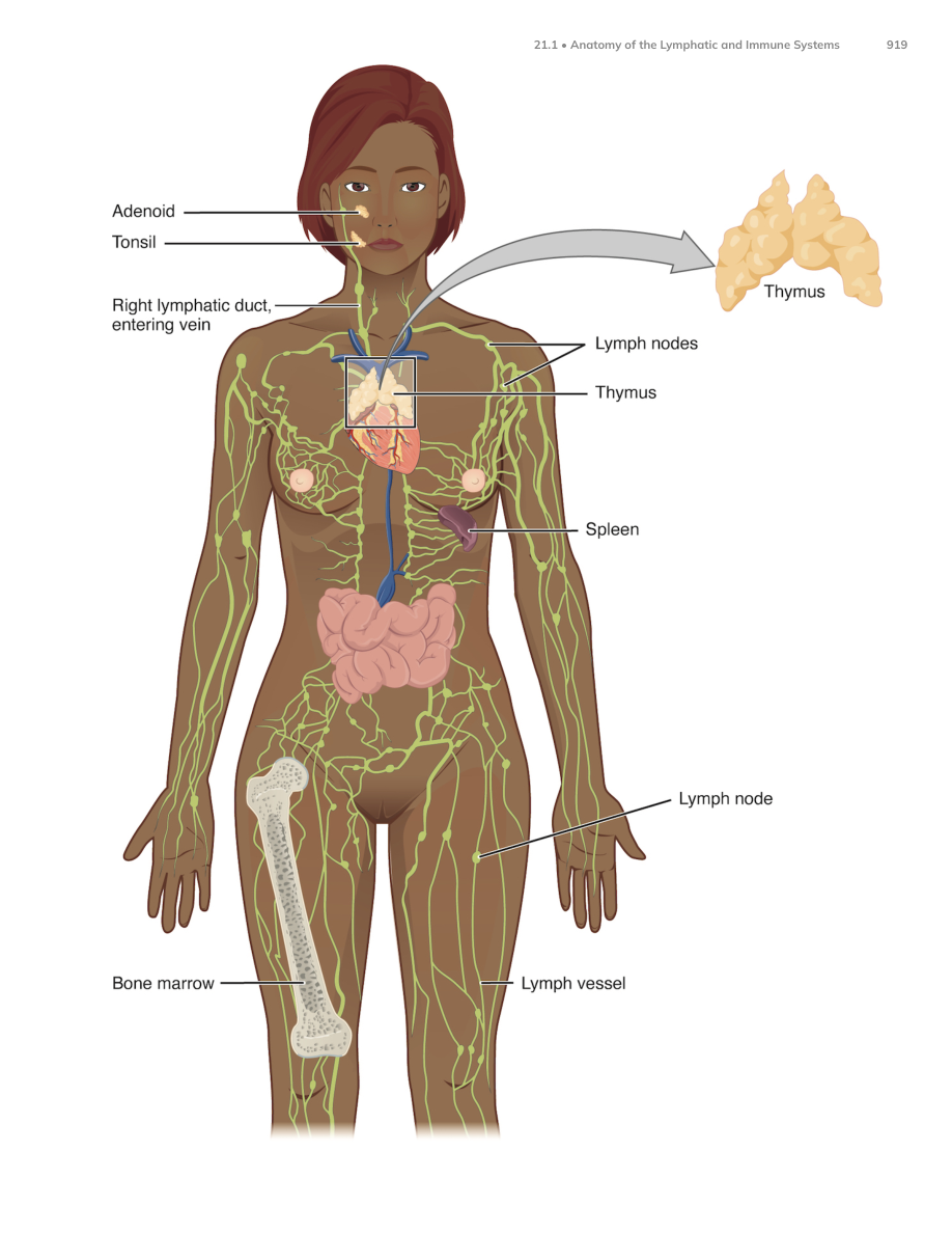
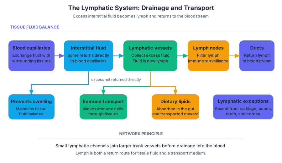
The lymphatic system begins in microscopic lymphatic capillaries, blind-ended vessels that collect excess interstitial fluid from tissues. Fluid leaves blood capillaries because of filtration forces and enters the spaces between cells. Most of it eventually returns directly to blood capillaries, but some remains in the tissues. Lymphatic capillaries reclaim this excess fluid and call it lymph once it enters the lymphatic vessels.
Lymphatic capillaries are especially suited to this task because their endothelial cells overlap like loosely fitted doors. When tissue fluid pressure rises, the overlapping edges separate and allow fluid, proteins, cellular debris, and sometimes pathogens to enter. Anchoring filaments connect the endothelial cells to surrounding tissue, helping hold the capillary open as swelling increases.
Lymphatic vessels resemble veins in having thin walls, valves, and low-pressure flow. Skeletal-muscle contraction, breathing movements, and nearby artery pulsations compress the vessels and push lymph forward. One-way valves prevent backflow. Unlike blood circulation, the lymphatic system has no central pump equivalent to the heart, so lymph flow depends on these mechanical forces.
The pathway is sequential:
- Excess interstitial fluid enters lymphatic capillaries.
- Lymph moves through collecting vessels and passes through one or more lymph nodes.
- Larger lymphatic trunks receive lymph from regions of the body.
- The trunks drain into the thoracic duct or the right lymphatic duct.
- These ducts return lymph to the venous blood near the junctions of the internal jugular and subclavian veins.
The right lymphatic duct drains the right upper limb and the right side of the head, neck, and thorax. The thoracic duct drains the rest of the body, including both lower limbs, the abdomen, the left upper limb, and the left side of the head, neck, and thorax. The thoracic duct begins in the abdomen at a dilated sac called the cisterna chyli, which receives lymph from intestinal and lumbar trunks.
A specialized lymphatic capillary called a lacteal lies inside each intestinal villus. Lacteals absorb dietary lipids and fat-soluble vitamins from the small intestine. The resulting lipid-rich lymph, called chyle, travels through intestinal lymphatic vessels before entering the bloodstream. This route allows large lipid particles to bypass ordinary blood capillaries in the intestinal lining.
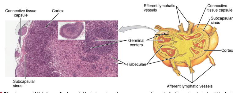
Lymph nodes are small, bean-shaped organs positioned along lymphatic vessels. Humans have approximately $500$–$600$ lymph nodes, especially concentrated in the groin, armpits, neck, chest, and abdomen. A node is enclosed by a connective-tissue capsule. Lymph enters through several afferent lymphatic vessels, flows through spaces called sinuses, and leaves through an efferent lymphatic vessel at the hilum.
The node’s structure slows lymph long enough for immune surveillance. Macrophages remove particles by phagocytosis, while lymphocytes inspect antigens carried in the lymph. The outer cortex contains lymphoid follicles rich in B lymphocytes; deeper regions contain many T lymphocytes. The medulla contains cords and sinuses populated by lymphocytes, macrophages, and antibody-secreting cells.
A swollen lymph node is therefore not simply a “clogged gland.” It often reflects increased immune-cell activity and cellular proliferation in response to antigens arriving from a nearby tissue. For example, an infection in the throat can enlarge cervical lymph nodes because lymph draining the head and neck carries microbial antigens into those nodes.
Lymphedema is persistent swelling caused by impaired lymph drainage. If lymphatic vessels are damaged, blocked, removed, or overwhelmed, protein-rich fluid accumulates in the interstitial spaces. Because the lymphatic system normally removes both fluid and proteins, lymphedema can become chronic and can make tissues vulnerable to infection.
A common example occurs after lymph-node removal or radiation treatment for breast cancer. If lymphatic drainage from the arm is disrupted, fluid can accumulate in the affected arm. Compression, movement, and medical treatment may improve drainage, but the underlying problem is mechanical failure of lymph transport rather than excess blood flow alone.
Lymphoid organs are divided into primary lymphoid organs and secondary lymphoid organs. Primary organs are where lymphocytes develop and mature. The red bone marrow produces blood cells and is the site where B lymphocytes mature. T lymphocytes begin in bone marrow but migrate to the thymus for maturation.
The thymus lies in the superior thorax, behind the sternum and above the heart. It has two lobes divided into lobules by connective-tissue septa. Each lobule has an outer cortex containing many immature T cells and an inner medulla containing more mature T cells and distinctive epithelial structures called thymic corpuscles or Hassall’s corpuscles.
T-cell maturation is a selection process. Developing T cells rearrange their antigen receptors, then undergo positive and negative selection. Cells must be able to recognize antigen presented by the body’s major histocompatibility complex, or MHC, molecules, but cells that react too strongly to self-antigens are eliminated or altered. This helps establish self-tolerance, the ability to avoid attacking normal body tissues.
The thymus is relatively large and active during childhood. Beginning around puberty, it undergoes thymic involution: functional thymic tissue gradually decreases and is replaced in part by adipose tissue. This does not eliminate all T-cell production, because immune memory and peripheral lymphoid tissues remain important, but it changes the balance of T-cell development across the lifespan.
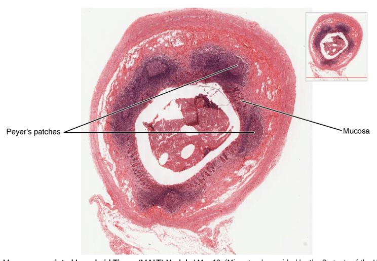
Secondary lymphoid organs and tissues are where mature lymphocytes encounter antigens and mount adaptive immune responses. They include lymph nodes, the spleen, tonsils, and mucosa-associated lymphoid tissue, or MALT. MALT is distributed in mucous membranes exposed to the outside environment, including the respiratory, digestive, and genitourinary tracts.
The spleen filters blood rather than lymph. Its white pulp contains lymphocytes and macrophages that monitor blood for pathogens and abnormal cells. Its red pulp removes worn-out red blood cells and stores or recycles components of blood. The tonsils are lymphoid nodules associated with the nasopharynx and oral cavity; they help detect pathogens entering through the nose and mouth.
The essential anatomical pattern is therefore: lymphatic vessels collect and transport material, lymph nodes filter lymph, the spleen filters blood, and primary and secondary lymphoid organs support the development or activation of lymphocytes. A common misconception is that lymph nodes manufacture lymph. They do not; lymph forms from interstitial fluid and is filtered as it passes through the nodes.
21.2 Barrier Defenses and the Innate Immune Response
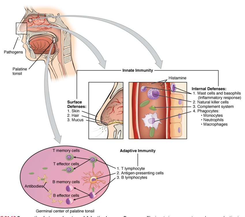
A pathogen does not begin by fighting antibodies. It first encounters the body’s innate defenses, rapid protective mechanisms that recognize broad features shared by groups of microbes. Innate immunity includes physical and chemical barriers, phagocytic cells, natural killer cells, inflammation, fever, and the complement system. Unlike adaptive immunity, it does not require prior exposure to respond.
The first barrier is the skin. Keratinized epithelial cells form a tough, continuously renewed surface that limits microbial entry. Sweat and sebum make the surface relatively acidic and contain antimicrobial substances. The normal microbiota also occupies attachment sites and consumes resources that might otherwise support pathogens.
Mucous membranes provide a second major barrier. Mucus traps particles and microbes, while cilia move mucus toward locations where it can be expelled or swallowed. Tears and saliva wash surfaces and contain lysozyme, an enzyme that damages bacterial cell walls. In the stomach, hydrochloric acid creates an environment hostile to many swallowed microbes.
Barrier defenses act in a sequence:
- A pathogen encounters intact skin or a mucous membrane.
- Mucus, fluid flow, cilia, acid, enzymes, and microbiota limit attachment and survival.
- If the barrier is broken, damaged cells and resident immune cells detect danger.
- Chemical signals recruit circulating immune cells and activate inflammation.
- Phagocytes and other innate mechanisms destroy the invading material.
Phagocytes are cells that engulf and digest particles. Important phagocytes include neutrophils, which are rapidly recruited from blood, and macrophages, which reside in tissues and also develop from circulating monocytes. Phagocytosis proceeds through recognition and attachment, engulfment into a vesicle called a phagosome, fusion with lysosomes, enzymatic digestion, and disposal of the remaining material.
Some phagocytes also release chemical signals called cytokines. Cytokines are short-range or circulating messenger proteins that alter the behavior of nearby cells, recruit additional leukocytes, and coordinate the transition from local recognition to systemic defense. Dendritic cells are particularly important because they capture antigens in tissues and help initiate adaptive immune responses.
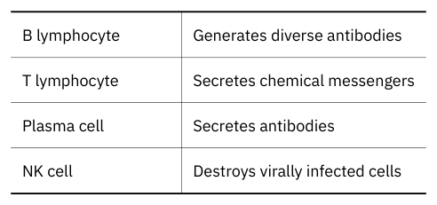
Natural killer cells, or NK cells, provide innate defense against infected or abnormal host cells. Rather than engulfing their targets, they release cytotoxic substances that create membrane damage and trigger programmed cell death. NK cells are especially important against virus-infected cells and some tumor cells, which may display abnormal patterns of MHC or other surface molecules.
Inflammation is a local protective response to tissue injury or infection. It isolates the damaged area, increases delivery of immune components, and begins tissue repair. Mast cells in connective tissue detect injury or chemical signals and release histamine, which changes nearby blood vessels.
The acute inflammatory response follows a recognizable sequence:
- Tissue damage or pathogen detection activates mast cells and other sentinel cells.
- Mast cells release histamine and other mediators.
- Nearby arterioles dilate, increasing local blood flow.
- Capillary permeability increases, allowing fluid and plasma proteins to enter the tissue.
- The tissue becomes warm, red, swollen, and painful.
- Neutrophils and other phagocytes adhere to the vessel wall, pass between endothelial cells, and migrate toward the damaged area.
- Phagocytes engulf pathogens and debris, while repair mechanisms begin.
The familiar signs of inflammation—warmth, redness, swelling, and pain—are not random side effects. Warmth and redness result largely from increased blood flow; swelling reflects fluid movement into tissues; pain results from pressure and chemical stimulation of sensory nerve endings. Inflammation is beneficial when controlled, but excessive or prolonged inflammation can damage healthy tissue.
Fever is a regulated rise in body temperature caused by an upward resetting of the hypothalamic temperature set point. Microbial products and immune cytokines can stimulate production of prostaglandins that alter hypothalamic regulation. Moderate fever may inhibit the growth of some pathogens and accelerate immune reactions, although very high or prolonged fever is harmful.
The complement system consists of plasma proteins that circulate in inactive forms and become activated in a cascade. Complement can be triggered by pathogen-surface features, by antibodies bound to antigens, or by other recognition pathways. Its effects include coating pathogens for phagocytosis, attracting immune cells, increasing inflammation, and forming membrane attack complexes that damage susceptible cell membranes.
A concrete example is a small cut contaminated with bacteria. Intact skin normally prevents entry. Once the cut breaks the barrier, mast cells release histamine, local vessels dilate, neutrophils leave the blood, complement proteins bind microbial surfaces, and macrophages engulf bacteria. If the infection remains localized, innate immunity may eliminate it before a strong adaptive response is needed.
21.3 The Adaptive Immune Response: T Lymphocytes and Their Functional Types
Innate immunity recognizes broad microbial patterns, but adaptive immunity recognizes particular molecular structures with remarkable specificity. The defining cells are lymphocytes. T lymphocytes, or T cells, mature in the thymus and use antigen-specific receptors to coordinate cellular immune responses.
An antigen is a substance recognized by an adaptive immune receptor. A large antigen, such as a protein, may contain several distinct antigenic determinants, or epitopes. Different T cells can recognize different epitopes on the same antigen. This explains how one microbial protein can activate multiple lymphocyte populations without every lymphocyte recognizing the entire molecule.
T cells generally do not recognize free antigen in solution. They recognize a processed antigen fragment displayed on the surface of another cell by an MHC molecule. This requirement is called antigen presentation. It allows the immune system to ask two questions at once: what fragment is present, and on what kind of cell is it being displayed?
Antigen presentation proceeds as follows:
- A pathogen, abnormal cell, or extracellular antigen is taken up or produces proteins inside a cell.
- Proteins are broken into peptide fragments.
- A peptide binds to an MHC molecule.
- The MHC–peptide complex moves to the cell surface.
- A T-cell receptor binds the displayed complex.
- Additional costimulatory signals determine whether the T cell becomes activated.
MHC class I molecules are found on nearly all nucleated cells. They present peptides derived from proteins made inside the cell, such as viral proteins. MHC class II molecules are found mainly on professional antigen-presenting cells, including dendritic cells, macrophages, and B cells. They present peptides derived from material taken up from outside the cell.
A T cell usually requires both antigen recognition and costimulation. This second signal helps prevent accidental activation by harmless or self-derived molecules. Once activated, the T cell undergoes clonal expansion, producing many genetically similar cells with the same antigen specificity. Some become active effector cells, while others become long-lived memory cells.
The major functional types of T cells are:
- Helper T cells, often identified as $CD4^+$ cells, coordinate immune responses by releasing cytokines. They stimulate B cells, activate macrophages, and help other T cells proliferate.
- Cytotoxic T cells, often identified as $CD8^+$ cells, kill infected, cancerous, or otherwise abnormal cells displaying antigen on MHC class I.
- Regulatory T cells suppress immune responses and help maintain self-tolerance.
- Memory T cells persist after an infection and respond more rapidly if the same antigen appears again.
Helper T cells do not usually kill infected cells directly. Instead, they act as coordinators. After recognizing antigen on an antigen-presenting cell, a helper T cell secretes cytokines that promote the activation and multiplication of B cells and cytotoxic T cells. Different helper-cell patterns support different kinds of immune defense.
Cytotoxic T-cell killing is direct but controlled. The cell binds its target, releases molecules that create pores or activate intracellular death pathways, and then detaches. The target cell undergoes apoptosis, limiting the release of intracellular contents that could cause additional inflammation.
A concrete example occurs during a viral respiratory infection. A virus enters epithelial cells and produces viral proteins. Peptides from those proteins appear on MHC class I molecules. Cytotoxic T cells recognizing the viral peptide kill infected cells, while helper T cells release cytokines that expand the antiviral response and support antibody production.
21.4 The Adaptive Immune Response: B Lymphocytes and Antibodies
B lymphocytes, or B cells, mature in red bone marrow and are central to humoral immunity, the adaptive defense mediated by antibodies in body fluids. Each B cell carries a specific membrane-bound antigen receptor. When the appropriate antigen binds, and when necessary helper-T-cell signals are present, that B cell can multiply and differentiate.
B-cell activation commonly follows this sequence:
- An antigen binds to a specific B-cell receptor.
- The B cell internalizes the antigen and processes it.
- The B cell presents an antigen fragment on MHC class II.
- A helper T cell recognizes the displayed fragment and provides costimulation and cytokines.
- The B cell undergoes clonal expansion.
- Some daughter cells become plasma cells, while others become memory B cells.
Plasma cells are antibody-secreting effector B cells. Antibodies, also called immunoglobulins, are soluble proteins shaped to bind particular epitopes. They do not generally destroy pathogens by themselves; instead, they neutralize toxins and viruses, block attachment, agglutinate particles, activate complement, and mark pathogens for phagocytosis.
A typical antibody has two identical heavy chains and two identical light chains joined by disulfide bonds. The variable regions at the tips form the antigen-binding sites. Their amino acid sequences differ among B-cell clones, producing specificity. The constant region forms the antibody’s Fc region, which interacts with complement proteins and receptors on immune cells.
The five major antibody classes are:
| Antibody class | Major role |
|---|---|
| IgM | Usually the first antibody class produced during a primary response; effective at activating complement and agglutinating antigens |
| IgG | The most abundant antibody in blood; supports neutralization, opsonization, and complement activation; can cross the placenta |
| IgA | Found in secretions such as saliva, tears, mucus, and breast milk; protects mucosal surfaces |
| IgE | Associated with allergic reactions and defense against some parasites; binds strongly to mast cells and basophils |
| IgD | Found mainly on the surface of naïve B cells, where it functions as an antigen receptor |
The first exposure to an antigen produces a primary immune response. It includes an initial delay while rare antigen-specific lymphocytes activate and expand. IgM commonly appears first, followed by increasing production of other classes such as IgG. Some activated cells become memory cells rather than immediate antibody producers.
A later exposure produces a secondary immune response. Memory B cells respond more quickly and strongly, often producing antibodies with greater affinity for the antigen. This is the biological basis of long-lasting adaptive immunity and vaccination.
Antibody action can be understood as a chain:
- An antibody binds a matching epitope.
- The bound antibody blocks a pathogen or toxin from attaching to host cells.
- Multiple antibodies may cross-link particles in agglutination.
- The Fc region recruits phagocytes or complement.
- The marked pathogen is removed or destroyed.
For example, after vaccination against a microbial toxin, memory B cells remain. If the toxin later enters the body, a rapid secondary response produces high concentrations of specific antibodies that bind the toxin before it can damage its target cells. The vaccine does not usually eliminate every future exposure; it prepares the adaptive system to respond efficiently.
21.5 The Immune Response against Pathogens
Different pathogens occupy different environments and therefore require different immune strategies. Bacteria may remain outside host cells or invade them; viruses depend on host cells for reproduction; fungi often grow as cells or filaments; and parasites may be large organisms that cannot be engulfed easily. Innate and adaptive defenses cooperate, but the dominant mechanisms differ.
Bacteria
Extracellular bacteria are attacked by complement, antibodies, neutrophils, and macrophages. Antibodies can neutralize bacterial toxins, block attachment, and coat bacterial surfaces in opsonization, making them easier for phagocytes to recognize. Complement can increase inflammation and, for some bacteria, form membrane attack complexes.
Intracellular bacteria are more difficult because antibodies have limited access inside host cells. Macrophages can engulf them, but some bacteria survive within phagosomes. Helper T cells can release cytokines that activate macrophages, strengthening their killing mechanisms. Cytotoxic T cells may destroy infected host cells when bacterial antigens appear on MHC class I.
For example, a bacterium entering through a skin wound may first trigger inflammation and complement activation. Neutrophils engulf it, antibodies later coat it, and macrophages clear the remaining organisms and debris. If the bacterium survives inside macrophages, T-cell cytokines may be required for effective elimination.
Viruses
Viruses reproduce inside host cells, so antibodies are most effective before cell entry or after virions leave infected cells. Interferons released by infected cells can induce antiviral states in neighboring cells and enhance NK-cell activity. NK cells provide early killing, while cytotoxic T cells later recognize viral peptides on MHC class I and destroy infected cells.
Antibodies contribute by neutralizing extracellular virions, preventing them from attaching to host-cell receptors. Helper T cells promote cytotoxic T-cell expansion and support B-cell responses. Memory cells make later responses faster and are the principal reason prior infection or vaccination can reduce disease severity.
A respiratory virus illustrates the division of labor: interferons slow spread through nearby epithelium, NK cells attack stressed infected cells, cytotoxic T cells remove established infected cells, and antibodies bind free viral particles before they infect additional cells.
Fungi
Fungi can cause superficial infections or invasive disease. Innate defenses are especially important because fungal cell walls and membranes contain structures recognized by pattern-recognition receptors. Neutrophils and macrophages attack fungal cells, while complement and inflammatory cytokines recruit additional leukocytes. T-cell responses help activate phagocytes and organize longer-lasting defense.
Fungal disease becomes particularly dangerous when immune defenses are suppressed. Pneumocystis jirovecii, for example, may be present in healthy lungs without causing disease but can produce serious pneumonia in people with severely weakened immune systems. This illustrates the difference between an organism’s presence and its ability to cause opportunistic disease.
Parasites
Parasites include protozoa and multicellular helminths. Large helminths cannot usually be engulfed by a single phagocyte, so antibodies and immune cells attack them from the outside. IgE binds to mast cells and other effector cells, while eosinophils release toxic substances onto parasite surfaces. Mucus, smooth-muscle contraction, and increased intestinal movement can help expel parasites.
Protozoan parasites may live inside host cells or in body fluids. Their control can require antibodies, complement, macrophage activation, and cytotoxic T cells, depending on the organism’s location. Parasites often persist because they change surface molecules, occupy protected tissues, or suppress host responses.
A concrete example is an intestinal helminth. The parasite stimulates a response involving helper T cells, IgE, mast cells, and eosinophils. Mast-cell mediators increase local fluid secretion and smooth-muscle activity, while eosinophils attach to the parasite and release damaging granules. The response is directed outward because the target is too large to engulf.
21.6 Diseases Associated with Depressed or Overactive Immune Responses
Immune protection fails in two broad ways: it may be too weak, producing immunodeficiency, or too strong or misdirected, producing hypersensitivity and autoimmunity. The same mechanisms that protect against infection can cause disease when they are absent, excessive, or aimed at the wrong target.
Immunodeficiency
Immunodeficiency is inadequate immune function. It may be primary, caused by genetic or developmental abnormalities, or secondary, acquired after birth because of infection, malnutrition, cancer, medical treatment, or other conditions. Immunodeficiency increases susceptibility to pathogens that healthy immune systems normally control.
Human immunodeficiency virus, or HIV, is a major example of acquired immunodeficiency. HIV infects important immune cells, especially $CD4^+$ helper T cells. As helper-T-cell numbers and function decline, coordination of cellular and humoral responses becomes progressively impaired. Opportunistic organisms that are usually controlled, including Pneumocystis jirovecii, can then cause severe disease.
A developmental example is severe combined immunodeficiency, in which both T-cell and B-cell functions are profoundly impaired. Affected individuals are vulnerable to many infections early in life because they cannot mount effective adaptive immune responses.
Hypersensitivity
Hypersensitivity is an excessive or inappropriate immune response to an antigen. The antigen may be harmless, such as pollen, or may be a substance that causes little reaction in most people. Hypersensitivity can be immediate or delayed and may involve antibodies, mast cells, complement, or T lymphocytes.
In an immediate allergic response, an initial exposure activates B cells to produce IgE. IgE binds to mast cells. On later exposure, the allergen cross-links mast-cell-bound IgE, causing release of histamine and other mediators. Vasodilation, increased permeability, mucus production, smooth-muscle contraction, itching, and inflammation follow.
A person with a pollen allergy may therefore develop sneezing, watery eyes, and nasal congestion even though pollen is not an invasive pathogen. The symptoms result from mast-cell mediators and inflammation, not from pollen reproducing inside the tissues.
Some hypersensitivities involve antibodies directed against cells or tissues, immune-complex deposition, or delayed T-cell responses. The clinical outcome depends on the location and duration of the response. Local reactions may remain limited to skin or airways, whereas systemic reactions can affect many organs.
Autoimmunity
Autoimmunity occurs when the immune system attacks the body’s own cells or extracellular substances. Normally, central and peripheral tolerance remove or restrain self-reactive lymphocytes. Autoimmune disease can arise when self-tolerance fails, self-antigens change, or immune regulation becomes inadequate.
Examples include systemic lupus erythematosus, in which antibodies and immune complexes can damage multiple tissues; rheumatoid arthritis, in which chronic immune inflammation damages joints; and type 1 diabetes mellitus, in which immune cells destroy pancreatic beta cells that produce insulin.
A concrete example is rheumatoid arthritis. Self-reactive lymphocytes and inflammatory mediators accumulate in joint tissues, producing swelling, pain, and progressive destruction of cartilage and bone. The immune system is active, but its target is the body’s own joint tissue rather than a pathogen.
21.7 Transplantation and Cancer Immunology
A transplanted organ is medically useful but immunologically unusual: it contains cells whose surface molecules may not match those of the recipient. The recipient’s immune system can recognize donor MHC molecules and other antigens as foreign. The resulting response is called rejection.
The risk of rejection is reduced by tissue typing, the comparison of MHC molecules between donor and recipient. A closer match usually reduces the number of donor antigens recognized as foreign, although matching does not guarantee acceptance. Blood-group compatibility and other biological factors also matter.
Transplant rejection develops through several interacting steps:
- Recipient immune cells encounter donor MHC or donor peptides.
- T cells become activated against the graft.
- Helper T cells release cytokines that recruit and activate other immune cells.
- Cytotoxic T cells damage donor cells, while antibodies and macrophages may contribute.
- Inflammation injures graft blood vessels and tissue, reducing organ function.
Immunosuppressive drugs reduce rejection by limiting lymphocyte activation or proliferation. Their unavoidable cost is weaker defense against infections and some abnormal cells. In a bone-marrow transplant, an additional problem can occur: donor immune cells may attack recipient tissues, a condition called graft-versus-host disease.
Cancer immunology begins with immune surveillance, the continual detection and removal of cells displaying abnormal antigens or abnormal patterns of cellular stress. NK cells, cytotoxic T cells, macrophages, and antibodies can all contribute. However, cancer cells arise from the body’s own cells and may resemble normal tissue closely enough to evade detection.
A concrete example is an organ transplant from a donor with a close MHC match. Matching may reduce the strength of the recipient’s T-cell response, but the recipient still requires monitoring and immunosuppressive treatment because minor antigen differences can activate rejection. The same immunosuppression that protects the graft can increase susceptibility to infection and reduce immune surveillance.
Across the entire system, the central balance is precise: barriers should keep pathogens out, innate responses should act quickly, adaptive responses should recognize specific targets and remember them, and regulatory mechanisms should stop defense once the threat is gone. Lymphatic vessels and organs make that coordination physically possible by moving fluid, filtering antigens, and bringing immune cells into contact with the information they need.
Content adapted from OpenStax Anatomy and Physiology 2e by J. Gordon Betts et al., licensed CC BY 4.0.

Source Materials
Study Anatomy and Physiology on Lykke
This course page is free to open: signed out, you can read its first two sections, and signing in, on any plan, opens the rest. The free Lykke Chrome extension syncs your own Canvas courses, so ChatGPT, Claude and Muse can answer from your classes and deadlines. It only reads Canvas.
Add the free extensionSign in to open every sectionAll sections of Anatomy and Physiology · Browse more course pages