From the free course Anatomy and Physiology on Lykke
Support, Movement, and Control
Key concepts: Skeletal system structure and functions · Muscular system and its role in movement · Bones, joints, and cartilage · Biomechanics: bones as levers and joints as fulcrums · Bone markings and projections for muscle attachment · Long-bone structure, including epiphyses, spongy bone, and red marrow · Endochondral bone growth and replacement of cartilage · Bone cells, osteoid, and matrix calcification · Skeletal disorders including scoliosis, kyphosis, osteoporosis, and Paget’s disease · Imaging and laboratory diagnosis of bone disorders
Orient the student to Support, Movement, and Control: what each chapter in it teaches and how the chapters build on one another (Chapter 5, The Integumentary System; Chapter 6, Bone Tissue and the Skeletal System; Chapter 7, Axial Skeleton; Chapter 8, The Appendicular Skeleton; Chapter 9, Joints; Chapter 10, Muscle Tissue; Chapter 11, The Muscular System; Chapter 12, The Nervous System and Nervous Tissue; Chapter 13, Anatomy of the Nervous System; Chapter 14, The Somatic Nervous System; Chapter 15, The Autonomic Nervous System; Chapter 16, The Neurological Exam; Chapter 17, The Endocrine System).
Support, Movement, and Control
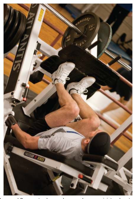
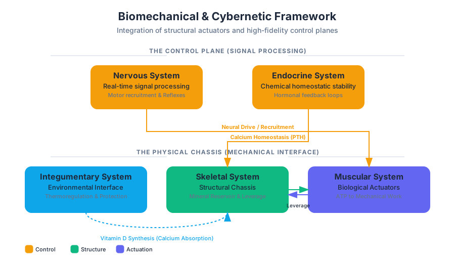
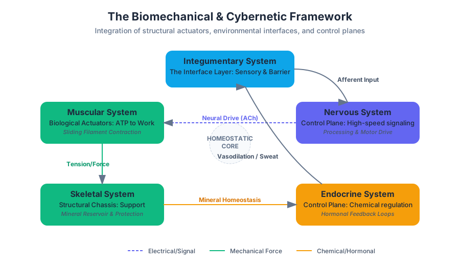
A leg press works because a muscle does not move the body by itself: it contracts, pulls on a tendon, and transmits force to a bone that pivots at a joint. The skeletal and muscular systems therefore operate as one mechanical system—one provides rigid levers and movable joints, while the other generates the forces that move them.
The skeletal system consists of bones and cartilage. It supports the body, protects internal organs, and enables movement together with the muscular system.
The skeletal system: support, protection, and movement
Bone, also called osseous tissue, is hard, dense connective tissue that forms most of the adult skeleton. Bone is not an inert framework like the mineralized material of a fossil; it is living tissue that grows, repairs itself, and continually renews its structure. This living quality allows the skeleton to change as the body grows and to respond to mechanical demands.
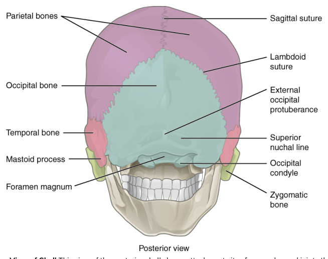
The skeletal system supports the body by providing a firm internal framework. It gives muscles stable attachment sites and maintains the overall shape of the body. It also protects delicate organs: the skull surrounds the brain, the vertebral column protects the spinal cord, and the ribs help shield organs within the thoracic cavity. Protection is therefore not a separate “extra” function; the same rigid structures that bear loads can also form barriers around vulnerable tissues.
Movement requires cooperation. Muscles generate force, but bones receive and transmit that force. A muscle that does not span a joint cannot move the bone on the other side of that joint. When a muscle spans a joint and contracts, its force is transmitted through connective attachments, especially tendons, to a bone. The resulting motion depends on the arrangement of the bones, the joint, and the muscle attachment.
The skeletal system also participates in broader body functions. Bone stores minerals, especially calcium, and the marrow inside certain bones contributes to blood-cell production. Red marrow produces blood cells through hematopoiesis, whereas yellow marrow stores fat. These functions reinforce the point that bone is a dynamic organ rather than merely a supporting scaffold.
Cartilage, joints, and smooth movement
Cartilage is semi-rigid connective tissue. It is firm enough to provide support but flexible enough to bend or compress slightly. Cartilage occurs in recognizable regions such as the nose, where it helps maintain shape while allowing flexibility, and at joints, where it helps create smooth, cushioned contact between bones.
At the surfaces of many joints, articular cartilage covers the ends of bones. Its smooth surface allows the bones to move against one another with less friction and helps distribute forces across the joint. The cartilage does not contract and cannot pull a bone; instead, it improves the mechanical conditions under which bones and muscles operate.
A joint is a place where bones meet. Joints vary in mobility, but movable joints are especially important for the lever system of the body. The joint acts as the pivot around which one or more bones rotate. Ligaments help hold bones together at movable joints and limit excessive movement, while cartilage helps cushion and guide contact between the joint surfaces.
A common misconception is that bones “push” the body into motion. Muscles primarily pull when they contract; the skeleton redirects those pulls through bones and joints. The movement we observe is therefore produced by a coordinated sequence: muscle contraction, force transmission through a tendon, bone movement, and rotation at a joint.
Bones as levers and joints as fulcrums
In biomechanics, a lever is a rigid structure that moves around a pivot, and a fulcrum is the pivot point. The body uses bones as levers and joints as fulcrums. When a muscle spans a joint and contracts, it applies force to a bone, causing the bone to rotate around the joint.
The leg-press exercise illustrates this relationship. The skeletal muscles of the lower limb contract against resistance. Their tendons transmit the force to bones, and the bones move at the joints of the lower limb. The equipment supplies external resistance, but the underlying biological mechanism remains the same: muscular force acts through skeletal levers that rotate around joints.
The mechanical advantage of a movement depends on the relative positions of the muscle attachment, the joint, and the load. A muscle may attach close to a joint and produce a large range of motion at the far end of a limb, even though the muscle must generate substantial force. Thus, the arrangement of the musculoskeletal system reflects a compromise between force, speed, stability, and range of motion.
Bone markings and muscle attachment
Bones are not smooth rods. Their surfaces contain ridges, bumps, depressions, and openings called bone markings. These markings reveal how bones interact with muscles, tendons, ligaments, blood vessels, and neighboring bones.
A projection is a part of a bone that extends above its surface. Some projections serve as attachment sites for muscles. When a muscle attaches to such a projection through a tendon, the projection provides a firm location where muscular force can be applied to the bone.
This arrangement is mechanically useful because the shape and position of a projection influence the direction of the muscle’s pull. A projection can increase the effective distance between the muscle’s line of action and the joint, changing the leverage of the muscle. In this way, bone shape is closely tied to movement: a marking is not merely a label on an anatomical diagram but evidence of repeated forces and functional attachments.
The structure of a long bone
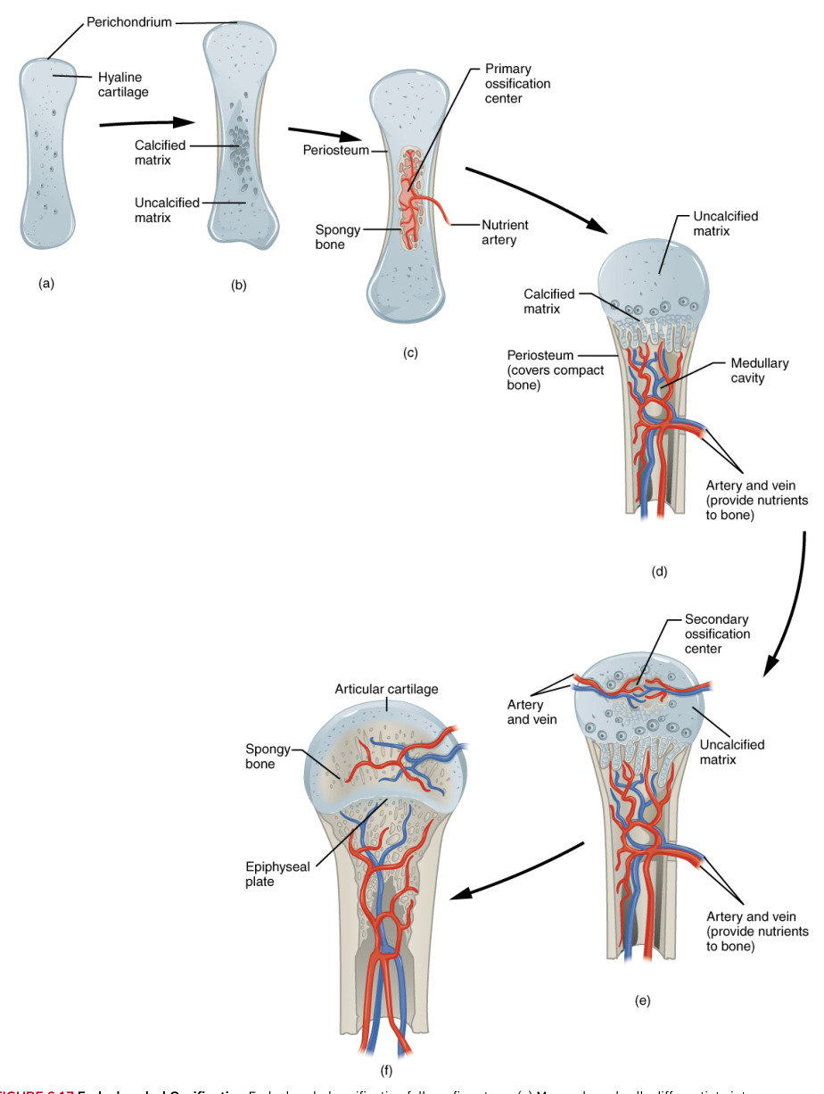
A long bone is organized around a central shaft and two expanded ends. The shaft is called the diaphysis, while each widened end is an epiphysis. The epiphyses are especially important because they contain substantial spongy bone and red marrow.
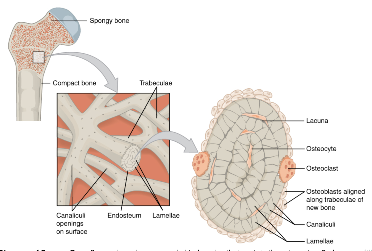
Spongy bone has a lattice-like internal structure rather than the dense appearance of compact bone. This arrangement reduces the amount of material needed while maintaining strength and creates spaces that can contain red marrow. The widened ends of long bones also form the regions that participate in joints, where articular cartilage covers the bone surfaces.
The shaft and ends are therefore specialized for different mechanical and physiological demands. The shaft provides a strong central support, while the epiphyses broaden the bone at its joints and contain spongy bone and red marrow. A growing long bone also contains a growth plate between the shaft and each epiphysis.
Growth at the epiphyseal plate
The epiphyseal plate, or growth plate, is a region of cartilage where a long bone increases in length. Its growth depends on an orderly progression of cellular zones. New cartilage is produced on one side, the cartilage enlarges and becomes calcified, and the calcified cartilage is then replaced by bone.
In the proliferative zone, cartilage cells continuously produce new cells through mitosis. Mitosis increases the number of cartilage cells and pushes the growing end of the bone farther away from the shaft. This zone is therefore responsible for generating new cartilage as longitudinal growth proceeds.
In the maturation zone, the cartilage cells enlarge. As they mature, the matrix surrounding them eventually calcifies. Calcification stiffens the matrix and prepares it for replacement by bone. The cartilage in this region is no longer functioning simply as flexible supporting tissue; it is being transformed into a temporary scaffold for ossification.
In the zone of ossification, blood vessels and osteoblasts invade the calcified cartilage. Osteoblasts are bone-forming cells. They replace the calcified cartilage with bone tissue, including spongy bone. This sequence explains how a long bone can lengthen without requiring the entire bone to grow uniformly at every surface.
As growth ends, the active growth plate is replaced by an epiphyseal line. The mature bone still retains the evidence of the former growth region, but it no longer lengthens through the same continuous cartilage-production process.
Clinical connection: abnormal spinal curvatures
The vertebral column normally has curves that help distribute body weight and maintain balance. Excessive curvature can interfere with posture, alignment, and movement. Scoliosis is an excessive lateral curvature of the spine, meaning the column bends abnormally from side to side.
Kyphosis is an excessive dorsoventral curvature. When pronounced, it can produce a hunchback appearance. The distinction matters because the two conditions describe different directions of abnormal spinal curvature: scoliosis is primarily lateral, whereas kyphosis is an exaggerated front-to-back curve.
These conditions also illustrate the interdependence of skeletal structure and muscular control. A change in spinal alignment affects how muscles attach, how forces pass through vertebrae and joints, and how the body maintains posture. Support, movement, and control are therefore not isolated functions: the living skeleton provides the framework, cartilage manages contact and flexibility, and muscles supply the force that turns anatomical structure into motion.
Content adapted from OpenStax Anatomy and Physiology 2e by J. Gordon Betts et al., licensed CC BY 4.0.
Source Materials
- Anatomy and Physiology 2e, Chapter 5: The Integumentary System
- Anatomy and Physiology 2e, Chapter 6: Bone Tissue and the Skeletal System
- Anatomy and Physiology 2e, Chapter 7: Axial Skeleton
- Anatomy and Physiology 2e, Chapter 8: The Appendicular Skeleton
- Anatomy and Physiology 2e, Chapter 9: Joints
- Anatomy and Physiology 2e, Chapter 10: Muscle Tissue
- Anatomy and Physiology 2e, Chapter 11: The Muscular System
- Anatomy and Physiology 2e, Chapter 12: The Nervous System and Nervous Tissue
- Anatomy and Physiology 2e, Chapter 13: Anatomy of the Nervous System
- Anatomy and Physiology 2e, Chapter 14: The Somatic Nervous System
- Anatomy and Physiology 2e, Chapter 15: The Autonomic Nervous System
- Anatomy and Physiology 2e, Chapter 16: The Neurological Exam
Study Anatomy and Physiology on Lykke
This course page is free to open: signed out, you can read its first two sections, and signing in, on any plan, opens the rest. The free Lykke Chrome extension syncs your own Canvas courses, so ChatGPT, Claude and Muse can answer from your classes and deadlines. It only reads Canvas.
Add the free extensionSign in to open every sectionAll sections of Anatomy and Physiology · Browse more course pages