From the free course Anatomy and Physiology on Lykke
Fluids, Transport, and Maintenance
Key concepts: Endocrine organs and hormone regulation · Cardiovascular system: heart and blood vessels · Adrenal medulla chromaffin cells · Erythropoietin production by the kidneys · Leukocyte production and lifespan · Leukopenia and impaired disease defense · Platelets and megakaryocytes · Hemostasis and platelet plug formation · Blood clotting factors and the common pathway · ABO and Rh blood-group classification
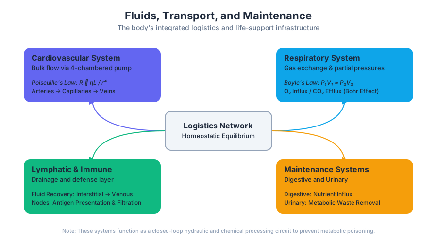
Orient the student to Fluids, Transport, and Maintenance: what each chapter in it teaches and how the chapters build on one another (Chapter 18, The Cardiovascular System: Blood; Chapter 19, The Cardiovascular System: The Heart; Chapter 20, The Cardiovascular System: Blood Vessels and Circulation; Chapter 21, The Lymphatic and Immune System; Chapter 22, The Respiratory System; Chapter 23, The Digestive System; Chapter 24, Metabolism and Nutrition; Chapter 25, The Urinary System; Chapter 26, Fluid, Electrolyte, and Acid-Base Balance).
Fluids, Transport, and Maintenance
The body stays alive because transport and regulation operate together: hormones coordinate distant organs, the cardiovascular system distributes materials, and blood continuously repairs damage and supports defense. A useful way to picture this is as a living city: endocrine organs send chemical messages, the heart drives circulation through a vessel network, and blood carries oxygen, nutrients, immune cells, and repair materials to neighborhoods that cannot exchange directly with the outside environment.
Endocrine organs and hormone regulation
An endocrine organ releases hormones—chemical messengers—into the surrounding fluid and ultimately into the bloodstream, allowing those hormones to influence target cells elsewhere in the body. The pancreas, adrenal glands, testes, and ovaries are endocrine organs. Their hormones regulate bodily processes such as metabolism, stress responses, fluid balance, and reproduction.
The pancreas lies in the abdomen. The adrenal glands sit above the kidneys, while the testes are located in the male groin area and the ovaries lie within the female pelvic region. Their locations are not merely landmarks: each organ releases hormones into the circulation so chemical signals can reach tissues throughout the body. Endocrine regulation therefore complements rapid nervous-system control with longer-lasting chemical coordination.
A common misconception is that an organ has only one role. The pancreas, for example, participates in digestion but also functions as an endocrine organ. Likewise, the kidneys are best known for filtering blood and producing urine, yet they also release the hormone erythropoietin. Many organs therefore have both their familiar primary functions and important endocrine functions.
The cardiovascular system: a transport network
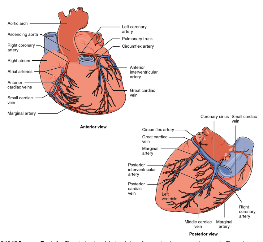
The cardiovascular system consists of the heart and a network of blood vessels. The heart is the muscular pump; the vessels are the routes through which blood travels. Together, they deliver oxygen and nutrients to tissues and help equalize temperature throughout the body.
In an anatomical diagram, arteries are conventionally shown in red and veins in blue. These colors are useful visual labels, not literal descriptions of blood color: oxygen-rich blood is typically bright red, whereas oxygen-poor blood is darker red. The essential distinction is directional. Arteries carry blood away from the heart, and veins return blood toward it; the color convention helps the reader follow the route.
The heart creates pressure by contracting. That pressure moves blood through vessels of different sizes, allowing exchange between the blood and tissues. Oxygen and nutrients leave the circulation where they are needed, while carbon dioxide and metabolic wastes enter the blood for transport to organs that can eliminate or process them. Circulating blood also distributes heat, helping reduce temperature differences between the body’s regions.
Adrenal medulla chromaffin cells
The adrenal glands contain an outer cortex and an inner adrenal medulla. The secretory cells of the adrenal medulla are called chromaffin cells. These cells release hormones associated with the body’s rapid stress response, especially epinephrine and norepinephrine.
Chromaffin cells are unusual because they are closely linked to nervous control: stimulation of the adrenal medulla causes them to secrete hormones into the blood, producing a body-wide response rather than a signal limited to one nerve ending. The cardiovascular system then distributes these hormones rapidly. This connection illustrates how the nervous, endocrine, and cardiovascular systems can cooperate: neural stimulation initiates the response, chromaffin cells provide the hormones, and blood carries them to target tissues.
Kidney control of erythrocyte production
The kidneys contribute to blood maintenance by producing erythropoietin, commonly abbreviated EPO. EPO is a glycoprotein hormone, meaning it is a protein-based hormone with attached carbohydrate groups. It is secreted by interstitial fibroblast cells of the kidney, particularly when oxygen levels are low.
Erythropoietin is a glycoprotein hormone secreted by kidney interstitial fibroblast cells in response to low oxygen levels; it stimulates the production of erythrocytes, or red blood cells.
EPO acts on blood-forming tissue to increase production of erythrocytes. More erythrocytes can improve the blood’s capacity to transport oxygen to tissues, correcting the problem that triggered the response. This is a negative-feedback relationship: reduced oxygen availability stimulates EPO release, EPO increases erythrocyte production, and the resulting increase in oxygen delivery reduces the original stimulus.
EPO also has clinical and ethical significance. Synthetic EPO can treat certain types of anemia, including anemia associated with some cancers. However, some athletes have used synthetic EPO for blood doping, an attempt to raise red blood cell numbers and improve oxygen delivery during performance. Increasing the concentration of cells can make blood more viscous, placing additional strain on the heart and increasing health risks. The same hormone can therefore be medically useful under supervision but dangerous when misused.
Leukocyte production and short lifespans
Leukocytes, or white blood cells, defend the body against disease. Unlike erythrocytes, which circulate primarily as oxygen-transporting cells, leukocytes participate in immune surveillance and responses to pathogens or tissue damage. Their production begins in the bone marrow.
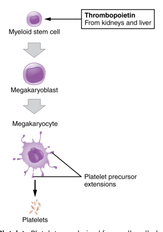
The formation of blood cells is called hemopoiesis. Bone-marrow precursor cells are directed toward mature blood-cell types by hemopoietic growth factors, including colony-stimulating factors and interleukins. These signaling molecules do not simply create cells automatically; they influence precursor-cell survival, division, differentiation, and maturation.
Leukocytes generally have short lifespans measured in hours or days, although the exact lifespan varies among leukocyte types and circumstances. Rapid turnover makes functional sense: immune cells must be produced in response to changing threats, and many are consumed, damaged, or deliberately removed after participating in defense. Bone marrow must therefore maintain a responsive supply rather than a fixed, permanent population.
Leukopenia and impaired disease defense
Leukopenia is a condition in which too few leukocytes are produced. Because leukocytes are central to defense, leukopenia may reduce the body’s ability to fight disease. A person with an inadequate leukocyte supply may be less able to contain pathogens that would otherwise be controlled.
Leukopenia means an abnormally low number of leukocytes; it can weaken disease defense because fewer immune cells are available to recognize and respond to threats.
The key reasoning is straightforward: impaired production reduces the number of available defenders, and fewer defenders can mean a weaker or slower immune response. This does not mean that every infection becomes inevitable, because defense also depends on barriers, immune-cell function, and other systems. It does mean that the blood-cell production system is part of immune protection, not merely a replacement service for worn-out cells.
Platelets and temporary vessel repair
Platelets, more accurately called thrombocytes, are not complete cells. They are cell fragments derived from very large bone-marrow cells called megakaryocytes. When a blood vessel is damaged, platelets gather at the injury and help form a temporary seal.
A platelet plug can temporarily close a small opening in a blood vessel. Its purpose is similar to placing a temporary plug in a leaking structure: it limits blood loss and buys time while a stronger repair develops. The plug is therefore an early response, not the final durable repair.
Hemostasis: from spasm to clot
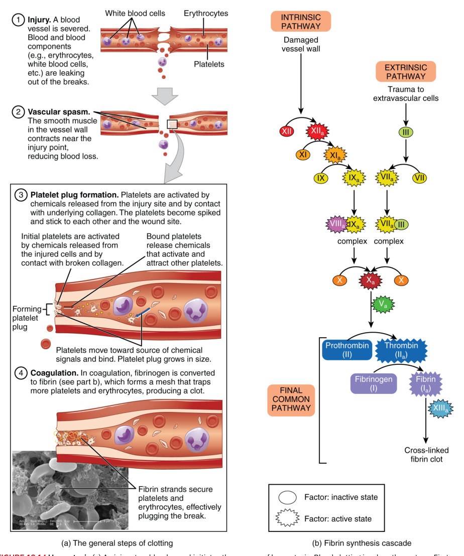
Hemostasis is the process that stops bleeding. It occurs in three major steps: vascular spasm, formation of the platelet plug, and coagulation. These steps overlap, but their order provides a useful framework for understanding how a damaged vessel is stabilized.
During a vascular spasm, the injured blood vessel constricts. This narrows the vessel and reduces blood flow through the damaged region. The response is especially valuable immediately after injury, because slowing the flow gives platelets and clotting proteins a better opportunity to act.
Next, platelets adhere to the damaged area and to one another, forming a platelet plug. This plug can temporarily seal a small opening. It is relatively quick but less durable than a clot, so the body must begin a more sophisticated repair process when the injury requires additional reinforcement.
That durable process is coagulation, the formation of a blood clot. Coagulation is often described as a cascade because one event activates the next through a series of steps. The process depends on known clotting factors, including calcium ions, and ultimately proceeds through a common pathway that produces the substances needed to stabilize the damaged region.
The practical sequence is therefore: constrict the vessel to reduce flow, assemble a platelet plug to cover the opening, and activate coagulation to produce a stronger clot. The next treatment of blood builds on this foundation by examining the blood cells, clotting machinery, and laboratory classifications that make these transport and maintenance functions measurable.
Content adapted from OpenStax Anatomy and Physiology 2e by J. Gordon Betts et al., licensed CC BY 4.0.
Source Materials
- Anatomy and Physiology 2e, Chapter 18: The Cardiovascular System: Blood
- Anatomy and Physiology 2e, Chapter 19: The Cardiovascular System: The Heart
- Anatomy and Physiology 2e, Chapter 20: The Cardiovascular System: Blood Vessels and Circulation
- Anatomy and Physiology 2e, Chapter 21: The Lymphatic and Immune System
- Anatomy and Physiology 2e, Chapter 22: The Respiratory System
- Anatomy and Physiology 2e, Chapter 23: The Digestive System
- Anatomy and Physiology 2e, Chapter 24: Metabolism and Nutrition
- Anatomy and Physiology 2e, Chapter 25: The Urinary System
- Anatomy and Physiology 2e, Chapter 26: Fluid, Electrolyte, and Acid-Base Balance
Study Anatomy and Physiology on Lykke
This course page is free to open: signed out, you can read its first two sections, and signing in, on any plan, opens the rest. The free Lykke Chrome extension syncs your own Canvas courses, so ChatGPT, Claude and Muse can answer from your classes and deadlines. It only reads Canvas.
Add the free extensionSign in to open every sectionAll sections of Anatomy and Physiology · Browse more course pages