From the free course High School Biology on Lykke
Eukaryotic Cell Structures and Organelles
Key concepts: Nucleus · Ribosomes · Endoplasmic Reticulum (ER) · Lysosomes · Cilia and Flagella
An exploration of the internal components of eukaryotic cells and their specialized functions.
Eukaryotic Cell Structures and Organelles: The Architecture of Cellular Complexity
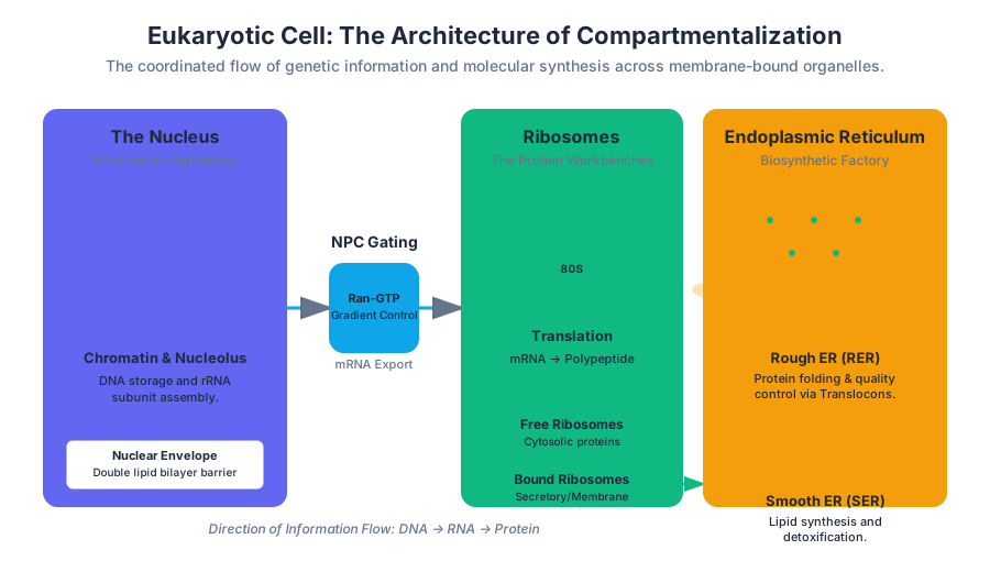
Eukaryotic cells represent a monumental leap in biological engineering. Unlike their prokaryotic counterparts, eukaryotes utilize compartmentalization—the segregation of biochemical processes into membrane-bound organelles. This spatial separation allows for the simultaneous execution of incompatible chemical reactions, such as the synthesis of proteins and their subsequent degradation, within the same cellular volume.
At its core, the eukaryotic cell is a high-throughput processing plant. It manages information (DNA), assembles components (ribosomes), performs quality control and logistics (ER and Golgi), manages waste (lysosomes), and facilitates movement (cilia and flagella). This article provides a deep-dive into the mechanics, thermodynamics, and structural biology of these critical systems.
The Nucleus: The Information Repository and Control Center
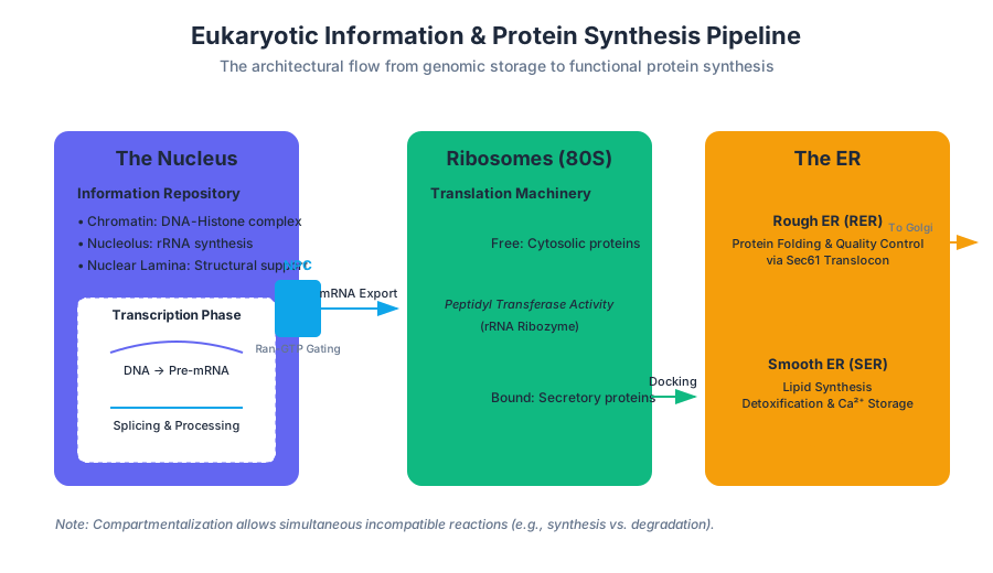
The nucleus is the defining feature of the eukaryotic cell. It serves as the central repository for the organism's genetic material and the site of transcription and RNA processing. By separating the genome from the cytoplasm, the nucleus allows for complex regulatory mechanisms, such as post-transcriptional splicing, which are unavailable to prokaryotes.
Structural Components
The nucleus is enclosed by the nuclear envelope, a double-membrane system. The outer membrane is continuous with the endoplasmic reticulum, while the inner membrane is lined by the nuclear lamina, a dense fibrillar network of intermediate filaments (lamins) that provides mechanical support.
| Component | Structure | Primary Function |
|---|---|---|
| Nuclear Envelope | Double lipid bilayer with perinuclear space | Physical barrier; regulates nucleocytoplasmic transport |
| Nuclear Pore Complex (NPC) | Octagonal protein scaffold (nucleoporins) | Selective gating of macromolecules (>40 kDa) |
| Nucleolus | Non-membrane bound sub-compartment | rRNA synthesis and ribosome subunit assembly |
| Chromatin | DNA-histone complex | Genomic packaging and epigenetic regulation |
Mechanics of Nuclear Transport
Transport across the nuclear envelope is governed by the Nuclear Pore Complex (NPC). While small molecules diffuse freely, larger proteins (>40-60 kDa) require a Nuclear Localization Signal (NLS) or a Nuclear Export Signal (NES).
The transport cycle is driven by the Ran-GTPase gradient. The concentration of Ran-GTP is high inside the nucleus (maintained by Ran-GEF) and low in the cytoplasm (maintained by Ran-GAP). This gradient provides the chemical potential necessary to drive cargo against a concentration gradient.
Ribosomes: The Protein Synthesis Machinery
Ribosomes are the "workbenches" of the cell, where the genetic code is translated into polypeptide chains. They are not technically organelles in the sense of being membrane-bound; rather, they are massive ribonucleoprotein (RNP) complexes.
Composition and Catalysis
A eukaryotic ribosome (80S) consists of a large subunit (60S) and a small subunit (40S). The catalytic heart of the ribosome is actually the ribosomal RNA (rRNA), making the ribosome a ribozyme. The rRNA facilitates the formation of the peptide bond through a process called peptidyl transfer.
The Central Dogma Constraint: Ribosomes can only read mRNA in the 5' to 3' direction. The rate of translation in eukaryotes is approximately 2–5 amino acids per second, significantly slower than prokaryotes (up to 20 aa/s), allowing for higher folding precision.
Free vs. Bound Ribosomes
- Free Ribosomes: Suspended in the cytosol. They synthesize proteins that function within the cytosol (e.g., glycolytic enzymes).
- Bound Ribosomes: Attached to the cytosolic side of the Endoplasmic Reticulum (ER). They synthesize proteins destined for insertion into membranes, packaging within organelles, or secretion.
The Endoplasmic Reticulum (ER): The Biosynthetic Factory
The Endoplasmic Reticulum (ER) is an extensive network of membranous tubules and sacs called cisternae. It accounts for more than half of the total membrane in many eukaryotic cells.
Rough ER (RER)
The RER is "rough" due to the presence of ribosomes on its surface. It is the primary site for the synthesis of secretory proteins. As a polypeptide is synthesized, it is threaded into the ER lumen through a protein pore called the translocon (Sec61 complex).
Smooth ER (SER)
The SER lacks ribosomes and performs diverse metabolic functions:
- Lipid Synthesis: Production of phospholipids, cholesterol, and steroid hormones.
- Detoxification: In liver cells, the SER contains enzymes (like Cytochrome P450) that add hydroxyl groups to drugs, making them more soluble and easier to flush from the body.
- Calcium Storage: In muscle cells, a specialized SER called the sarcoplasmic reticulum pumps $Ca^{2+}$ ions from the cytosol into the lumen.
Protein Folding and Quality Control
The ER lumen provides a unique oxidizing environment that favors the formation of disulfide bonds ($S-S$), which stabilize protein structures. Chaperone proteins, such as BiP, assist in folding. If a protein fails to fold correctly, it is subject to ER-Associated Degradation (ERAD), where it is exported back to the cytosol and destroyed by proteasomes.
\text{Protein Folding Success Rate } (\eta) \approx \frac{k_{fold}}{k_{fold} + k_{agg}}
Where $k_{fold}$ is the rate constant of native folding and $k_{agg}$ is the rate constant of non-specific aggregation. The ER environment is optimized to maximize $\eta$ via chaperones.
Lysosomes: The Digestive and Recycling Center
Lysosomes are membrane-bound sacs of hydrolytic enzymes used to digest macromolecules. They represent the cell's "stomach" and recycling plant.
The Acidic Microenvironment
Lysosomal enzymes (acid hydrolases) function optimally at a pH of approximately 4.5–5.0. The lysosome maintains this gradient by utilizing a V-type ATPase (proton pump) that actively transports $H^+$ ions from the cytosol (pH ~7.2) into the lysosomal lumen.
| Enzyme Category | Substrate | Resulting Product |
|---|---|---|
| Proteases | Proteins | Amino acids |
| Nucleases | Nucleic Acids | Nucleotides |
| Glycosidases | Polysaccharides | Monosaccharides |
| Lipases | Lipids | Fatty acids & Glycerol |
Mechanisms of Degradation
- Phagocytosis: Digestion of external materials (e.g., bacteria) engulfed by the cell.
- Autophagy: "Self-eating." The lysosome recycles the cell's own damaged organelles. This is a critical survival mechanism during nutrient deprivation.
Clinical Insight: Lysosomal Storage Diseases (LSDs), such as Tay-Sachs disease, occur when a specific lysosomal enzyme is missing or defective. This leads to the toxic accumulation of undigested substrates, eventually causing cellular death and organ failure.
Cilia and Flagella: Cellular Locomotion
Many eukaryotic cells possess surface appendages for movement. While they differ in length and beating patterns, cilia and flagella share a nearly identical internal ultrastructure.
The 9+2 Axoneme
The core of these structures is the axoneme, which consists of nine doublets of microtubules arranged in a ring around two central single microtubules. This is the "9+2" arrangement.
- Microtubules: Polymers of $\alpha$- and $\beta$-tubulin.
- Dynein Arms: Motor proteins attached to the outer doublets. They use ATP hydrolysis to "walk" along the adjacent microtubule.
- Nexin: Cross-linking proteins that limit the sliding of microtubules, causing the entire structure to bend instead of slide apart.
Comparison of Movement
| Feature | Cilia | Flagella |
|---|---|---|
| Length | Short (~5–10 $\mu m$) | Long (~50–200 $\mu m$) |
| Number | Numerous per cell | Usually 1 or 2 per cell |
| Motion | Oar-like (power stroke and recovery) | Undulatory (snake-like) |
| Function | Movement of cell or fluid over surface | Propulsion of the entire cell |
The Physics of the Power Stroke
The bending of a cilium is a result of the coordinated activation of dynein motors. Because the microtubules are anchored at the basal body (a structure similar to a centriole), the sliding force is converted into a bending moment.
Integration: The Endomembrane System
None of these organelles operate in isolation. They are linked via the endomembrane system, a flow of membrane and proteins mediated by vesicles.
- Nucleus exports mRNA to the Cytoplasm.
- Ribosomes on the Rough ER translate mRNA into proteins.
- Proteins are folded in the ER and sent via transport vesicles to the Golgi Apparatus (not covered in detail here, but acts as the shipping center).
- The Golgi sorts proteins for delivery to the Plasma Membrane, Lysosomes, or secretion.
Common Pitfalls and Misconceptions
- "Ribosomes are organelles": While often called organelles, they are not membrane-bound. In rigorous technical contexts, they are "macromolecular machines."
- "Smooth ER only makes lipids": It is also crucial for carbohydrate metabolism (gluconeogenesis) and calcium sequestration.
- "Cilia are only for swimming": Many human cells have a primary cilium, a non-motile 9+0 structure that acts as a sensory antenna for chemical and mechanical signals.
- "The Nucleus is in the center": The position of the nucleus is highly regulated by the cytoskeleton and varies by cell type (e.g., it is pushed to the periphery in adipocytes).
Article Metadata
- Status: Peer Reviewed
- Technical Level: Level 400 (Advanced Undergraduate / Graduate)
- Domain: Molecular Cell Biology
- Last Updated: 2023.10.27
Source Materials
Study High School Biology on Lykke
This course page is free to open: signed out, you can read its first two sections, and signing in, on any plan, opens the rest. The free Lykke Chrome extension syncs your own Canvas courses, so ChatGPT, Claude and Muse can answer from your classes and deadlines. It only reads Canvas.
Add the free extensionSign in to open every sectionAll sections of High School Biology · Browse more course pages