From the free course Introductory Biology on Lykke
Course Introduction and Biological Foundations
Key concepts: Prokaryotes vs. Eukaryotes · Domains of Life · Model Organisms · Central Dogma · Gene Expression
Introduction to the domains of life, model organisms, and the central dogma of molecular biology.
Course Introduction and Biological Foundations
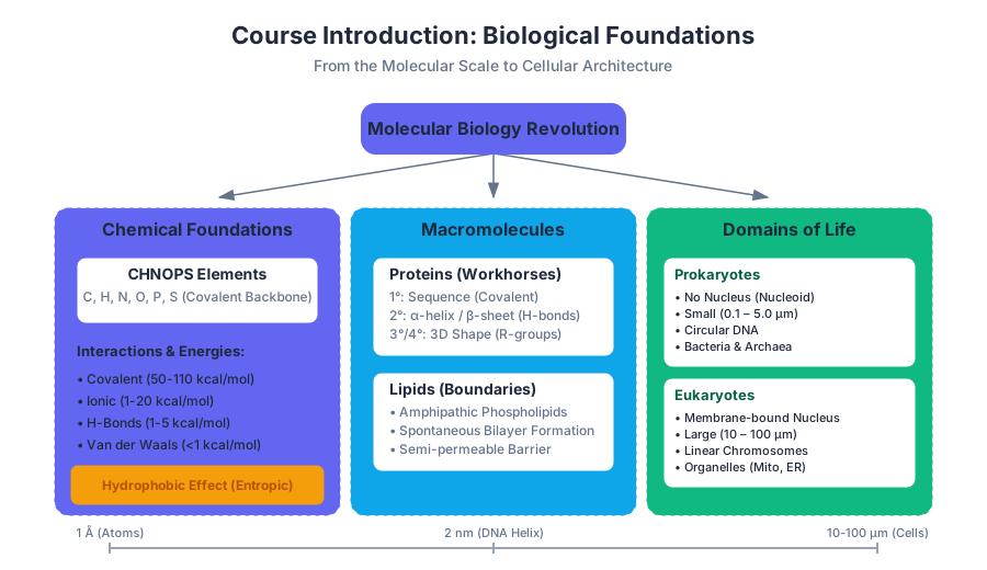
The study of biology has undergone a radical transformation over the last century, evolving from a purely descriptive discipline into a rigorous, molecular, and information-based science. This "Molecular Biology Revolution" has integrated principles from chemistry, physics, and engineering to decode the logic of living systems. At its core, modern biology seeks to understand how the interaction of inanimate molecules—lipids, proteins, nucleic acids, and carbohydrates—gives rise to the emergent property of life.
The Molecular Scale and Chemical Foundations
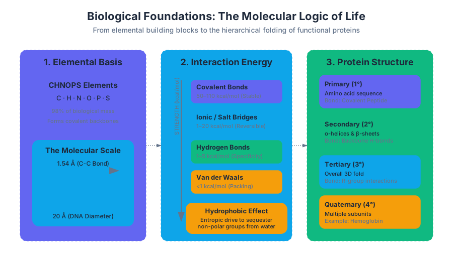
To understand biological systems, one must first appreciate the scale at which they operate. Biological interactions occur primarily at the nanometer ($10^{-9}$ m) and Angstrom ($\text{\AA}$, $10^{-10}$ m) scales. For context, a typical carbon-carbon covalent bond is approximately $1.54\ \text{\AA}$, while a DNA double helix is about $20\ \text{\AA}$ (2 nm) in diameter.
Life is built from a specific subset of the periodic table, often summarized by the acronym CHNOPS: Carbon, Hydrogen, Nitrogen, Oxygen, Phosphorus, and Sulfur. These elements form the covalent backbone of all biological macromolecules.
Covalent vs. Non-Covalent Interactions
The stability and dynamism of life depend on the balance between strong covalent bonds and weak non-covalent interactions.
- Covalent Bonds: Formed by the sharing of electron pairs. These are the "permanent" links that define the primary structure of molecules (e.g., the peptide bond in proteins).
- Non-Covalent Interactions: These are reversible and much weaker, yet they are responsible for the three-dimensional folding of proteins, the hybridization of DNA strands, and the formation of cell membranes.
| Interaction Type | Typical Energy (kcal/mol) | Basis of Interaction | Biological Example |
|---|---|---|---|
| Covalent Bond | 50 – 110 | Electron sharing | Backbone of DNA |
| Ionic Interaction | 1 – 20 | Electrostatic attraction | Salt bridges in proteins |
| Hydrogen Bond | 1 – 5 | Dipole-dipole (H to O/N) | Base pairing in DNA |
| Van der Waals | < 1 | Transient dipoles | Packing of lipid tails |
| Hydrophobic Effect | Variable (Entropic) | Exclusion of water | Protein core folding |
The Hydrophobic Effect: This is perhaps the most important non-covalent "force" in biology. It is not an attractive force in the traditional sense, but an entropic drive. Water molecules form highly ordered "clathrate" cages around non-polar solutes. By sequestering non-polar groups together, the system minimizes the surface area in contact with water, releasing water molecules into the bulk phase and increasing the entropy of the universe ($\Delta S > 0$).
The Domains of Life and Cellular Architecture
Biological diversity is categorized into three primary domains: Bacteria, Archaea, and Eukarya. This classification is based on ribosomal RNA sequences and fundamental differences in cellular machinery.
Prokaryotes vs. Eukaryotes
The most significant structural divide is between prokaryotes (Bacteria and Archaea) and eukaryotes.
| Feature | Prokaryotes | Eukaryotes |
|---|---|---|
| Nucleus | Absent (Nucleoid region) | Present (Membrane-bound) |
| Organelles | Generally absent | Present (Mitochondria, ER, Golgi) |
| DNA Structure | Circular, usually single | Linear, multiple chromosomes |
| Size | Small (0.1 – 5.0 $\mu$m) | Large (10 – 100 $\mu$m) |
| Transcription/Translation | Coupled in cytoplasm | Spatially separated |
| Cell Wall | Peptidoglycan (Bacteria) | Cellulose (Plants) or Chitin (Fungi) |
Macromolecules I: Lipids and the Membrane Boundary
Lipids are unique among macromolecules because they are not polymers in the traditional sense. Instead, they aggregate through non-covalent interactions to form the phospholipid bilayer, which defines the boundary of the cell and its internal compartments.
Amphipathic Nature
A typical phospholipid consists of a hydrophilic (polar) "head" group and two hydrophobic (non-polar) fatty acid "tails." In an aqueous environment, these molecules spontaneously organize into bilayers.
- Self-Healing: Because the bilayer is held together by non-covalent hydrophobic interactions, it is dynamic. If the membrane is punctured, the hydrophobic tails' exposure to water is energetically unfavorable, causing the membrane to spontaneously reseal.
- Semi-permeability: The hydrophobic core acts as a barrier to ions and large polar molecules (e.g., $Na^+$, Glucose), while allowing small non-polar molecules (e.g., $O_2$, $CO_2$) to diffuse freely.
Macromolecules II: Proteins and Amino Acids
Proteins are the "workhorses" of the cell, serving as catalysts (enzymes), structural components, and signaling molecules. They are polymers of alpha-amino acids.
Amino Acid Structure
Every amino acid shares a common backbone: a central carbon ($\alpha$-carbon) bonded to an amino group ($-NH_2$), a carboxyl group ($-COOH$), a hydrogen atom, and a variable Side Chain (R-group). There are 20 standard amino acids, categorized by the chemical properties of their R-groups:
- Non-polar (Hydrophobic): e.g., Leucine, Valine, Phenylalanine.
- Polar Uncharged (Hydrophilic): e.g., Serine, Glutamine.
- Charged: e.g., Aspartic Acid (negative), Lysine (positive).
Protein Folding and Hierarchy
Protein structure is described in four levels:
- Primary ($1^\circ$): The linear sequence of amino acids linked by covalent peptide bonds.
- Secondary ($2^\circ$): Local folding patterns like $\alpha$-helices and $\beta$-sheets, stabilized by hydrogen bonds between backbone atoms.
- Tertiary ($3^\circ$): The overall 3D shape of a single polypeptide, driven by R-group interactions.
- Quaternary ($4^\circ$): The assembly of multiple polypeptide subunits (e.g., Hemoglobin).
Case Study: Sickle Cell Anemia
Sickle cell anemia provides a classic example of how a microscopic change in primary structure yields a macroscopic disease state. A single point mutation in the gene for $\beta$-globin causes the substitution of Glutamic Acid (polar/charged) with Valine (non-polar) at position 6.
- Mechanism: In the deoxygenated state, the hydrophobic Valine residue on one hemoglobin molecule associates with a hydrophobic pocket on another. This leads to the polymerization of hemoglobin into long fibers, distorting the red blood cell into a "sickle" shape, which clogs capillaries and reduces oxygen transport.
Macromolecules III: Enzymes and Metabolism
Enzymes are biological catalysts that accelerate chemical reactions by lowering the Activation Energy ($E_a$). They do not change the equilibrium of a reaction ($\Delta G$), only the rate at which equilibrium is reached.
Thermodynamics of Life
Biological reactions are governed by the Gibbs Free Energy equation: $$\Delta G = \Delta H - T\Delta S$$
- Exergonic Reactions ($\Delta G < 0$): Spontaneous reactions that release energy.
- Endergonic Reactions ($\Delta G > 0$): Non-spontaneous reactions that require energy input.
Reaction Coupling: Cells perform endergonic reactions (like building DNA) by coupling them to highly exergonic reactions, most commonly the hydrolysis of ATP to ADP.
\begin{aligned}
\text{Reaction 1 (Endergonic): } & A \rightarrow B \quad (\Delta G = +5 \text{ kcal/mol}) \\
\text{Reaction 2 (Exergonic): } & ATP \rightarrow ADP + P_i \quad (\Delta G = -7.3 \text{ kcal/mol}) \\
\hline
\text{Coupled Reaction: } & A + ATP \rightarrow B + ADP + P_i \quad (\Delta G = -2.3 \text{ kcal/mol})
\end{aligned}
Macromolecules IV: Nucleic Acids and the Central Dogma
Nucleic acids (DNA and RNA) are the information-carrying molecules of the cell. They are polymers of nucleotides, each consisting of a sugar, a phosphate group, and a nitrogenous base (A, T, C, G, or U).
The Central Dogma
The Central Dogma describes the directional flow of genetic information:
- Replication: DNA makes copies of itself.
- Transcription: DNA is used as a template to synthesize messenger RNA (mRNA).
- Translation: mRNA is read by ribosomes to synthesize a specific protein.
Gene Expression: While almost every cell in a multicellular organism contains the same genome, they look and function differently because they express different subsets of genes. This is regulated by transcription factors and epigenetic modifications.
Model Organisms in Biological Research
Because the fundamental mechanisms of life (the genetic code, metabolic pathways) are conserved across evolution, biologists use model organisms to study complex processes in simpler, more controllable systems.
| Organism | Common Name | Primary Use |
|---|---|---|
| Escherichia coli | Bacteria | DNA replication, gene expression, protein production |
| Saccharomyces cerevisiae | Yeast | Eukaryotic cell cycle, protein trafficking |
| Arabidopsis thaliana | Thale cress | Plant genetics and development |
| Drosophila melanogaster | Fruit fly | Genetics, embryonic development |
| Caenorhabditis elegans | Roundworm | Cell lineage, apoptosis (cell death) |
| Mus musculus | Mouse | Immunology, human disease modeling |
Common Pitfalls and Misconceptions
- "Spontaneous" means "Fast": In thermodynamics, a spontaneous reaction ($\Delta G < 0$) is one that can occur without external energy, but it might take millions of years (e.g., the breakdown of a diamond). Enzymes are required to make these reactions occur on a biological timescale.
- The "Blueprint" Metaphor: DNA is often called a blueprint, but it's more like a recipe or an executable program. A blueprint is a static 1:1 map; DNA contains conditional instructions (e.g., "if temperature > 37°C, produce Heat Shock Proteins").
- Lipid Bilayer Rigidity: Students often imagine the cell membrane as a static wall. In reality, it is a "Fluid Mosaic." Lipids and proteins diffuse laterally within the plane of the membrane quite rapidly.
Source Materials
Study Introductory Biology on Lykke
This course page is free to open: signed out, you can read its first two sections, and signing in, on any plan, opens the rest. The free Lykke Chrome extension syncs your own Canvas courses, so ChatGPT, Claude and Muse can answer from your classes and deadlines. It only reads Canvas.
Add the free extensionSign in to open every sectionAll sections of Introductory Biology · Browse more course pages