From the free course Introductory Biology on Lykke
Chemical Bonding and Biological Macromolecules
Key concepts: Covalent and Non-covalent Bonds · Hydrophobic Interactions · Lipids and Membranes · Protein Hierarchy · Carbohydrates
Exploration of the chemical interactions and structural properties of lipids, proteins, and carbohydrates.
Chemical Bonding and Biological Macromolecules
The Molecular Foundation of Life
Biology has transitioned from a descriptive science of "what we see" to a precise engineering discipline of "how it works" at the molecular scale. To understand the cell, one must understand the forces governing the interactions of the CHNOPS elements (Carbon, Hydrogen, Nitrogen, Oxygen, Phosphorus, and Sulfur). These six elements constitute approximately 98% of the mass of most living organisms.
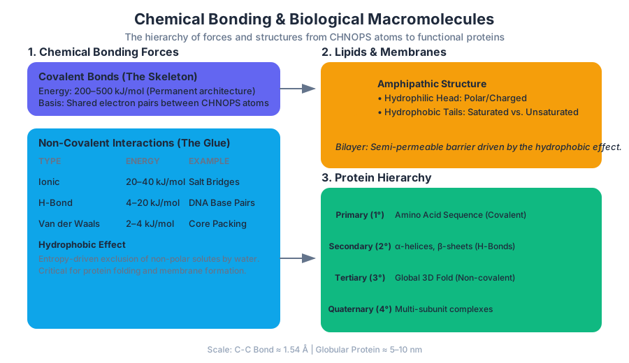
The scale of these interactions is measured in Angstroms ($\text{\AA}$, $10^{-10}$ m) and Nanometers (nm, $10^{-9}$ m). A typical C-C covalent bond is roughly $1.54\text{\AA}$, while the diameter of a globular protein may span 5–10 nm. At this scale, the distinction between "strong" and "weak" forces dictates the difference between a molecule's permanent architecture and its dynamic, functional movements.
Chemical Bonding: The Architecture of the Cell
Biological structures are held together by a continuum of forces, ranging from the robust sharing of electrons to transient induced dipoles.
Covalent Bonds: The "Hard-Wired" Skeleton
A covalent bond occurs when two atoms share a pair of valence electrons. In biological systems, these are the "permanent" connections that define the identity of a molecule.
- Bond Energy: Typically ranges from 200 to 500 kJ/mol. This is significantly higher than the thermal energy available at physiological temperature ($RT \approx 2.5$ kJ/mol), meaning covalent bonds do not break spontaneously.
- Polarity: When electrons are shared unequally due to differences in electronegativity (e.g., O-H or N-H bonds), the bond becomes polar, creating partial charges ($\delta+$ and $\delta-$) that are crucial for non-covalent interactions.
Non-Covalent Interactions: The Dynamic Glue
While covalent bonds build the molecules, non-covalent interactions determine how those molecules fold, associate, and signal.
| Interaction Type | Basis of Interaction | Energy (kJ/mol) | Biological Example |
|---|---|---|---|
| Ionic Bond | Attraction between opposite full charges | 20–40 | Salt bridges in protein interiors |
| Hydrogen Bond | Dipole-dipole interaction involving H and O, N, or F | 4–20 | DNA base pairing; $\alpha$-helices |
| Van der Waals | Transient induced dipoles | 2–4 | Packing of non-polar side chains |
| Hydrophobic Effect | Entropy-driven exclusion of non-polar solutes | Variable | Protein folding; Membrane formation |
The Hydrophobic Effect
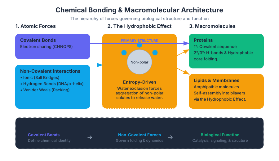
The hydrophobic effect is perhaps the most misunderstood "force" in biology. It is not an attractive force between non-polar molecules, but rather an entropy-driven exclusion of non-polar substances by water.
Definition: When a non-polar molecule is placed in water, the water molecules are forced to form a highly ordered "clathrate" cage around it to maintain hydrogen bonding. This decrease in the entropy of water ($\Delta S < 0$) is energetically unfavorable. By aggregating together, non-polar molecules minimize their surface area, releasing water molecules back into the bulk phase and increasing the total entropy of the system ($\Delta S > 0$).
Lipids and Biological Membranes
Lipids are unique among the four macromolecule classes because they are not polymers in the traditional sense. Instead, they are defined by their solubility: they are hydrophobic or amphipathic (possessing both hydrophilic and hydrophobic regions).
Phospholipids and the Bilayer
The fundamental unit of the cell membrane is the phospholipid. It consists of a glycerol backbone, two fatty acid "tails," and a phosphate-containing "head" group.
- The Head: Polar and hydrophilic, interacting with the aqueous environment.
- The Tails: Non-polar hydrocarbon chains.
- Saturated: No double bonds; straight chains that pack tightly (solid at room temp).
- Unsaturated: Contain one or more cis-double bonds; "kinks" prevent tight packing (liquid at room temp).
Membrane Dynamics and Permeability
The lipid bilayer is a semi-permeable barrier. It allows the passage of small, non-polar molecules (like $O_2$ and $CO_2$) but acts as a formidable barrier to ions and large polar molecules (like glucose).
Protein Hierarchy: From Sequence to Function
Proteins are the "workhorses" of the cell, executing the instructions encoded in DNA. They are polymers of $\alpha$-amino acids.
The Amino Acid Building Block
Every amino acid shares a common structure: a central carbon ($C_\alpha$), an amino group ($-NH_2$), a carboxyl group ($-COOH$), and a variable R-group (side chain). There are 20 standard amino acids, categorized by the chemical nature of their R-groups.
| Category | Properties | Examples |
|---|---|---|
| Non-polar | Hydrophobic; found in protein cores | Leucine, Valine, Phenylalanine |
| Polar Uncharged | Can form H-bonds; hydrophilic | Serine, Threonine, Glutamine |
| Charged (Acidic) | Negatively charged at pH 7 | Aspartic Acid, Glutamic Acid |
| Charged (Basic) | Positively charged at pH 7 | Lysine, Arginine, Histidine |
The Four Levels of Protein Structure
- Primary ($1^\circ$): The linear sequence of amino acids linked by peptide bonds. The peptide bond has double-bond character due to resonance, making it planar and rigid.
- Secondary ($2^\circ$): Local spatial arrangement of the backbone, stabilized by H-bonds between the backbone carbonyl oxygen and amide hydrogen. Common motifs include the $\alpha$-helix and $\beta$-pleated sheet.
- Tertiary ($3^\circ$): The overall 3D fold of a single polypeptide chain, driven by R-group interactions (hydrophobic packing, ionic bridges, disulfide bonds).
- Quaternary ($4^\circ$): The association of multiple polypeptide subunits into a functional complex (e.g., the four subunits of Hemoglobin).
Case Study: Sickle Cell Anemia
Sickle cell anemia is a profound example of how a change in the $1^\circ$ sequence dictates $4^\circ$ pathology. A single mutation (Glu6 $\rightarrow$ Val6) in the $\beta$-globin chain replaces a polar, charged residue with a non-polar, hydrophobic one. Under low oxygen conditions, this "hydrophobic patch" causes hemoglobin tetramers to polymerize into long fibers, distorting the red blood cell into a sickle shape.
\text{Gibbs Free Energy of Folding:} \\
\Delta G_{folding} = \Delta H_{folding} - T \Delta S_{folding} \\
\\
\text{Where:} \\
\Delta H \text{ (Enthalpy): Sum of H-bonds and VdW interactions.} \\
\Delta S \text{ (Entropy): Includes conformational entropy (negative) } \\
\text{and the hydrophobic effect (positive).}
Carbohydrates: Energy and Identity
Carbohydrates (saccharides) serve two primary roles: fuel storage and structural/recognition markers.
Monosaccharides and the Glycosidic Bond
Monosaccharides like glucose ($C_6H_{12}O_6$) can exist in linear or ring forms. When two monosaccharides link, they form a glycosidic bond via a dehydration reaction.
- $\alpha$-Linkage: The -OH group on Carbon-1 is below the plane. Found in Starch and Glycogen (easily digested for energy).
- $\beta$-Linkage: The -OH group on Carbon-1 is above the plane. Found in Cellulose (highly stable, structural component of plant cell walls). Humans lack the enzymes to hydrolyze $\beta$-1,4 linkages.
Glycoproteins and Blood Groups
Carbohydrates often conjugate to proteins (glycoproteins) on the cell surface. These "sugar coats" are essential for cell-cell recognition. The ABO blood group system is defined by specific carbohydrate antigens attached to lipids and proteins on the red blood cell surface.
| Blood Type | Terminal Sugar | Enzyme Present |
|---|---|---|
| O | H-antigen (fucose) | None (functional) |
| A | N-acetylgalactosamine | A-transferase |
| B | Galactose | B-transferase |
| AB | Both A and B sugars | Both transferases |
Common Pitfalls and Misconceptions
- The "Bond" Misnomer: Students often think of the "hydrophobic bond." There is no such thing. It is an exclusion principle driven by the entropy of water, not an attraction between lipids.
- Equilibrium vs. Rate: Enzymes (proteins) change the rate of a reaction by lowering activation energy ($E_a$), but they do not change the equilibrium constant ($K_{eq}$) or the $\Delta G$ of the reaction.
- Denaturation: Heating a protein breaks non-covalent interactions (secondary, tertiary, and quaternary structure) but usually leaves the covalent peptide bonds (primary structure) intact.
Source Materials
Study Introductory Biology on Lykke
This course page is free to open: signed out, you can read its first two sections, and signing in, on any plan, opens the rest. The free Lykke Chrome extension syncs your own Canvas courses, so ChatGPT, Claude and Muse can answer from your classes and deadlines. It only reads Canvas.
Add the free extensionSign in to open every sectionAll sections of Introductory Biology · Browse more course pages