From the free course Introductory Biology on Lykke
Cell Structure and Organelle Dynamics
Key concepts: Endosymbiotic Theory · Mitochondria and Chloroplasts · Protein Trafficking · Endoplasmic Reticulum · Golgi Apparatus
The organization of eukaryotic cells and the evolutionary origins of organelles.
Cell Structure and Organelle Dynamics
The eukaryotic cell is not merely a "bag of enzymes" but a highly partitioned chemical reactor. The transition from the relatively simple architecture of prokaryotes to the complex, compartmentalized nature of eukaryotes represents one of the most significant "phase transitions" in biological history. By isolating specific biochemical reactions within membrane-bound organelles, eukaryotes circumvent the diffusion limits that constrain bacterial size, allowing for a massive increase in genomic complexity and metabolic efficiency.
This section explores the mechanics of cellular compartmentalization, the evolutionary origins of these compartments via endosymbiosis, and the sophisticated logistics system—protein trafficking—that ensures the right molecular "cargo" reaches the correct "address."
The Evolutionary Genesis: Endosymbiotic Theory
The Endosymbiotic Theory posits that the defining organelles of eukaryotic cells—mitochondria and chloroplasts—originated as independent prokaryotic organisms that were engulfed by a proto-eukaryotic host. Rather than being digested, these bacteria entered into a mutualistic relationship, eventually becoming obligate organelles.
The Mechanics of Integration
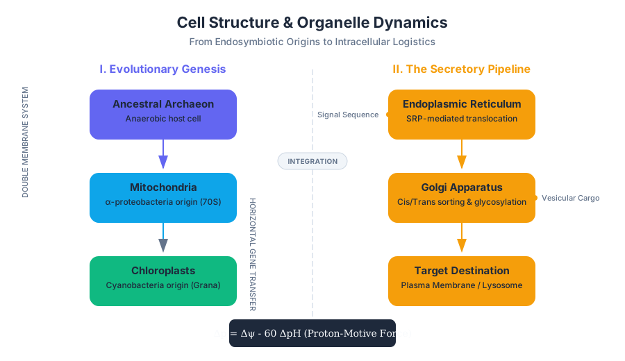
The theory, championed by Lynn Margulis in the 1960s, explains several "anomalies" in cell biology. Mitochondria are derived from α-proteobacteria, while chloroplasts are derived from cyanobacteria. The integration process involved a massive horizontal gene transfer (HGT), where the majority of the endosymbiont's genome was migrated to the host nucleus, leaving only a vestigial "mitogenome" or "plastid genome" behind.
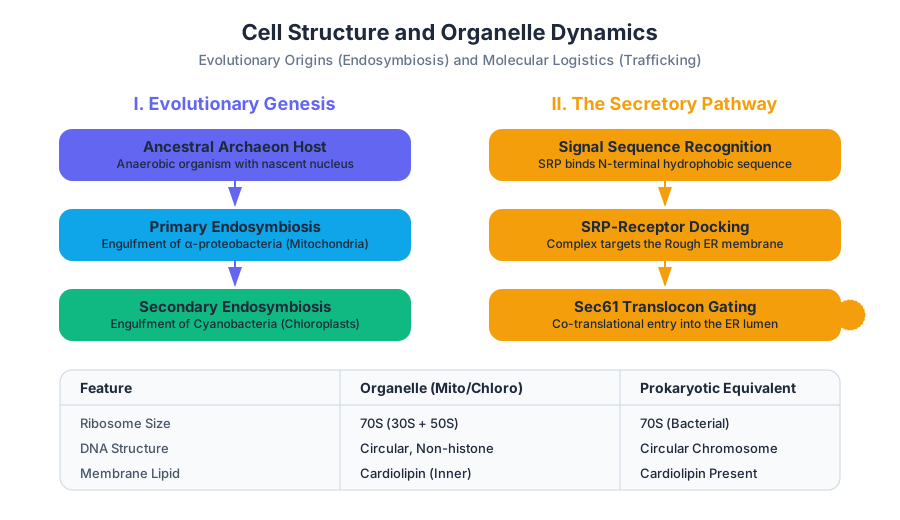
| Feature | Mitochondria / Chloroplasts | Prokaryotic Equivalent | Eukaryotic (Nuclear/Cytosolic) |
|---|---|---|---|
| DNA Structure | Circular, non-histone bound | Circular | Linear, histone-bound |
| Ribosome Size | 70S (30S + 50S) | 70S | 80S (40S + 60S) |
| Division | Binary Fission | Binary Fission | Mitosis/Meiosis |
| Membrane | Double (Inner/Outer) | Single (plus peptidoglycan) | Single (Plasma membrane) |
| Initiator tRNA | fMet (formylmethionine) | fMet | Methionine |
Evidence and Proofs
The strongest evidence for this theory lies in the double membrane system. The inner membrane of a mitochondrion contains cardiolipin, a phospholipid found almost exclusively in bacterial membranes and not in the eukaryotic plasma membrane. Furthermore, the sensitivity of mitochondrial ribosomes to antibiotics like streptomycin (which targets bacterial 70S ribosomes) but not to eukaryotic inhibitors provides a functional "smoking gun" for their bacterial ancestry.
The Serial Endosymbiosis Theory (SET): Evolution did not happen in a single step. First, an anaerobic archaeon likely engulfed an aerobic proteobacterium (forming the mitochondrion). Later, a subset of these now-aerobic eukaryotes engulfed a photosynthetic cyanobacterium, leading to the lineage of plants and algae.
Mitochondria and Chloroplasts: The Energy Transducers
Mitochondria and chloroplasts are the primary sites of energy conversion. While they share an endosymbiotic origin, their thermodynamic roles are inverse: mitochondria perform oxidative phosphorylation (catabolic), while chloroplasts perform photosynthesis (anabolic).
Mitochondrial Dynamics and the Chemiosmotic Gradient
The mitochondrion is structured to maximize the surface area of its inner membrane through folds called cristae. This is where the Electron Transport Chain (ETC) resides. The fundamental "algorithm" of the mitochondrion is the establishment of a proton-motive force ($\Delta p$).
The electrochemical potential gradient ($\Delta \mu_{H^+}$) is defined by the Nernst-Planck equation, but in biological systems, we simplify it to:
\Delta p = \Delta \psi - 60 \Delta pH
Where:
- $\Delta p$ is the proton-motive force (mV).
- $\Delta \psi$ is the electrical membrane potential.
- $\Delta pH$ is the pH gradient across the membrane.
Chloroplast Architecture
Chloroplasts contain a third membrane system: the thylakoids. These are stacked into grana, where the light-dependent reactions occur. The stroma (the fluid surrounding the thylakoids) contains the enzymes for the Calvin Cycle. Unlike mitochondria, which pump protons out of the matrix into the intermembrane space, chloroplasts pump protons from the stroma into the thylakoid lumen.
The Secretory Pathway and Protein Trafficking
If the cell is a factory, the Endoplasmic Reticulum (ER) and Golgi Apparatus constitute the assembly line and shipping department. Most proteins destined for the plasma membrane, secretion, or lysosomes must pass through this system.
The Signal Hypothesis
How does a protein "know" it belongs in the ER? This is governed by the Signal Hypothesis, for which Günter Blobel won the Nobel Prize. Proteins destined for the secretory pathway possess an N-terminal signal sequence (typically 16–30 hydrophobic amino acids).
- Recognition: As the protein is translated, the Signal Recognition Particle (SRP) binds to the signal sequence.
- Arrest: Translation pauses to prevent the protein from folding prematurely in the cytosol.
- Targeting: The SRP-ribosome complex binds to the SRP receptor on the ER membrane.
- Translocation: The ribosome is handed off to the Sec61 translocon, a protein-conducting channel. Translation resumes, and the protein is "threaded" into the ER lumen (co-translational translocation).
The Endoplasmic Reticulum (ER): Quality Control
The ER is divided into the Rough ER (studded with ribosomes) and the Smooth ER (site of lipid synthesis and detoxification).
Protein Folding and N-linked Glycosylation
Once inside the ER lumen, proteins undergo critical modifications. The most important is N-linked glycosylation, where a pre-assembled 14-sugar oligosaccharide is transferred to an Asparagine (Asn) residue within the motif Asn-X-Ser/Thr.
This sugar tag acts as a "quality control" marker. Chaperone proteins like Calnexin and Calreticulin bind to these sugars to ensure the protein folds correctly. If a protein fails to fold after multiple attempts, it is ejected back into the cytosol for degradation via the ER-Associated Degradation (ERAD) pathway.
| Signal Sequence Motif | Destination | Recognition Factor |
|---|---|---|
| N-terminal Hydrophobic | ER Lumen / Membrane | SRP (Signal Recognition Particle) |
| KDEL (C-terminal) | ER Retention | KDEL Receptor (Golgi-to-ER) |
| SKL (C-terminal) | Peroxisome | Pex5 |
| Arg/Lys Rich (NLS) | Nucleus | Importin α/β |
| Mannose-6-Phosphate | Lysosome | M6P Receptor |
The Golgi Apparatus: The Sorting Hub
The Golgi is a series of flattened stacks called cisternae. It is polarized, with a cis face (receiving) and a trans face (shipping).
The Cisternal Maturation Model
There are two competing theories on how cargo moves through the Golgi. The current consensus is the Cisternal Maturation Model, which suggests that the cisternae themselves "age" and move from cis to trans, changing their enzymatic composition over time, while the processing enzymes are recycled backward via COPI vesicles.
Post-Translational Modification
In the Golgi, the N-linked oligosaccharides added in the ER are trimmed and modified. Some proteins receive O-linked glycosylation (sugars added to Oxygen on Ser/Thr). The Golgi also performs "proteolytic processing"—for example, converting proinsulin into active insulin by snipping out the C-peptide.
Vesicular Transport: The Logistics of "Bud and Fuse"
Movement between the ER, Golgi, and plasma membrane is mediated by small, membrane-bound vesicles. This process requires two distinct steps: budding (forming the vesicle) and fusion (merging with the target).
Vesicle Coats
Vesicles do not form spontaneously; they are sculpted by "coat proteins" that deform the membrane into a sphere and select the cargo.
| Coat Type | Direction | Major Components |
|---|---|---|
| COPII | ER $\rightarrow$ cis-Golgi (Anterograde) | Sar1 GTPase, Sec23/24, Sec13/31 |
| COPI | Golgi $\rightarrow$ ER (Retrograde) | Arf GTPase, Coatomer |
| Clathrin | Golgi $\rightarrow$ Lysosome / Plasma Membrane | Clathrin, AP-1/AP-2 adaptors |
The SNARE Hypothesis: Ensuring Specificity
How does a vesicle from the Golgi know to fuse with the plasma membrane and not a mitochondrion? This is controlled by SNARE proteins.
- v-SNAREs (vesicle) bind to specific t-SNAREs (target).
- The interaction forms a highly stable four-helix bundle that "zips" the two membranes together, overcoming the electrostatic repulsion between phospholipid headgroups.
Mathematical Derivation: The Energy of Membrane Fusion
The energy required to bring two membranes close enough to fuse (the "hydration barrier") is significant. The SNARE complex must release enough free energy ($\Delta G$) to overcome this.
\Delta G_{fusion} \approx \gamma \cdot \Delta A + W_{hydration}
Where:
- $\gamma$ is the surface tension of the membrane.
- $\Delta A$ is the change in surface area.
- $W_{hydration}$ is the work required to remove water molecules between bilayers.
SNARE zipping provides approximately 35 $k_BT$ of energy, which is sufficient to drive the formation of a fusion pore.
Real-World Application: Analyzing Signal Peptides
In bioinformatics, identifying these trafficking signals is crucial for predicting protein function.
Lysosomes and Peroxisomes: The Waste Management System
While the ER and Golgi are for synthesis, the lysosomes and peroxisomes are for degradation.
Lysosomes: The Acidic Stomach
Lysosomes contain acid hydrolases—enzymes that break down polymers (proteins, lipids, nucleic acids). This is a "fail-safe" mechanism: if a lysosome ruptures, the enzymes will denature in the neutral pH (~7.2) of the cytosol, preventing the cell from digesting itself.
Peroxisomes: Oxidative Detox
Peroxisomes handle long-chain fatty acid oxidation and the detoxification of compounds like alcohol. They produce hydrogen peroxide ($H_2O_2$) as a byproduct, which is immediately neutralized by the enzyme catalase.
Clinical Correlation: Zellweger Syndrome is a genetic disorder where peroxisomes fail to assemble correctly. This leads to the accumulation of very-long-chain fatty acids, causing severe neurological damage, as the brain relies heavily on peroxisomal lipid processing for myelin formation.
Summary of Organelle Dynamics
The eukaryotic cell functions as a distributed system. The "state" of the cell is maintained by constant flux—vesicles budding from one compartment and fusing with another.
| Organelle | Primary Metabolic / Structural Role | Industrial Analogue |
|---|---|---|
| Nucleus | Genomic storage, RNA synthesis | Headquarters / Blueprint Archive |
| ER (Rough) | Protein synthesis and folding | Factory Floor (Assembly) |
| ER (Smooth) | Lipid synthesis, $Ca^{2+}$ storage | Chemical Processing Plant |
| Golgi | Protein modification and sorting | Distribution Center / Mailroom |
| Mitochondria | ATP production (Aerobic) | Power Plant |
| Lysosome | Macromolecule degradation | Recycling Center |
| Peroxisome | Redox signaling, Lipid oxidation | Hazardous Waste Treatment |
Edge Case: Retrograde Transport and Toxins
Some pathogens hijack the secretory pathway in reverse. Ricin (from castor beans) and Shiga toxin enter the cell via endocytosis, travel backward through the Golgi to the ER, and then "escape" into the cytosol by mimicking unfolded proteins. They then deactivate ribosomes, halting all protein synthesis and killing the cell.
Source Materials
Study Introductory Biology on Lykke
This course page is free to open: signed out, you can read its first two sections, and signing in, on any plan, opens the rest. The free Lykke Chrome extension syncs your own Canvas courses, so ChatGPT, Claude and Muse can answer from your classes and deadlines. It only reads Canvas.
Add the free extensionSign in to open every sectionAll sections of Introductory Biology · Browse more course pages