From the free course Introductory Biology on Lykke
Cell Signaling and Communication
Key concepts: Ligand-Receptor Interaction · G-Protein Coupled Receptors (GPCR) · Kinase Cascades · Signal Amplification · Second Messengers
Mechanisms of signal transduction, including GPCR and MAPK pathways.
Cell Signaling and Communication
In the complex landscape of multicellular life, the ability of a cell to perceive and respond to its environment is not merely a feature—it is the fundamental requirement for homeostasis, development, and survival. Cell signaling is the biochemical "language" used by cells to coordinate their behavior. At its core, this process involves the conversion of an extracellular signal (a ligand) into a specific intracellular response through a sequence of molecular events known as signal transduction.
This article provides a deep dive into the mechanics of cellular communication, from the thermodynamics of ligand binding to the sophisticated logic gates of kinase cascades.
Ligand-Receptor Interaction: The Initiation of Signal
The signaling process begins with Reception, where a signaling molecule, or ligand, binds to a specific receptor protein. This interaction is governed by the principles of molecular recognition and thermodynamics. Most receptors are transmembrane proteins, possessing an extracellular binding domain and an intracellular signaling domain.
Thermodynamics and Kinetics of Binding
The interaction between a ligand ($L$) and its receptor ($R$) is typically reversible and can be described by the following equilibrium:
R + L \rightleftharpoons RL
The affinity of this interaction is quantified by the dissociation constant ($K_d$), defined as:
$$K_d = \frac{[R][L]}{[RL]}$$
A lower $K_d$ indicates a higher affinity, meaning the receptor can be saturated even at low ligand concentrations. This is crucial in endocrine signaling, where hormones circulate in the blood at nanomolar or picomolar concentrations.
Modes of Signaling
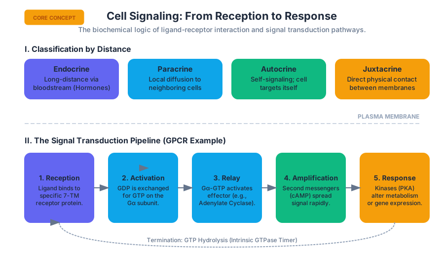
Cells communicate across different distances and through various media. The classification of signaling is based on the distance the ligand travels to reach its target.
| Signaling Type | Distance | Mechanism | Example |
|---|---|---|---|
| Endocrine | Long | Ligands (hormones) travel through the bloodstream. | Insulin, Adrenaline |
| Paracrine | Short | Ligands diffuse through the interstitial fluid to nearby cells. | Neurotransmitters, Growth Factors |
| Autocrine | Self | The cell secretes a ligand that binds to its own receptors. | T-cell proliferation signals |
| Juxtacrine | Contact | Signal is transmitted via direct physical contact between cell membranes. | Notch signaling, Gap junctions |
Key Insight: The specificity of a cellular response is determined not just by the ligand, but by the specific repertoire of receptors expressed by the target cell and the downstream machinery coupled to those receptors.
G-Protein Coupled Receptors (GPCR): The Molecular Switch
GPCRs represent the largest and most diverse group of membrane receptors in eukaryotes. They are characterized by a conserved structure of seven transmembrane (7-TM) alpha-helices. GPCRs act as Guanine Nucleotide Exchange Factors (GEFs) for heterotrimeric G-proteins.
The G-Protein Cycle
A heterotrimeric G-protein consists of three subunits: $\alpha$, $\beta$, and $\gamma$. In its inactive state, the $\alpha$-subunit is bound to GDP.
- Activation: Ligand binding induces a conformational change in the GPCR, allowing it to bind the G-protein.
- Exchange: The GPCR triggers the release of GDP from the $G\alpha$ subunit and the binding of GTP.
- Dissociation: The $G\alpha$-GTP complex dissociates from the $G\beta\gamma$ dimer. Both components can then modulate the activity of downstream effector proteins.
- Hydrolysis: The $G\alpha$ subunit has intrinsic GTPase activity, eventually hydrolyzing GTP back to GDP, which leads to the re-association of the heterotrimer and termination of the signal.
Second Messengers: Intracellular Relays
Once a receptor is activated, the signal must be propagated inside the cell. This is often achieved through second messengers—small, non-protein, water-soluble molecules or ions that spread rapidly throughout the cell by diffusion.
Cyclic AMP (cAMP) and the PKA Pathway
One of the most common effectors for GPCRs is Adenylate Cyclase, which converts ATP into cAMP. cAMP typically activates Protein Kinase A (PKA), which then phosphorylates various target proteins to alter cellular metabolism or gene expression.
Phospholipase C (PLC) and Calcium Signaling
Another major pathway involves the activation of Phospholipase C (PLC), which cleaves the membrane phospholipid $PIP_2$ into two distinct second messengers:
- Inositol trisphosphate ($IP_3$): Diffuses to the Endoplasmic Reticulum (ER) and opens ligand-gated $Ca^{2+}$ channels.
- Diacylglycerol (DAG): Remains in the membrane and, together with $Ca^{2+}$, activates Protein Kinase C (PKC).
| Second Messenger | Origin | Primary Target | Typical Effect |
|---|---|---|---|
| cAMP | ATP (via Adenylate Cyclase) | Protein Kinase A (PKA) | Glycogen breakdown, gene transcription |
| $IP_3$ | $PIP_2$ (via PLC) | $IP_3$-gated $Ca^{2+}$ channels | Release of $Ca^{2+}$ from ER |
| DAG | $PIP_2$ (via PLC) | Protein Kinase C (PKC) | Cell growth and differentiation |
| $Ca^{2+}$ | ER or extracellular space | Calmodulin, PKC | Muscle contraction, exocytosis |
Receptor Tyrosine Kinases (RTK) and Kinase Cascades
While GPCRs act as switches, Receptor Tyrosine Kinases (RTKs) function as high-fidelity integrators of growth and differentiation signals. Unlike GPCRs, RTKs have intrinsic enzymatic activity.
Mechanism of RTK Activation
- Ligand Binding: Two ligands bind to two adjacent RTK monomers.
- Dimerization: The binding causes the monomers to associate, forming a dimer.
- Trans-autophosphorylation: The kinase domain of one monomer phosphorylates the tyrosine residues on the cytoplasmic tail of the other monomer.
- Docking: These phosphorylated tyrosines serve as high-affinity binding sites for intracellular signaling proteins containing SH2 domains.
The MAPK Cascade: A Multi-Stage Amplifier
A classic downstream pathway for RTKs is the Mitogen-Activated Protein Kinase (MAPK) cascade. This involves a sequence of phosphorylation events:
Ras (G-protein) -> Raf (MAPKKK) -> MEK (MAPKK) -> ERK (MAPK).
Mathematical Derivation: Signal Amplification Factor
The power of a cascade lies in its ability to amplify a signal exponentially. If each enzyme in a 3-tier cascade activates $n$ molecules of the next tier, the total amplification $A$ is:
A = \prod_{i=1}^{k} n_i
Where:
- $k$ is the number of stages (tiers).
- $n_i$ is the catalytic turnover (amplification) at stage $i$.
Example Derivation: If 1 receptor activates 100 molecules of Enzyme A, and each Enzyme A activates 100 molecules of Enzyme B, and each Enzyme B produces 1000 molecules of product:
- Stage 1: $1 \times 10^2 = 100$
- Stage 2: $100 \times 10^2 = 10,000$
- Stage 3: $10,000 \times 10^3 = 10,000,000$ Total Amplification: $10^7$ (10 million-fold increase from a single binding event).
Signal Amplification and Integration
Signal amplification ensures that a minute concentration of an extracellular ligand can trigger a massive cellular response. This is best exemplified by the "Fight or Flight" response mediated by epinephrine.
Quantitative Breakdown of Epinephrine Signaling
The following table illustrates how a single molecule of epinephrine binding to a $\beta$-adrenergic receptor leads to the release of millions of glucose molecules.
| Stage | Molecule | Number of Molecules |
|---|---|---|
| 1 | Epinephrine (Ligand) | 1 |
| 2 | Activated GPCR | 1 |
| 3 | Activated $G\alpha_s$ | $10^2$ |
| 4 | Adenylate Cyclase (Effector) | $10^2$ |
| 5 | cAMP (Second Messenger) | $10^4$ |
| 6 | Protein Kinase A (PKA) | $10^4$ |
| 7 | Phosphorylase Kinase | $10^5$ |
| 8 | Glycogen Phosphorylase | $10^6$ |
| 9 | Glucose-1-Phosphate | $10^8$ |
Theorem of Signal Integration: Cells rarely respond to a single signal in isolation. Crosstalk between pathways (e.g., a GPCR pathway inhibiting an RTK pathway) allows the cell to perform complex logic, such as "Only divide if Growth Factor A is present AND Cell Stress B is absent."
Termination and Homeostasis
A signal that cannot be turned off is as dangerous as a signal that cannot be turned on (e.g., oncogenic Ras mutations in cancer). Cells employ several mechanisms to terminate signaling:
- Ligand Dissociation: As extracellular ligand concentration drops, receptors revert to inactive states.
- GTP Hydrolysis: $G\alpha$ subunits return to the GDP-bound state.
- Phosphodiesterases (PDE): Enzymes that break down cAMP into AMP, quenching the second messenger signal.
- Phosphatases: Enzymes that remove phosphate groups from proteins, reversing the action of kinases.
- Receptor Desensitization: Phosphorylation of the receptor itself can lead to the binding of arrestins, which block further G-protein activation and trigger endocytosis of the receptor.
Common Pitfalls and Misconceptions
- "Ligands enter the cell": A common mistake is thinking all ligands enter the cell to cause an effect. While steroid hormones (lipophilic) do cross the membrane, the vast majority of ligands (peptides, neurotransmitters) never enter the cytoplasm; they act strictly as "messengers" that knock on the door.
- "Kinases only activate": Phosphorylation is a covalent modification that changes protein conformation. While it often activates enzymes, it can also inhibit them (e.g., phosphorylation of Glycogen Synthase inhibits its activity).
- "One receptor, one effect": A single receptor type can trigger different effects in different tissues depending on the G-proteins and downstream effectors present in those specific cells (e.g., Acetylcholine slows the heart but stimulates skeletal muscle contraction).
Source Materials
Study Introductory Biology on Lykke
This course page is free to open: signed out, you can read its first two sections, and signing in, on any plan, opens the rest. The free Lykke Chrome extension syncs your own Canvas courses, so ChatGPT, Claude and Muse can answer from your classes and deadlines. It only reads Canvas.
Add the free extensionSign in to open every sectionAll sections of Introductory Biology · Browse more course pages