Anatomy and Physiology
Institution: MIT
1 study materials · 4 sections
OpenStax Anatomy and Physiology 2e is a comprehensive, open-access textbook designed for two-semester life science and allied health courses. The course is organized by body systems, moving from microscopic levels of organization to complex physiological interactions. It emphasizes inclusive instruction and utilizes interactive digital resources, such as surgical videos and histology, to enhance the learning experience for all students.
Course Sections
Foundations of Anatomy and Physiology
Key concepts: Homeostasis · Levels of Organization · Cell Biology · Histology
Covers the fundamental levels of organization in the human body, from chemical and cellular structures to the various types of tissues.
Foundations of Anatomy and Physiology
The study of the human body is traditionally bifurcated into two interdependent disciplines: Anatomy, the study of biological structure, and Physiology, the study of biological function. In the modern synthesis, these are viewed as inseparable; structure dictates function, and function, over evolutionary time, shapes structure. This "Form-Function Complementarity" serves as the bedrock for clinical medicine and biological engineering.
To understand a complex organism, we must apply a reductionist approach to its components while maintaining a systems-level view of their integration. This article explores the fundamental pillars of the human body: the hierarchical levels of organization, the mechanisms of homeostatic regulation, the functional unit of life (the cell), and the specialized architectures of tissues (histology).
Levels of Organization
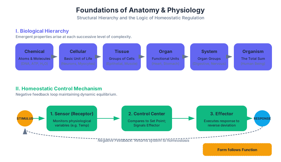
The human body is not a monolithic entity but a nested hierarchy of increasing complexity. Each level represents an emergent property—a phenomenon where the whole exhibits characteristics that its individual parts do not possess.
1. The Chemical Level
At the most fundamental level, atoms (such as Carbon, Hydrogen, Oxygen, and Nitrogen) bond to form molecules. These include simple inorganic molecules like $H_2O$ and complex organic macromolecules like DNA, proteins, and lipids.
2. The Cellular Level
The cell is the smallest independently functioning unit of a living organism. Even at this level, specialized functions emerge, such as contractility in muscle cells or signal transduction in neurons.
3. The Tissue Level
A tissue is a group of similar cells that work together to perform a specific function. For example, a layer of epithelial cells acts as a barrier, while a bundle of muscle fibers generates force.
4. The Organ Level
An organ is a structure composed of two or more tissue types that work together to perform a complex task. The stomach, for instance, utilizes epithelial tissue for secretion, smooth muscle for churning, and nervous tissue for coordination.
5. The Organ System Level
An organ system is a group of organs that cooperate to meet a major physiological need. The digestive system (stomach, intestines, liver, pancreas) processes nutrients, while the cardiovascular system distributes them.
6. The Organismal Level
The organism is the highest level of organization—a living human being representing the total sum of all structural levels working in concert to maintain life.
| Level | Definition | Representative Example | Emergent Property |
|---|---|---|---|
| Chemical | Atomic and molecular building blocks | Adenosine Triphosphate (ATP) | Energy storage/transfer |
| Cellular | The basic unit of life | Hepatocyte (Liver cell) | Metabolic detoxification |
| Tissue | Groups of similar cells | Myocardium (Cardiac muscle) | Rhythmic contraction |
| Organ | Multiple tissues working as a unit | Heart | Fluid propulsion (Pumping) |
| System | Groups of organs with shared goals | Cardiovascular System | Systemic perfusion |
| Organism | The integrated whole | Homo sapiens | Consciousness and reproduction |
Implementation: Modeling Biological Hierarchy
In computational biology, we often model these levels as a nested object-oriented structure to simulate systemic responses to localized stimuli.
/*
 * Low-level representation of biological hierarchy in C.
 * Demonstrates the composition of an Organ from Tissues and Cells.
 */

#include <stdio.h>
#include <stdlib.h>

typedef enum { EPITHELIAL, CONNECTIVE, MUSCLE, NERVOUS } TissueType;

typedef struct Cell {
    unsigned long id;
    double metabolic_rate;
    void (*function_ptr)(void);
} Cell;

typedef struct Tissue {
    TissueType type;
    Cell* cells;
    size_t cell_count;
} Tissue;

typedef struct Organ {
    char name[32];
    Tissue* components;
    size_t tissue_count;
    void (*organ_logic)(struct Organ*);
} Organ;

void pump_heart(Organ* self) {
    printf("Organ [%s] is executing systemic perfusion logic.\n", self->name);
}

int main() {
    Organ heart = {"Heart", NULL, 4, pump_heart};
    heart.organ_logic(&heart);
    return 0;
}
Homeostasis: The Logic of Life
Homeostasis is the state of steady internal, physical, and chemical conditions maintained by living systems. It is not a static state but a dynamic equilibrium, where continuous adjustments are made to keep variables within a narrow, life-sustaining range.
Definition: Homeostasis is the process by which biological systems maintain stability while adjusting to changing external conditions. It is governed by feedback loops that minimize the "error signal" between the actual state and the desired set point.
The Control Mechanism
Every homeostatic system consists of three fundamental components:
- Sensor (Receptor): Monitors a physiological value (e.g., blood glucose levels).
- Control Center (Integrator): Compares the value to the set point. If the value deviates significantly, it signals the effector.
- Effector: Causes a change to reverse the situation and return the value to the set point.
Negative vs. Positive Feedback
Most homeostatic mechanisms rely on negative feedback, which reverses a deviation from the set point. In contrast, positive feedback intensifies a change, moving the system further away from the normal state to achieve a specific endpoint (e.g., childbirth or blood clotting).
| Feature | Negative Feedback | Positive Feedback |
|---|---|---|
| Goal | Stability and Maintenance | Completion of a specific event |
| Response | Opposes the stimulus | Reinforces the stimulus |
| Frequency | Constant/Frequent | Rare/Episodic |
| Example | Thermoregulation, Blood Pressure | Oxytocin release in labor, Action potentials |
Mathematical Representation of Feedback
The efficiency of a homeostatic system can be described by its Gain. Gain is the ratio of the correction to the remaining error.
Gain = \frac{\text{Correction}}{\text{Residual Error}}
If a system has a high gain, it is highly effective at maintaining the set point. For instance, the baroreceptor system for blood pressure has a gain of approximately 2, meaning it corrects about 2/3 of a sudden pressure change.
Common Pitfalls: Homeostasis vs. Equilibrium
A common misconception is that homeostasis is synonymous with chemical equilibrium. In reality, a body at equilibrium is dead. Homeostasis requires a constant input of energy (ATP) to maintain gradients (e.g., the sodium-potassium pump). This is more accurately described as a steady state.
Cell Biology: The Functional Module
The cell is the "atom" of physiology. To understand how the body functions, one must understand the internal machinery of the cell and the properties of the plasma membrane.
The Plasma Membrane and Selective Permeability
The plasma membrane is an amphipathic lipid bilayer that acts as a gatekeeper. It maintains the Membrane Potential ($V_m$), an electrical gradient essential for the function of nerve and muscle cells.
Organelle Specialization
Organelles are membrane-bound compartments that allow incompatible biochemical reactions to occur simultaneously.
| Organelle | Industrial Analogue | Primary Physiological Function |
|---|---|---|
| Nucleus | Administrative Office | DNA storage, transcription, and coordination |
| Mitochondria | Power Plant | ATP production via aerobic respiration |
| Ribosomes | Assembly Line | Protein synthesis (translation) |
| Endoplasmic Reticulum | Manufacturing & Transport | Protein folding (Rough) and lipid synthesis (Smooth) |
| Golgi Apparatus | Shipping & Receiving | Post-translational modification and packaging |
| Lysosomes | Waste Management | Enzymatic breakdown of cellular debris |
Implementation: Calculating Membrane Potential
The Nernst Equation allows us to calculate the equilibrium potential for a specific ion, providing insight into how cells maintain electrical excitability.
import math

def calculate_nernst_potential(concentration_out, concentration_in, charge, temperature_c=37):
    """
    Calculates the Nernst equilibrium potential for a given ion.
    Formula: E = (RT / zF) * ln([ion]_out / [ion]_in)
    """
    # Constants
    R = 8.314  # Ideal gas constant (J/mol·K)
    F = 96485  # Faraday constant (C/mol)
    T = temperature_c + 273.15  # Convert C to Kelvin

    # RT/F constant at 37C is approx 26.7 mV
    # We multiply by 1000 to get the result in millivolts
    potential = ( (R * T) / (charge * F) ) * math.log(concentration_out / concentration_in)
    return potential * 1000

# Example for Potassium (K+)
# Typical values: [K+]out = 5mM, [K+]in = 150mM
k_potential = calculate_nernst_potential(5, 150, 1)
print(f"Equilibrium potential for K+: {k_potential:.2f} mV")
Histology: The Study of Tissues
Histology bridges the gap between cell biology and organ physiology. There are four primary tissue types, each defined by its cellular morphology and the composition of its Extracellular Matrix (ECM).
1. Epithelial Tissue
Epithelial tissues are sheets of cells that cover exterior surfaces, line internal cavities, and form glands. They are characterized by high cellularity, polarity (apical vs. basal surfaces), and avascularity.
- Simple Squamous: Optimized for diffusion (e.g., lung alveoli).
- Stratified Squamous: Optimized for protection (e.g., skin).
- Columnar: Optimized for absorption and secretion (e.g., intestine).
2. Connective Tissue
Connective tissue is the most abundant and diverse tissue type. Unlike epithelium, it is defined more by its ECM than by the cells themselves. The ECM consists of ground substance and fibers (collagen, elastic, reticular).
3. Muscle Tissue
Muscle tissue is specialized for contraction and thermogenesis.
- Skeletal: Voluntary, striated, multinucleated.
- Cardiac: Involuntary, striated, intercalated discs.
- Smooth: Involuntary, non-striated, found in hollow organs.
4. Nervous Tissue
Nervous tissue is specialized for the rapid communication of information via electrochemical impulses. It consists of neurons (signal transmitters) and neuroglia (support cells).
| Tissue Category | Cell Density | Extracellular Matrix | Primary Function |
|---|---|---|---|
| Epithelial | Very High | Minimal (Basal Lamina) | Barrier, Secretion, Absorption |
| Connective | Low to Moderate | Abundant (Fibers + Ground Sub.) | Support, Binding, Transport |
| Muscle | High | Moderate | Movement, Heat Production |
| Nervous | Moderate | Minimal | Communication, Control |
Data Management in Histology
In a clinical pathology setting, histology data is often managed via structured databases to track staining protocols (e.g., H&E, PAS) and sample origins.
-- Schema for a Histology Sample Tracking System
CREATE TABLE TissueSamples (
    sample_id INT PRIMARY KEY AUTO_INCREMENT,
    patient_id INT NOT NULL,
    tissue_type ENUM('Epithelial', 'Connective', 'Muscle', 'Nervous'),
    collection_site VARCHAR(100),
    fixative_used VARCHAR(50) DEFAULT '10% Neutral Buffered Formalin',
    stain_protocol VARCHAR(50),
    pathologist_notes TEXT,
    collection_timestamp TIMESTAMP DEFAULT CURRENT_TIMESTAMP
);

-- Query to find all cardiac muscle samples stained with H&E
SELECT sample_id, collection_site, collection_timestamp
FROM TissueSamples
WHERE tissue_type = 'Muscle'
AND stain_protocol = 'Hematoxylin and Eosin'
AND collection_site LIKE '%Heart%';
Synthesis: The Integrated Organism
The foundations of anatomy and physiology reveal a system of profound elegance. The Levels of Organization provide the structural blueprint; Cell Biology provides the functional machinery; Histology provides the specialized materials; and Homeostasis provides the control logic that keeps the entire system operational.
When these foundations are understood, pathology becomes clearer. A "disease" is rarely a random event; it is typically a failure of a specific level of organization or a breakdown in a homeostatic feedback loop. For example:
- Diabetes Mellitus: A failure of the homeostatic loop regulating blood glucose (Sensor/Effector failure).
- Cancer: A failure of cellular-level regulation of the cell cycle.
- Scurvy: A chemical-level deficiency leading to defective connective tissue (ECM) synthesis.
By mastering these foundations, the student of human biology moves from memorizing facts to understanding the engineering principles of life.
Support, Movement, and Control
Key concepts: Integumentary System · Skeletal System · Muscular System · Nervous System · Endocrine System
Explores the systems responsible for the body's structure, movement, and internal communication, including the skeletal, muscular, nervous, and endocrine systems.
Support, Movement, and Control: The Biomechanical and Cybernetic Framework
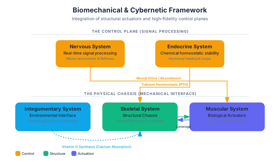
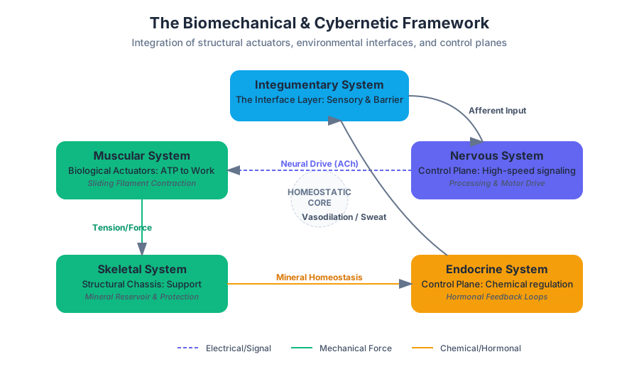
In the architecture of the human body, the systems governing support, movement, and control represent a sophisticated integration of structural engineering and high-fidelity signal processing. While the Integumentary, Skeletal, and Muscular systems provide the physical chassis and actuators required for interaction with the physical world, the Nervous and Endocrine systems function as the control plane, managing real-time responses and long-term homeostatic stability.
This article decomposes these systems into their functional components, examining the mechanics of movement, the chemistry of signaling, and the feedback loops that maintain biological integrity.
### The Integumentary System: The Interface Layer
The Integumentary System is the body's primary interface with the external environment. Far from being a passive container, it is a dynamic organ system comprising the skin, hair, nails, and exocrine glands. It serves as the first line of defense (innate immunity), a sensory receptor array, and a critical thermoregulatory heat exchanger.
1. What it is
The skin is organized into two primary layers: the Epidermis (the epithelial outer layer) and the Dermis (the connective tissue layer), resting upon the Hypodermis (subcutaneous fascia).
The Acid Mantle: A very thin, slightly acidic film on the surface of human skin acting as a barrier to bacteria, viruses, and other potential contaminants. It is formed by the mix of sebum (oil) and sweat.
2. Why it matters
Without the integumentary system, the body would succumb to rapid dehydration (desiccation), uncontrolled infection, and thermal failure. It also facilitates the synthesis of Vitamin D, which is essential for calcium absorption in the skeletal system—showing the first major link between these "separate" systems.
3. How it works: Thermoregulation Pipeline
When core temperature rises, the hypothalamus triggers two primary mechanisms:
- Vasodilation: Dermal blood vessels dilate to increase heat loss via radiation.
- Sudoriferous Activation: Eccrine sweat glands secrete water and electrolytes; evaporation provides latent heat cooling.
| Layer | Primary Tissue Type | Key Cell Types | Functional Role |
|---|---|---|---|
| Epidermis | Stratified Squamous Epithelium | Keratinocytes, Melanocytes | Waterproofing, UV protection, Barrier |
| Dermis | Dense Irregular Connective | Fibroblasts, Macrophages | Structural integrity, Sensation, Blood supply |
| Hypodermis | Adipose / Areolar | Adipocytes | Insulation, Energy storage, Shock absorption |
### The Skeletal System: The Structural Chassis
The Skeletal System provides the rigid framework required for locomotion and the protection of soft-tissue "payloads" (organs). It is a metabolically active system that undergoes constant remodeling in response to mechanical stress.
1. What it is
The system is divided into the Axial Skeleton (central axis: skull, vertebral column, thoracic cage) and the Appendicular Skeleton (limbs and girdles).
2. How it works: Bone Remodeling (Wolff’s Law)
Bones are not static. They operate under Wolff’s Law, which states that bone grows or remodels in response to the forces or demands placed upon it. This is managed by two competing cell populations:
- Osteoblasts: Bone-forming cells (the "builders").
- Osteoclasts: Bone-resorbing cells (the "demolition crew").
3. Concrete Example: Calcium Homeostasis
The skeletal system acts as a mineral reservoir. If blood calcium levels drop, the Parathyroid Hormone (PTH) stimulates osteoclasts to break down bone matrix, releasing $Ca^{2+}$ into the bloodstream.
4. Common Pitfalls
A common misconception is that bone is "dead" material like wood or stone. In reality, bone is highly vascularized. If you break a bone, the "healing" is a rapid cellular response involving hematoma formation and fibrocartilaginous callus production—processes impossible in non-living structures.
### The Muscular System: Biological Actuators
The Muscular System converts chemical energy (ATP) into mechanical work. It is the only tissue in the body capable of active contraction.
1. What it is
Muscles are categorized by their control (voluntary vs. involuntary) and their cellular morphology (striated vs. smooth).
| Muscle Type | Control | Location | Histology |
|---|---|---|---|
| Skeletal | Voluntary | Attached to bones | Striated, Multinucleated |
| Cardiac | Involuntary | Heart wall | Striated, Intercalated discs |
| Smooth | Involuntary | Walls of hollow organs | Non-striated, Spindle-shaped |
2. How it works: The Sliding Filament Theory
Contraction occurs at the Sarcomere level. When a motor neuron releases Acetylcholine (ACh), it triggers an action potential in the muscle fiber, releasing $Ca^{2+}$ from the sarcoplasmic reticulum.
3. Code Implementation: Simulating Muscle Fiber Recruitment
In a "senior engineer" view, muscle recruitment follows the Henneman's Size Principle: smaller motor units are recruited first to allow for fine motor control, with larger units added as force requirements increase.
/*
 * Low-level simulation of Motor Unit Recruitment
 * Language: C (Systems-level logic)
 */

#include <stdio.h>

typedef struct {
    int id;
    float force_output; // Newtons
    int threshold;      // Activation threshold (0-100)
    int is_active;
} MotorUnit;

void update_recruitment(MotorUnit units[], int count, int neural_drive) {
    for (int i = 0; i < count; i++) {
        if (neural_drive >= units[i].threshold) {
            units[i].is_active = 1;
        } else {
            units[i].is_active = 0;
        }
    }
}

int main() {
    MotorUnit biceps_brachii[3] = {
        {1, 5.0, 10, 0},  // Small, slow-twitch
        {2, 20.0, 40, 0}, // Medium
        {3, 50.0, 80, 0}  // Large, fast-twitch
    };

    int input_signal = 45; // Moderate effort
    update_recruitment(biceps_brachii, 3, input_signal);

    printf("Active Units at Drive %d:\n", input_signal);
    for(int i=0; i<3; i++) {
        if(biceps_brachii[i].is_active)
            printf("Unit %d producing %.1fN\n", biceps_brachii[i].id, biceps_brachii[i].force_output);
    }
    return 0;
}
### The Nervous System: The High-Speed Data Bus
The Nervous System is the body's rapid-response control center. It processes sensory input, integrates information, and initiates motor output via electrochemical signals.
1. The Protocol: The Action Potential
The fundamental unit of communication is the Action Potential. This is a binary "all-or-none" event where the membrane potential of a neuron rapidly rises and falls.
2. Mathematical Representation
The equilibrium potential for a specific ion is calculated using the Nernst Equation:
E_{ion} = \frac{RT}{zF} \ln \frac{[ion]_{outside}}{[ion]_{inside}}
Where:
- $R$ is the universal gas constant.
- $T$ is absolute temperature.
- $z$ is the valence of the ion.
- $F$ is Faraday's constant.
3. Pseudocode: Synaptic Transmission Logic
FUNCTION Synaptic_Transfer(PreSynaptic_Signal):
    IF PreSynaptic_Signal.Voltage > Threshold THEN
        OPEN Voltage_Gated_Ca_Channels()
        RELEASE Neurotransmitter(Vesicles)
        DIFFUSE Neurotransmitter across Synaptic_Cleft
        BIND Neurotransmitter to PostSynaptic_Receptors

        IF Receptor_Type == EXCITATORY:
            DEPOLARIZE PostSynaptic_Membrane
        ELSE IF Receptor_Type == INHIBITORY:
            HYPERPOLARIZE PostSynaptic_Membrane
    END IF
END FUNCTION
### The Endocrine System: The Global Broadcast System
If the Nervous System is a fiber-optic network (point-to-point, high speed), the Endocrine System is a wireless broadcast system (global signal, slower latency, high persistence). It uses Hormones secreted into the blood to reach distant target cells.
1. Mechanisms of Action
Hormones act only on cells with specific receptors. They are categorized by chemical structure:
- Amino Acid Derivatives: (e.g., Epinephrine) - Fast acting, membrane-bound receptors.
- Lipid-Derived (Steroids): (e.g., Testosterone, Cortisol) - Slow acting, cross the cell membrane to alter gene expression directly.
2. Feedback Loops: The PID Controller of Biology
Most endocrine functions are regulated by Negative Feedback Loops. For example, the regulation of blood glucose by Insulin and Glucagon acts like a thermostat.
| Parameter | High Blood Glucose | Low Blood Glucose |
|---|---|---|
| Sensor | Pancreatic Beta Cells | Pancreatic Alpha Cells |
| Hormone Released | Insulin | Glucagon |
| Target Action | Glucose uptake by cells, Glycogenesis | Glycogenolysis, Gluconeogenesis |
| Result | Glucose levels decrease | Glucose levels increase |
3. Real-World Usage: Data Analysis of Endocrine Response
Using Python to visualize the "Dose-Response" curve of a hormone, which typically follows a sigmoidal (Log-Logistic) pattern.
import numpy as np
import matplotlib.pyplot as plt

# Hill Equation for Hormone Binding
def dose_response(concentration, max_effect, ec50, hill_coefficient):
    return max_effect / (1 + (ec50 / concentration)**hill_coefficient)

concentrations = np.logspace(-10, -6, 100) # Molar concentration
response = dose_response(concentrations, 100, 1e-8, 1.2)

plt.semilogx(concentrations, response)
plt.title("Hormone Receptor Saturation Curve")
plt.xlabel("Hormone Concentration (M)")
plt.ylabel("Biological Response (%)")
plt.grid(True)
plt.show()
### Synthesis: The Integrated Control Loop
The true complexity of "Support, Movement, and Control" is found in the intersection of these systems.
Case Study: The "Fight or Flight" Response
- Nervous System: Amygdala perceives a threat and signals the Hypothalamus.
- Endocrine System: The Adrenal Medulla releases Epinephrine (Adrenaline) into the blood.
- Muscular System: Increased blood flow and glucose availability prepare muscles for explosive contraction.
- Integumentary System: Sudoriferous glands activate (cold sweat) to prepare for the heat of exertion.
- Skeletal System: Acts as the lever system for the muscles to execute the escape.
Comparison of Control Systems
| Feature | Nervous System | Endocrine System |
|---|---|---|
| Signal Type | Electrical (Impulses) & Chemical (NTs) | Chemical (Hormones) |
| Transmission Path | Neurons / Synapses | Bloodstream |
| Speed of Onset | Milliseconds | Seconds to Days |
| Duration of Effect | Very Short (Transient) | Long-lasting |
| Specificity | High (Targeted to specific cells) | Broad (Any cell with receptors) |
### Common Pitfalls in Systemic Integration
- The "Isolation" Fallacy: Students often study the Skeletal system as a dry collection of bones. However, bones are the primary site of Hematopoiesis (blood cell production). A failure in the skeletal system (e.g., leukemia in bone marrow) immediately compromises the immune and respiratory systems.
- Confusing "Control" with "Consciousness": Most "Control" is autonomic. The Nervous system manages heart rate, digestion, and pupil dilation without any CPU cycles from the conscious mind (Prefrontal Cortex).
- Hormonal Latency: Engineers often forget that endocrine signals have a "washout" period. Unlike a light switch (Nervous), an endocrine signal is like a dye in a river; even after the source stops, the signal persists until cleared by the liver or kidneys.
End of Article
Fluids, Transport, and Maintenance
Key concepts: Cardiovascular System · Lymphatic and Immune Systems · Respiratory System · Digestive System · Urinary System
Examines the systems that transport nutrients and oxygen while removing waste, including the cardiovascular, respiratory, digestive, and urinary systems.
Fluids, Transport, and Maintenance
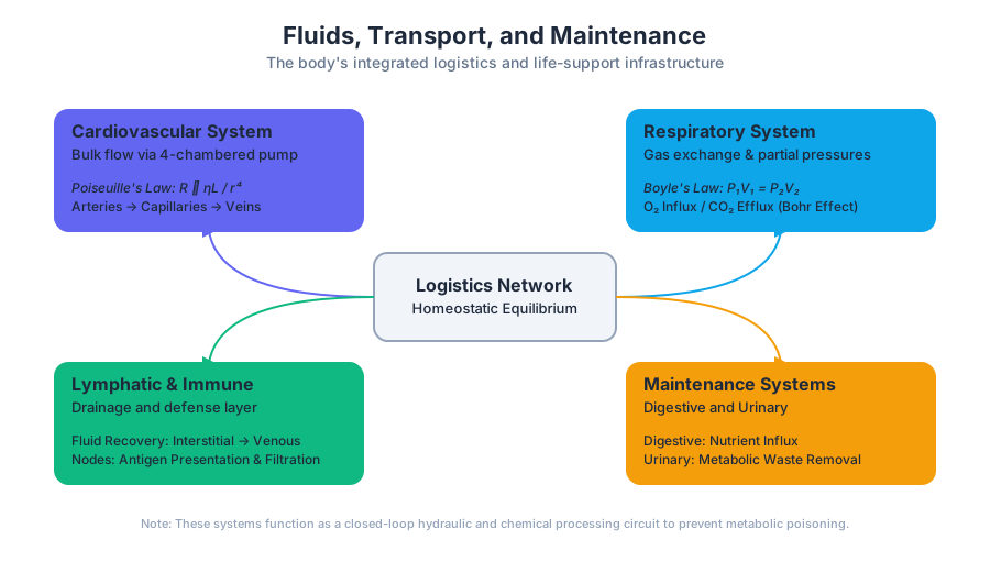
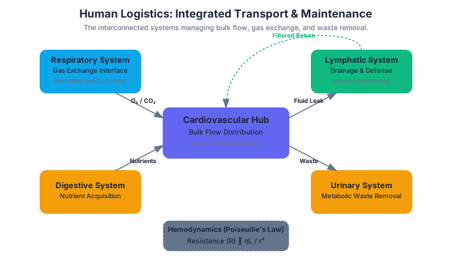
The human body is a complex biological machine that operates far from thermodynamic equilibrium. To maintain this state, it requires a constant influx of energy and nutrients, a robust transport mechanism to distribute these resources, and a sophisticated waste-management infrastructure to prevent metabolic poisoning. This section explores the Fluids, Transport, and Maintenance systems—comprising the Cardiovascular, Lymphatic, Respiratory, Digestive, and Urinary systems—which collectively function as the body's logistics and life-support network.
The Cardiovascular System: The Central Transport Hub
The Cardiovascular System is a closed-loop hydraulic circuit powered by a four-chambered muscular pump: the heart. Its primary objective is the bulk flow of blood, a specialized fluid connective tissue, to ensure that every cell remains within a few micrometers of a capillary for efficient diffusion.
Hemodynamics and Vessel Architecture
The physics of blood flow is governed by Poiseuille's Law, which states that resistance ($R$) is proportional to the viscosity of the blood ($\eta$) and the length of the vessel ($L$), but inversely proportional to the fourth power of the radius ($r^4$). This means that small changes in vessel diameter (vasoconstriction or vasodilation) have a massive impact on blood pressure and flow distribution.
| Vessel Type | Primary Function | Structural Feature | Pressure Level |
|---|---|---|---|
| Arteries | Pressure Reservoirs | Thick Tunica Media (Elastic/Muscular) | High |
| Arterioles | Resistance Vessels | High Smooth Muscle content | Variable (Control) |
| Capillaries | Exchange Vessels | Single layer of Endothelium (Tunica Intima) | Low |
| Venules | Collection | Thin walls, porous | Very Low |
| Veins | Volume Reservoirs | Presence of Valves; High Compliance | Lowest |
The Cardiac Cycle
The heart operates via a rhythmic alternation between Systole (contraction/ejection) and Diastole (relaxation/filling). This cycle is coordinated by an intrinsic conduction system, starting at the Sinoatrial (SA) Node, often called the natural pacemaker.
Key Insight: The Frank-Starling Law of the Heart The heart possesses an intrinsic ability to adapt to changing volumes of inflowing blood. Specifically, the force of cardiac contraction is directly proportional to the initial length of the muscle fiber (Preload). This ensures that the output of the left and right ventricles remains balanced over time.
/*
 * Low-level simulation of Poiseuille's Law for Hemodynamics
 * Calculating Resistance (R) and Flow Rate (Q) in a vessel segment.
 */

#include <stdio.h>
#include <math.h>

#define PI 3.14159265358979323846

typedef struct {
    double radius;    // meters
    double length;    // meters
    double viscosity; // Pascal-seconds (Pa·s)
} BloodVessel;

double calculate_resistance(BloodVessel v) {
    // R = (8 * eta * L) / (pi * r^4)
    return (8.0 * v.viscosity * v.length) / (PI * pow(v.radius, 4));
}

double calculate_flow_rate(double pressure_drop, double resistance) {
    // Q = DeltaP / R
    return pressure_drop / resistance;
}

int main() {
    BloodVessel arteriole = {0.000015, 0.002, 0.0035}; // 15um radius, 2mm length
    double delta_p = 4000.0; // 4000 Pa pressure drop

    double R = calculate_resistance(arteriole);
    double Q = calculate_flow_rate(delta_p, R);

    printf("Vessel Resistance: %.2e Pa*s/m^3\n", R);
    printf("Flow Rate (Q): %.2e m^3/s\n", Q);

    return 0;
}
Lymphatic and Immune Systems: The Drainage and Defense Layer
While the cardiovascular system is a closed loop, it is "leaky." At the capillary level, Hydrostatic Pressure forces fluid out into the interstitial space. Not all of this fluid returns via Osmotic Pressure.
Fluid Recovery and Filtration
The Lymphatic System acts as a secondary circulatory route. It picks up excess interstitial fluid (now called lymph), filters it through lymph nodes, and returns it to the venous circulation via the subclavian veins.
- Lymphatic Capillaries: Highly permeable, "blind-ended" vessels.
- Lymph Nodes: Biological "checkpoints" packed with lymphocytes (B and T cells) that scan for pathogens.
- Spleen: The largest lymphatic organ, responsible for filtering blood and recycling aged red blood cells.
The Immune Response
The immune system is categorized into Innate Immunity (non-specific, immediate) and Adaptive Immunity (specific, memory-based). The lymphatic system provides the physical infrastructure where these two systems interact, particularly during Antigen Presentation.
The Respiratory System: The Gas Exchange Interface
The respiratory system's primary function is to maintain the partial pressure gradients of Oxygen ($O_2$) and Carbon Dioxide ($CO_2$) between the atmosphere and the blood.
Mechanics of Ventilation
Breathing is driven by pressure changes in the thoracic cavity, governed by Boyle's Law ($P_1V_1 = P_2V_2$). When the diaphragm contracts, thoracic volume increases, pressure drops below atmospheric pressure, and air rushes in.
Gas Transport and the Bohr Effect
Oxygen is transported primarily bound to Hemoglobin in red blood cells. The affinity of hemoglobin for $O_2$ is not static; it changes based on the local environment—a phenomenon known as the Bohr Effect.
\text{pH and Hemoglobin Affinity (Simplified)} \\
\uparrow [H^+] \text{ (lower pH)} \rightarrow \text{Right shift in dissociation curve} \rightarrow \downarrow \text{Affinity for } O_2 \\
\text{This ensures } O_2 \text{ is dropped off at metabolically active (acidic) tissues.}
| Parameter | Definition | Typical Value (Adult Male) |
|---|---|---|
| Tidal Volume (TV) | Volume of air moved in/out during normal breath | 500 mL |
| Inspiratory Reserve (IRV) | Extra air that can be forcibly inhaled | 3000 mL |
| Residual Volume (RV) | Air remaining in lungs after maximal exhalation | 1200 mL |
| Vital Capacity (VC) | Total exchangeable air (TV + IRV + ERV) | 4800 mL |
The Digestive System: The Nutrient Extraction Engine
The digestive system is a continuous tube (the Alimentary Canal) that processes raw materials into chemical energy and structural building blocks.
The Processing Pipeline
- Ingestion and Mechanical Digestion: Mastication (chewing) in the mouth and churning in the stomach.
- Chemical Digestion: The enzymatic breakdown of macromolecules.
- Absorption: The movement of nutrients from the lumen of the small intestine into the blood or lymph.
- Defecation: The elimination of indigestible substances.
Enzymatic Breakdown
Different regions of the GI tract specialize in different macromolecules:
| Enzyme | Source | Substrate | Optimal pH |
|---|---|---|---|
| Salivary Amylase | Salivary Glands | Polysaccharides | 6.7 - 7.0 |
| Pepsin | Stomach (Chief Cells) | Proteins | 1.5 - 2.0 |
| Pancreatic Lipase | Pancreas | Triglycerides | 7.0 - 8.0 |
| Trypsin | Pancreas | Proteins/Peptides | 7.0 - 8.0 |
Key Concept: The Hepatic Portal System All blood draining from the digestive organs (except the lower rectum) is diverted to the Liver via the hepatic portal vein. This allows the liver to process nutrients, detoxify metabolic byproducts, and store glycogen before the blood reaches the rest of the body.
The Urinary System: The Homeostatic Filter
The Urinary System is the body's master chemist. While often thought of as just "making urine," its primary role is the precise regulation of blood volume, blood pressure, and blood pH.
The Nephron: The Functional Unit
Each kidney contains approximately one million nephrons. The process of urine formation occurs in three distinct stages:
- Glomerular Filtration: Non-selective "bulk" filtration of water and small solutes out of the blood.
- Tubular Reabsorption: Reclaiming useful substances (glucose, ions, water) back into the blood.
- Tubular Secretion: Active transport of wastes (H+, K+, drugs) from the blood into the filtrate.
The RAAS Pathway
The Renin-Angiotensin-Aldosterone System (RAAS) is the primary hormonal mechanism for long-term blood pressure regulation.
# Pseudocode logic for RAAS Activation
def check_blood_pressure(bp_sensor):
    if bp_sensor.detected_drop():
        kidney.release_renin()

def blood_chemistry_cascade():
    # Renin converts Angiotensinogen to Angiotensin I
    # ACE (Angiotensin Converting Enzyme) converts I to II
    angiotensin_II = ACE_conversion(angiotensin_I)

    if angiotensin_II.is_present():
        vasoconstrict_systemic_arteries()
        adrenal_cortex.release_aldosterone()
        pituitary.release_ADH()

    # Result: Increased Na+ reabsorption, increased water retention,
    # increased peripheral resistance -> BP rises.
System Integration: The Maintenance Loop
These systems do not operate in isolation. They are linked by feedback loops that maintain Homeostasis.
Case Study: pH Buffering
When you exercise, your muscles produce $CO_2$ and Lactic Acid.
- Respiratory Response: The brainstem detects rising $CO_2$ (via pH drop) and increases ventilation rate to "blow off" $CO_2$.
- Urinary Response: The kidneys secrete $H^+$ ions into the urine and reabsorb Bicarbonate ($HCO_3^-$) to buffer the blood.
- Cardiovascular Response: Heart rate increases to transport $CO_2$ to the lungs and $O_2$ to the muscles faster.
Common Pitfalls in Understanding
- Arteries vs. Veins: A common mistake is defining arteries as "carrying oxygenated blood." While usually true, the Pulmonary Artery carries deoxygenated blood to the lungs. The correct definition is based on direction: Arteries go Away from the heart; Veins return to the heart.
- Filtration vs. Secretion: In the kidney, filtration is a passive process driven by blood pressure in the glomerulus. Secretion is an active, selective process occurring later in the tubule.
- Lymph Flow: Unlike the cardiovascular system, the lymphatic system has no central pump. Lymph moves via the "skeletal muscle pump" and respiratory pressure changes.
Summary of Maintenance Functions
| System | Primary Maintenance Role | Key Regulatory Variable |
|---|---|---|
| Cardiovascular | Convective Transport | Mean Arterial Pressure (MAP) |
| Lymphatic | Fluid Balance / Defense | Interstitial Fluid Volume |
| Respiratory | Gas Exchange | Partial Pressure of $CO_2$ ($PCO_2$) |
| Digestive | Nutrient Acquisition | Blood Glucose / Amino Acid levels |
| Urinary | Waste Removal / Osmolarity | Plasma Osmolarity / pH |
This integrated network ensures that despite massive fluctuations in external environment and internal activity, the "internal sea" (the extracellular fluid) remains stable enough for life to persist.
Reproduction and Human Development
Key concepts: Reproductive Systems · Embryology · Genetics · Human Development
Covers the biological processes of the reproductive systems, human development from conception to birth, and the principles of inheritance.
Reproduction and Human Development
The biological imperative of any species is the successful transmission of genetic material to the next generation. In humans, this process is an intricate orchestration of anatomical specialization, hormonal signaling, and molecular precision. This article explores the mechanisms of human reproduction, the trajectory of embryological development, and the foundational principles of genetics that govern inheritance.
Reproductive Systems: The Architecture of Continuity
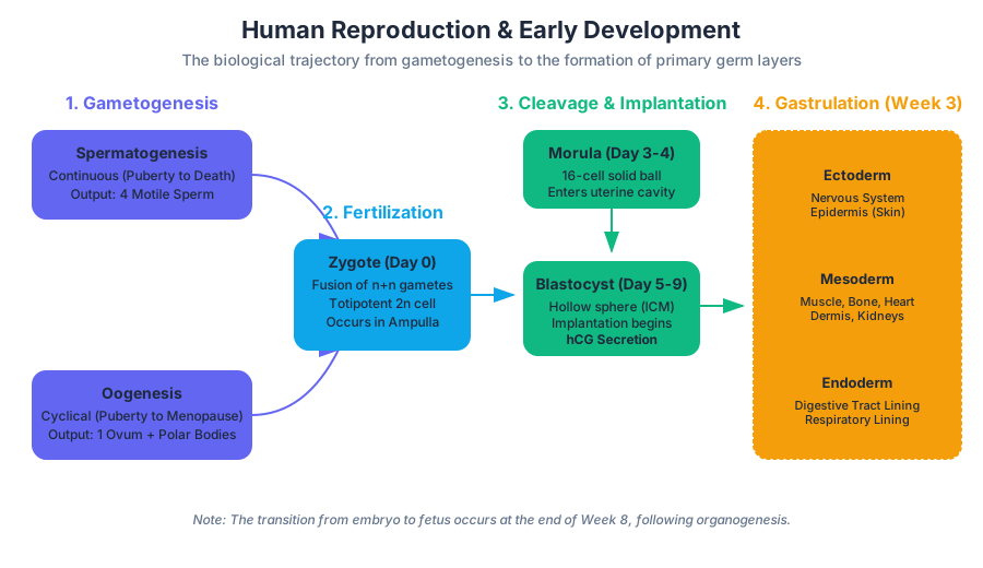
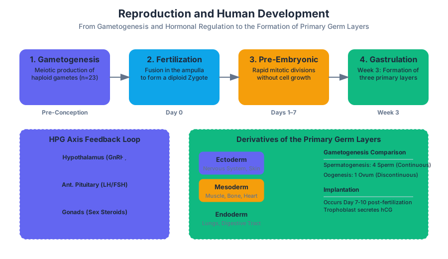
Human reproduction relies on anisogamy, the fusion of two morphologically distinct gametes: the small, motile spermatozoon and the large, nutrient-rich ovum. The systems responsible for producing these gametes are regulated by the Hypothalamic-Pituitary-Gonadal (HPG) axis, a complex feedback loop involving Gonadotropin-Releasing Hormone (GnRH), Luteinizing Hormone (LH), and Follicle-Stimulating Hormone (FSH).
Comparative Physiology of Gametogenesis
While both sexes utilize meiosis to produce haploid cells ($n=23$), the timing and output of these processes differ significantly.
| Feature | Spermatogenesis | Oogenesis |
|---|---|---|
| Location | Seminiferous tubules (Testes) | Ovaries |
| Timing | Puberty until death | Begins in utero; arrested until puberty; ends at menopause |
| Meiotic Continuity | Continuous process | Discontinuous (arrested in Prophase I and Metaphase II) |
| Output | 4 functional sperm cells | 1 functional ovum + 2-3 polar bodies |
| Cycle Duration | ~64–72 days | ~28 days (monthly cycle) |
The HPG Axis Feedback Loop
The regulation of reproductive function is a classic example of a homeostatic control system. In males, testosterone provides negative feedback to the hypothalamus and anterior pituitary. In females, the feedback is more complex, switching from negative to positive feedback to trigger the LH surge required for ovulation.
/*
 * HPG_Axis_Simulation.c
 * A low-level simulation of hormonal feedback loops in the male reproductive system.
 * This demonstrates the negative feedback mechanism of testosterone on GnRH/LH.
 */

#include <stdio.h>

typedef struct {
    double gnrh;        // Hypothalamus
    double lh_fsh;      // Pituitary
    double testosterone; // Gonads
} HormonalState;

void update_hpg_axis(HormonalState *state, double metabolic_clearance_rate) {
    // GnRH secretion is inhibited by high testosterone (Negative Feedback)
    double gnrh_inhibition = state->testosterone * 0.5;
    state->gnrh = (10.0 - gnrh_inhibition) > 0 ? (10.0 - gnrh_inhibition) : 0.1;

    // LH/FSH responds to GnRH levels
    state->lh_fsh = state->gnrh * 1.2;

    // Testosterone production stimulated by LH
    state->testosterone += (state->lh_fsh * 0.8) - (state->testosterone * metabolic_clearance_rate);
}

int main() {
    HormonalState body = {5.0, 6.0, 20.0};
    printf("Cycle | GnRH | LH/FSH | Testosterone\n");
    for (int i = 0; i < 10; i++) {
        update_hpg_axis(&body, 0.15);
        printf("%5d | %4.2f | %6.2f | %12.2f\n", i, body.gnrh, body.lh_fsh, body.testosterone);
    }
    return 0;
}
Embryology: From Zygote to Neonate
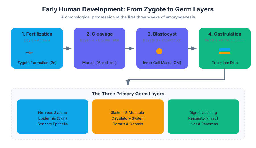
Embryology is the study of the first eight weeks of development following fertilization. This period is characterized by rapid cellular proliferation, migration, and differentiation.
Fertilization and the Pre-Embryonic Stage
Fertilization typically occurs in the ampulla of the uterine tube. It is not a single event but a sequence of biochemical hurdles:
- Capacitation: Sperm undergo membrane changes in the female tract to become fertile.
- Acrosomal Reaction: Enzymes are released to penetrate the zona pellucida.
- Cortical Reaction: Once a sperm penetrates, the oocyte releases granules to prevent polyspermy (fertilization by multiple sperm).
Definition: The Zygote The diploid cell ($2n=46$) resulting from the fusion of a haploid spermatozoon and a haploid secondary oocyte. It represents the first stage of a unique genetic individual.
Cleavage and Implantation
Following fertilization, the zygote undergoes cleavage, a series of rapid mitotic divisions without intervening growth. This leads to the formation of a morula (16-cell solid ball) and eventually a blastocyst.
| Stage | Time Post-Fertilization | Key Characteristics |
|---|---|---|
| Zygote | Day 0 | Single diploid cell; totipotent. |
| Morula | Day 3-4 | Solid ball of cells; enters the uterine cavity. |
| Blastocyst | Day 5-7 | Hollow sphere; contains Inner Cell Mass (ICM) and Trophoblast. |
| Implantation | Day 7-10 | Trophoblast invades the endometrium; hCG production begins. |
Gastrulation and Germ Layer Formation
During the third week, the process of gastrulation transforms the two-layered embryonic disc into a three-layered structure. These three germ layers are the precursors to all adult tissues:
- Ectoderm: Becomes the nervous system and epidermis.
- Mesoderm: Becomes muscle, bone, circulatory system, and dermis.
- Endoderm: Becomes the epithelial lining of the digestive and respiratory tracts.
Fetal Development and Placentation
The transition from embryo to fetus occurs at the end of week 8. The fetal period (weeks 9–40) is primarily focused on growth and functional maturation of organ systems.
The Placenta: The Life-Support Interface
The placenta is a unique fetomaternal organ. It performs respiratory, nutritive, excretory, and endocrine functions without ever allowing the direct mixing of maternal and fetal blood.
- Maternal Side: Decidua basalis (modified endometrium).
- Fetal Side: Chorionic villi containing fetal capillaries.
- Diffusion Barrier: Oxygen, glucose, and antibodies (IgG) cross into fetal circulation; $CO_2$ and urea cross into maternal circulation.
Trimester Milestones
| Trimester | Duration | Primary Focus | Critical Events |
|---|---|---|---|
| First | Weeks 1–12 | Organogenesis | Heart begins beating (Wk 4); most vulnerable to teratogens. |
| Second | Weeks 13–28 | Growth & Detail | Fetal movement (quickening); vernix caseosa and lanugo form. |
| Third | Weeks 29–Birth | Weight Gain & Prep | Surfactant production in lungs; rapid brain development. |
Genetics and Human Inheritance
Genetics is the study of how traits are transmitted via DNA. The human genome consists of approximately 20,000–25,000 genes distributed across 23 pairs of chromosomes.
Mendelian vs. Non-Mendelian Inheritance
While Gregor Mendel’s laws of segregation and independent assortment provide the foundation, human genetics often involves more complex patterns.
- Autosomal Dominant/Recessive: Traits on non-sex chromosomes (e.g., Cystic Fibrosis is recessive).
- Incomplete Dominance: The phenotype is an intermediate (e.g., Familial Hypercholesterolemia).
- Codominance: Both alleles are expressed equally (e.g., ABO blood groups).
- Polygenic Inheritance: Traits determined by multiple genes (e.g., skin color, height).
Logic of the Punnett Square
To predict the probability of offspring genotypes, we use a matrix approach. For a dihybrid cross (two traits), the complexity increases to a $4 \times 4$ grid.
import numpy as np

def generate_punnett_square(parent1_alleles, parent2_alleles):
    """
    Generates a Punnett square for a single-gene trait.
    Example: parent1 = 'Aa', parent2 = 'Aa'
    """
    p1 = list(parent1_alleles)
    p2 = list(parent2_alleles)

    square = []
    for a1 in p1:
        row = []
        for a2 in p2:
            # Sort to ensure 'Aa' instead of 'aA'
            genotype = "".join(sorted([a1, a2]))
            row.append(genotype)
        square.append(row)

    return np.array(square)

# Example: Heterozygous cross for Albinism (a = recessive)
result = generate_punnett_square("Aa", "Aa")
print("Punnett Square Matrix:")
print(result)

# Calculate probabilities
flat_res = result.flatten()
unique, counts = np.unique(flat_res, return_counts=True)
probabilities = dict(zip(unique, counts / len(flat_res)))

print("\nGenotype Probabilities:")
for geno, prob in probabilities.items():
    print(f"{geno}: {prob * 100:.1f}%")
Epigenetics and Environmental Interaction
The phenotype is not merely the result of the genotype ($P = G + E$). Epigenetics refers to heritable changes in gene expression that do not involve changes to the underlying DNA sequence. Mechanisms include:
- DNA Methylation: Adding methyl groups to cytosine bases, typically silencing genes.
- Histone Acetylation: Modifying the proteins DNA wraps around to make genes more or less accessible.
Clinical Applications and Data Management
In modern medicine, reproductive data and genetic screening are vital for prenatal care and treating infertility.
IVF and Assisted Reproductive Technology (ART)
In Vitro Fertilization involves the hormonal stimulation of ovaries, oocyte retrieval, fertilization in a lab setting, and embryo transfer. Managing the data for these procedures requires rigorous tracking.
-- Schema for an Embryology Lab Database
CREATE TABLE Patients (
    patient_id INT PRIMARY KEY,
    name VARCHAR(100),
    dob DATE,
    blood_type ENUM('A+', 'A-', 'B+', 'B-', 'AB+', 'AB-', 'O+', 'O-')
);

CREATE TABLE Oocyte_Retrieval (
    retrieval_id INT PRIMARY KEY,
    patient_id INT,
    retrieval_date DATE,
    oocytes_collected INT,
    protocol_type VARCHAR(50),
    FOREIGN KEY (patient_id) REFERENCES Patients(patient_id)
);

CREATE TABLE Embryo_Development (
    embryo_id INT PRIMARY KEY,
    retrieval_id INT,
    fertilization_status BOOLEAN,
    cleavage_grade INT CHECK (cleavage_grade BETWEEN 1 AND 4),
    blastocyst_score VARCHAR(5), -- e.g., '4AA'
    is_transferred BOOLEAN,
    FOREIGN KEY (retrieval_id) REFERENCES Oocyte_Retrieval(retrieval_id)
);
Common Pitfalls in Understanding
- The "Master Plan" Fallacy: Development is often viewed as a blueprint being followed. In reality, it is a self-organizing system where local cellular interactions and chemical gradients (morphogens) drive the process.
- Genetic Determinism: The belief that "genes are destiny." Most human traits are multifactorial, involving significant environmental and stochastic (random) components.
- Identical vs. Fraternal Twins: Monozygotic (identical) twins come from one zygote that splits; Dizygotic (fraternal) twins come from two separate eggs fertilized by two separate sperm. They are genetically no more similar than regular siblings.
Inclusive Instruction: Diversity in Development
Modern biological education recognizes that "male" and "female" represent the ends of a biological spectrum. Intersex variations (such as Androgen Insensitivity Syndrome or Klinefelter Syndrome) demonstrate that chromosomal sex ($XX/XY$), gonadal sex (ovaries/testes), and phenotypic sex (external genitalia) do not always align in a binary fashion. Understanding these variations is essential for equitable healthcare and a complete scientific understanding of human development.
Summary of Developmental Timeline
- Week 1: Fertilization, Cleavage, Blastocyst formation.
- Week 2: Implantation, Bilaminar disc formation.
- Week 3: Gastrulation (Ecto, Meso, Endoderm), Neurulation begins.
- Weeks 4-8: Organogenesis; all major systems established.
- Weeks 9-Birth: Fetal period; growth, maturation, and preparation for extrauterine life.
The study of reproduction and development is a study of ourselves—how we begin as a single cell and, through the precise execution of genetic instructions and environmental cues, become the most complex structures in the known universe.
Source Materials
Study Anatomy and Physiology with AI — Free on Lykke
Sign up for free to generate personalized flashcards, quizzes, and study guides from this course. Chat with an AI tutor that knows the material.
Get Started FreeView this course wiki on Lykke · Browse all public course wikis